
Abstract
The structure and functions of the central nervous system are influenced by environmental stimuli, which also play an important role in brain diseases. Enriched environment (EE) consists of producing modifications in the environment of standard laboratory animals to induce an improvement in their biological conditions. This paradigm promotes transcriptional and translational effects that result in ameliorated motor, sensory, and cognitive stimulation. EE has been shown to enhance experience-dependent cellular plasticity and cognitive performance in animals housed under these conditions compared with animals housed under standard conditions. In addition, several studies claim that EE induces nerve repair by restoring functional activities through morphological, cellular, and molecular adaptations in the brain that have clinical relevance in neurological and psychiatric disorders. In fact, the effects of EE have been studied in different animal models of psychiatric and neurological diseases, such as Alzheimer’s disease, Parkinson’s disease, schizophrenia, ischemic brain injury, or traumatic brain injury, delaying the onset and progression of a wide variety of symptoms of these disorders. In this review, we analyze the action of EE focused on diseases of the central nervous system and the translation to humans to develop a bridge to its application.
Keywords
Impact Statement
This work proposes a noninvasive method for the treatment of different pathologies, including neurodegenerative diseases, schizophrenia, and ischemia, among others, which have a great impact on society. This review offers an alternative to the use of the usual drugs, which are often ineffective or develop resistance. The use of enriched environment (EE) or combined treatment with conventional drugs has shown promising results in animal models of various brain diseases. In addition, it gathers information on the impact of EE in humans and on drugs that are able to mimic the beneficial effects of EE for patients with physical limitations that prevent them, for example, from exercising. For this reason, EE could be a promising tool for the treatment of various pathologies, both on its own and in combination with other treatments.
Introduction
The enriched environment (EE) paradigm addresses the role of external stimuli in brain development. Donald Hebb, comparing rats living as pets to laboratory rats, found a significant improvement for cognitive abilities in the homebred animals. 1 Following these findings, Rosenzweig established the definition of EE as “the combination of inanimate and social stimulation” which is always multifactorial and multimodal. 2 Since then, the effects of enrichment on brain development have been studied for their lasting cognitive and learning effects.
Enriched environment consists of increasing somatosensory, motor, and social stimulation in the environment of laboratory animals that result in an improvement of their biological functions. 3 Therefore, cages used for this paradigm are larger than standard ones, allowing a higher number of animals inside them (6–8), thus providing more social stimulation. Somatosensory stimulation is achieved by adding small objects of different colors, sizes, textures, and shapes. In addition, the cages are equipped with wheels, tunnels, and exercise ropes. 4 Objects’ location as well as food and drink are changed from time to time. 5
All of these stimuli increase the animal activity inducing changes from the cognitive to the molecular level. 6 Each of the previously mentioned EE components has different effects on the brain, being necessary to separate them to understand their own beneficial effect (Figure 1). Increased physical exercise alone, encouraged with access to exercise wheels, leads to changes throughout the brain. 7 The social and somatosensory components, in contrast, have been less well studied separately.
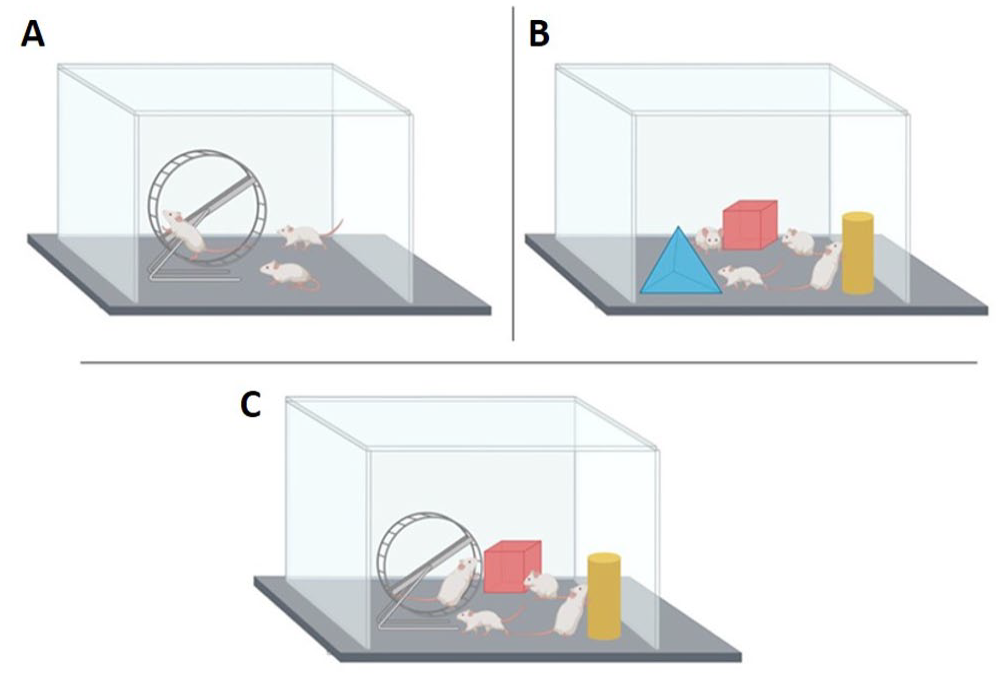
Examples of enriched environment (EE) conditions. (A) Home cage in which a running wheel has been included to enhance physical exercise. (B) Home cage in which different facilitators have been included to induce EE. (C) Home cage in which toys promote exploration and stimulating behaviors, and a running wheel to promote physical exercise, both components of EE.
Nevertheless, the beneficial effects of rearing in an EE have been observed under both normal and pathological conditions. These changes can be divided into two groups. On one hand, short-term changes, also known as molecular effects, are mainly neuroprotective. They are characterized by selective changes in gene expression, showing cellular and spatiotemporal specificity in several classes of gene ontology, growth factor action, and neural transmission.8,9 In fact, EE promotes neuroprotective responses, mainly by increasing neurotrophic factors in different brain areas. 10 On the other hand, long-term changes, known as cellular effects, produce alterations in gene expressions in neurons and glial cells. 11 Specifically, these changes encompass synaptic plasticity,12,13 increase synaptogenesis 14 and adult neurogenesis,15,16 in addition to changes in metabolism, 17 the immune system 18 and the hypothalamic–pituitary–adrenal axis. 19
Therefore, all these modifications induce synaptic structural changes that result in improved learning and memory.20,21 Thus, rearing in EE would be beneficial as a neuroprotective strategy in different brain pathologies. Similarly, in humans, the maintenance of mental health in elderly is promoted by an intellectually, physically, and socially active lifestyle. 22
Brain disorders and enriched environment
Once the beneficial effect of the EE on healthy animals was established, the neuroprotective role of EE has been studied in various animal models of brain diseases over the last two decades.
Regarding neurodegenerative diseases, Alzheimer’s disease (AD) is the leading cause of dementia worldwide. It is caused by both environmental and genetic factors, with neurodegeneration being the ultimate outcome. 23 The pathophysiology of AD includes the formation of both soluble and aggregated forms of amyloid-beta (Ab). These forms of Ab produce neuronal stress and death. 24 Neuronal activity is responsible for modulating beta-amyloid production, and it occurs due to the amyloidogenic processing of amyloid precursor protein (APP). In fact, amyloid plaques and neurofibrillary tangles are the main markers related to AD neuropathology. 25 Several studies have shown that EE-induced cognitive and physical stimulation can slow down the progression of AD and other forms of dementia.26,27 Exposure to EE during 16 weeks has shown beneficial cognitive effects related to recognition, working memory, reference learning, and strategy switching in APPswe (having a mixed C57B6/SJL/SW/B6D2 background) mice model of AD. 28 In addition, it has also been demonstrated that rearing in EE leads to a reduction in amyloid deposits and Ab levels compared with standard housing control mice in APPswe/PS1DE9 transgenic mice (double-transgenic for mutant APP and presenilin 1, a key component of c-secretase). 29
The second most common neurodegenerative disease after AD is Parkinson’s disease (PD) which is characterized by tremors and bradykinesia. 30 The pathophysiology of PD comprise the loss of dopaminergic neurons in the substantia nigra pars compacta (SNpc), triggering reduced dopamine levels in the basal ganglia and the formation of Lewy bodies and Lewy neurites. 31 In this case, the neuroprotective effects of the EE have been studied in different animal models that reproduce PD. Studies based on MPTP (1-methyl-4-phenyl, 6-tetrahydropyridine neurotoxin) PD model have shown that mice exposed to EE are more resistant to MPTP neurotoxicity than control mice. 32 Furthermore, after 28 days of rearing in an EE, increased tyrosine hydroxylase (TH)-positive cells were observed in this mouse model. These results indicate that EE could decrease the loss of dopaminergic neuronal cells in the SNpc and striatum. 33 In 6-hydroxydopamine (6-OHDA) PD models, increased cell proliferation has been observed in the SN after EE rearing. 34 In addition, decreased striatal dopaminergic loss and increased expression of cells positive for ionized calcium binding adaptor molecule 1 (Iba-1), nerve antigen 2 (NG2), and glial fibrillary acidic protein (GFAP) were also described. These results were accompanied by improved motor behavior. 35 Finally, mouse models based on overexpression of α-synuclein have also shown improvements after breeding on EE. Seo et al. 36 described a reduction in oxidative stress and apoptosis, in addition to an improvement of olfactory functions after two weeks of EE exposure.
Despite the fact that translationally to humans is not as simple as in the case of pharmacological treatments, in humans EE is associated with an active lifestyle. Thus, healthy way of life and environmental factors have a direct impact on the cognitive health of the human brain. 37 Activities such as painting, reading, writing, and aerobic physical exercise (running or walking) promote the generation of cognitive reserves in the human brain. 38 In fact, it has been observed that cognitive stimulation generates a greater cognitive reserve, increasing synaptic connections in areas involved in learning and memory, compensating for the pathophysiological mechanisms of dementia before its clinical manifestation 39 and producing a delay in the onset of AD of approximately five years. 40 In addition to cognitive and physical factors, the social component is highly relevant to the risk of suffering from AD. It has been observed that whereas isolation increases the risk of suffering from AD, having a long social life is associated with protection from cognitive decline.41,42
EE can also act as a neuroprotective strategy on those patients who already suffer from dementia. Recently, it has been described that the use of enriched gardens, which are spaces that stimulate both cognition and motor tasks, in addition to giving patients independence in their daily activities, is capable of improving the cognitive and motor skills of these patients. 43
In PD human patients, the active lifestyle produces an improvement in motor, 44 nonmotor and cognitive symptoms. 45 In fact, exercise accompanied by regular physical activity increases the release of neuroprotective factors, 46 reducing the risk of suffering PD.44,47
The EE has also been used as a nonpharmacological neuroprotective strategy in psychiatric disorders. Schizophrenia is a neurodevelopmental disorder that results in abnormal brain development, and it can be caused by both environmental factors and genetic predisposition. 48 Although cognitive and social dysfunctions are observed during childhood, it is in adolescence when the ultra-high risk (UHR) period occurs, and it is during this period that psychosis appears. 49 Several studies have suggested that the cognitive deficits observed in schizophrenia appear due to impaired development of GABAergic systems. 50 Gamma-aminobutyric acid (GABA) maintains the excitatory/inhibitory balance and, therefore, optimizes synapses. 51 This integration of GABAergic activity occurs in the course of adolescence, during the functional remodeling of medial prefrontal cortex (mPFC).51,52 A reduction in GAD67, which is responsible for the synthesis of 90% of GABA, 53 has been described in the hippocampus of animal models of schizophrenia. This reduction was observed especially in Parvalbumin (PV)-positive interneurons, the main subset of GABAergic cells.54,55 In contrast, an increase in GAD67 cells has been found after exposure to EE in MK-801 rat model mimicking schizophrenia. In addition, increased brain derived neurotrophic factor (BDNF) and its receptor TrkB 56 were detected after EE exposure, and therefore, it can be affirmed that EE improves glutamatergic neurotransmission. 57 Other studies have also shown that exposure to stimulating environments after weaning prevents the behavioral and cognitive deficits produced by chronic administration of MK-801.58,59 In our own studies, the exposure to EE in early adulthood for a short period of time (18 days) was found to ameliorate cognitive dysfunction and GABAergic markers deficits in a rat model of schizophrenia. 60 In addition, a shorter exposure to EE (10 days) already showed an improvement in vascular network, neurogenesis, and dendritic complexity in the hippocampus. 61
As previously mentioned, the risk of suffering from schizophrenia because of genetic susceptibility may be enhanced by environmental factors. 62 However, it has been observed that aerobic physical exercise is capable of improving the severity of positive, negative, and general symptoms of patients with schizophrenia, improving their quality of life.63,64 Specifically, there is an improvement on working memory, social cognition, and attention. 65 In addition, after physical exercise also has been reported an increase in peripheral BDNF levels in schizophrenic patients compared with patients receiving regular medication 66 or healthy controls, 67 showing cognitive improvements after this increase. 68
EE has also been studied as a therapy against stroke. Ischemic brain injury is the leading cause for morbidity and mortality worldwide. This pathology is mainly caused by both reduced metabolic capacity and reduced oxygen supply (hypoxia) in the brain. Alteration of mitochondrial membrane potential, formation of free radicals and reactive oxygen species (ROS), and activation of inflammatory pathways are the main pathological processes involved during ischemia. 69 Inflammation occurs because of increased ROS production, leading to oxidative damage of proteins and DNA, and peroxidation of membrane lipids.70,71 Likewise, neuronal injury caused by ischemia is aggravated by mitochondrial dysfunction, as neurons require a high-energy demand. 72 Consequently, neurological dysfunction occurs.70,71 Different studies have demonstrated that rearing in EE mitigates the deleterious effects of ischemia. In fact, it induces neuroprotection through the reduction in oxidative stress 73 and brain inflammation, 74 attenuation of blood–brain barrier dysfunction, 75 and enhanced induction of angiogenesis 76 and neurogenesis. 77 Related to mitochondrial dysfunction, it has been described that EE exposure produces an increase in mitochondrial cytochrome C oxidase subunit IV (COX IV) protein levels in the cortex, showing an indirect measure of mitochondrial biogenesis. 78 Therefore, EE could be beneficial for recovery from ischemia through regulation of mitochondrial biogenesis. 78 Regarding cognitive effects, an improvement in learning and visuospatial memory was also observed after EE breeding for three weeks in a rat model of four-vessel occlusion ischemia. This behavioral improvement is accompanied by a reduction in degeneration and a decrease in caspase-3 expression in the hippocampus, so that EE would be mediating a reduction in apoptosis. 79 On the contrary, increased expression of vascular endothelial growth factor (VEGF) and its receptor, whose main function is angiogenesis, 80 has also been described after breeding in an EE for 14 days in an animal model of ischemia. Increased angiogenesis leads to improved neuronal function. 81 Furthermore, rearing in EE also increases the expression of endothelial nitric oxide synthase (eNOS) through CD34-positive endothelial progenitor cells in middle cerebral artery occlusion (MCAO) rat model. 82 eNOS, together with VEGF, is related to the regulation of angiogenesis and vascular function through nitric oxide synthesis. 83 Therefore, EE breeding has neuroprotective effects by improving local microcirculation after ischemic brain injury. 84
In human patients, the practice of physical exercise after stroke produces axonal growth and improved activity of the pyramidal and extrapyramidal systems in both hemispheres, through functional compensation in unaffected areas of the brain involved in limb activity. 85 In addition, the effect of intense training of the lower extremities promoting an improvement and recovery of the corresponding brain areas in patients with acute ischemic stroke has also been described. 86 Finally, physical exercise induces an improvement in motor function, improving both balance and gait in the subacute phase of stroke patients. 87
Another cause of brain pathology is traumatic brain injury (TBI), which affects 10 million people a year worldwide. 88 TBI can be caused by numerous different events, such as motorbike accidents, falls, firearms, and high-impact sports, among others. Consequently, neurobehavioral and cognitive dysfunction occurs 89 which, in turn, leads to psychiatric comorbidities. 90 The pathophysiology of TBI consists mainly of anxiety, depression, and memory problems, 91 produced by hypoxia, neuroinflammation, edema, metabolic changes, and gliosis. 92 EE also has been found to achieve favorable effects after TBI. 93 Twenty-one days of EE exposure improved motor function and spatial learning acquisition, in addition to reduce the volume of cortical lesion in a controlled cortical impact (CCI) rat model of TBI. 94 Other authors have described an improvement in recognition memory and short-term special memory after six weeks of breeding in EE in a mice model of TBI. 95 Similar results compared with continuous rearing were described in motor behavior and spatial learning after rearing in EE for 6 h per day for 19 days. 96 Therefore, this result is consistent with clinical rehabilitation performed in TBI patients. 96
Neurorehabilitation is currently the most promising approach to recover some functions and autonomy in patients who have suffered TBI. 97 Thus, it was observed that after performing aerobic exercise for 12 weeks, TBI patients showed improvements in cognitive function, especially in processing speed, executive functioning, and general cognitive function. 98
Enviromimetics: drugs that mimic or enhance the beneficial effects of cognitive activity and physical exercise
The use of EE as a rehabilitation therapy in combination with treatments has been studied for its clinical approach. Physical activity-induced cellular and molecular processes can be enhanced by an “exercise mimetic.” The most popular molecular target for enviromimetic drugs is BDNF which mediates experience-dependent cellular plasticity in both the developing and adult nervous system. 99 Increased expression of this neurotrophin has been observed following physical exercise, 100 learning, 101 and EE. 102 In addition, BDNF is responsible for synaptic plasticity.
Related to this idea, researchers have found that drugs that up-regulate BDNF also induce positive effects on cellular plasticity. Thus, the most commonly used antidepressants are selective serotonin reuptake inhibitors (SSRIs), and it is known that elevate hippocampal BDNF levels. 103 In animal models of neurodegenerative diseases such as AD and PD, beneficial effects have been described after an increase in BDNF levels. 104 In addition, specific histone deacetylase (HDAC) inhibitors also improve cellular plasticity, increase BDNF expression, and show advantageous effects in animal models of neurodegenerative diseases. 105 Thus, HDACs and SSRIs are drugs that act as enviromimetics and enhance endogenous brain repair, neuroprotection, boost cell survival and differentiation when co-administered with cell therapies. Therefore, by combining these drugs with EE, a synergistic brain-enhancing effect can be achieved for maximum therapeutic boost. 106
On the contrary, Magnesium-L-threonate (MgT) administration is able to improve spatial memory and recognition in an APPswe/PS1 mouse model of AD. 107 AD is characterized by synaptic loss in the hippocampus 108 and reduced activity of the Ca2+/calmodulin-dependent protein kinase II (CaMKII) molecule, which is important in the N-methyl-D-aspartate receptor (NMDAR) signaling pathway and is involved in memory formation. 109 After the synergistic combination of EE with MgT, a reduction in synaptic loss of the hippocampus was observed, accompanied by increased CaMKII activity. Therefore, MgT could increase the effects of breeding in EE to prevent and reverse neuronal deficits in mice with AD. 110 Another alternative is the use of memantine, which is a drug that blocks NMDA receptors. 111 The combined therapy of this drug with EE showed an improvement in the spatial memory of SAMP8 mice compared with any individual treatment, in addition to a reduction in the expression of APP. 112
In PD animal models, Cerebrolysin (CBL) has been tested, a drug capable of improving motor alterations, maintaining dopaminergic levels, and reducing oxidative stress. 113 The combined administration of this drug with EE showed improvements at the motor, morphological, and molecular level in 6-OHDA PD model. 114 Specifically, there was a 14% reduction in the retraction of the injured striatal hemisphere,114,115 diminished reduction in cell density in the SN, and increased release of neurotrophins related to cell survival. 114
Furthermore, resveratrol 116 or caffeine and theanine 117 were administered in combination with EE showing improvements in mobility and Bederson scale in a rat cerebral ischemia model induced by MCAO. These effects were improved when rats were exposed to EE immediately after MCAO injury, rather than two days after surgery, enhancing the EE-induced neuroprotective effect. In addition, researchers have found that combined therapy with EE and resveratrol increases the number and activity of antioxidants, improving oxidative stress during ischemia. 69 This combined therapy also leads to a reduction in hydrogen peroxide. 118
In TBI, drugs targeting the serotonin 1A (5-HT1A) receptor are used since they generate motor and cognitive improvement, accompanied by a neuroprotective effect, and decreased volume of the lesion. 119 It has been described that the combination of these drugs with EE in a rat model of TBI achieved cognitive and motor recovery, attenuated hippocampal injury (conferring histological protection), and loss of medial septal cells. 120 In addition, acetylcholinesterase inhibitor drugs increase the availability of acetylcholine in postsynaptic receptors, mitigating the memory problems suffered by these patients. 121 On the contrary, combination of galantamine (an acetylcholinesterase inhibitor) and EE resulted in an additive effect that improved functional motor skills and the acquisition of spatial learning in a rat model of TBI. 122 In addition, these patients present agitation and aggressiveness, 123 for which they are treated with antipsychotics that generate deleterious effects on cognitive function. 124 It has been observed that the combination with EE minimizes the negative effects of the drug in a rat model of TBI. 125
Considering all the abovementioned results, future work should continue the search for drugs that mimic the effects observed after physical exercise or exposure to EE to obtain a greater synergistic effect of the neuroprotective effects induced by EE.
Conclusions
Both laboratory animal experiments and clinical studies have shown that the environment is crucial in brain plasticity for normal neural development of the brain and, in turn, is highly relevant in the repair of neurological disorders. EE not only modifies basic brain anatomy, but also modulates electrophysiology, neurotrophic factors, and cognitive capacity. At the same time, the multidimensional effect of EE on nerve repair in central nervous system (CNS) lesions has allowed researchers to search for therapeutic approaches that do not rely on drugs. However, these positive effects of EE can be implemented with other therapies such as pharmacological therapies to see possible synergies. Several studies mentioned in this work have shown that these beneficial effects are enhanced in the case of dual treatment. Although the exact mechanisms by which EE shows beneficial effects have not been fully elucidated, its neuroprotective effect is becoming increasingly well-known in both physiological conditions and brain pathologies. Further study of EE-induced mechanisms from a basic point of view will allow a better understanding for their future use in human application.
Footnotes
Acknowledgements
Andrea Vaquero-Rodríguez is financed by a predoctoral fellowship of the University of the Basque Country (UPV/EHU).
Authors’ Contributions
JVL designed the project. AV-R with the collaboration of HB and NO researched data for the article and bibliography. Figure was designed by AV-R in collaboration with NO and HB. AV-R wrote the first draft. NO, HB, and JVL contributed to discussion of the content, reviewed, and edited this paper before submission. All authors have read and agreed to the published version of the manuscript due to the knowledge and experience in the study of the depressive disorders.
Declaration of Conflicting Interests
The author(s) declared no potential conflicts of interest with respect to the research, authorship, and/or publication of this article.
Funding
The author(s) disclosed receipt of the following financial support for the research, authorship, and/or publication of this article: Grant PID2 021-126434OB-I00 funded by MCIN/AEI/ 10.13039/501100011033 and ERDF A way of making Europe. It has also been funded by the Basque Government (IT1706-22, PUE21- 03, PIBA_2020_1_0048) and the UPV/EHU (COLAB20/07). This research was conducted in the scope of the Transborder Joint Laboratory (LTC) “non-motor Comorbidities in Parkinson’s Disease (CoMorPD)”.