



Abstract
Marin-Valencia I, Kocabas A, Rodriguez-Navas C, Miloushev VZ, González-Rodríguez M, Lees H, Henry KE, Vaynshteyn J, Longo V, Deh K, Eskandari R, Mamakhanyan A, Berishaj M, Keshari KR. Cell Metab. 2024 Jun 4;36(6):1394-1410.e12. doi: 10.1016/j.cmet.2024.05.002. PMID: 38838644; PMCID: PMC11187753. A vexing problem in mitochondrial medicine is our limited capacity to evaluate the extent of brain disease in vivo. This limitation has hindered our understanding of the mechanisms that underlie the imaging phenotype in the brain of patients with mitochondrial diseases and our capacity to identify new biomarkers and therapeutic targets. Using comprehensive imaging, we analyzed the metabolic network that drives the brain structural and metabolic features of a mouse model of pyruvate dehydrogenase deficiency (PDHD). As the disease progressed in this animal, in vivo brain glucose uptake and glycolysis increased. Propionate served as a major anaplerotic substrate, predominantly metabolized by glial cells. A combination of propionate and a ketogenic diet extended lifespan, improved neuropathology, and ameliorated motor deficits in these animals. Together, intermediary metabolism is quite distinct in the PDHD brain—it plays a key role in the imaging phenotype, and it may uncover new treatments for this condition.
Commentary
Until now, it was debated whether propionate, a metabolite produced by the gut microbiome, was metabolized in brain, 1 though it had been shown to be neuroprotective and anticonvulsant (see references by Marin-Valencia et al 2 ). Using mice deficient in brain pyruvate dehydrogenase (PDH), which have an increased need for auxiliary brain fuel, Marin-Valencia et al (2024) elegantly showed that propionate is indeed metabolized and anaplerotic in brain.
Mice lacking PDH in most glial and neuronal cells were employed, which similar to human PDH deficiency, 3 exhibited neuronal cell death, astrogliosis, neuroinflammation, and seizures.2,4 Metabolic analysis of PDH-deficient brain showed several expected changes, including high lactate levels and impaired entry of glucose-derived carbons into the Krebs cycle (Figure 1, red arrows). However, in PDH-deficient brains, unlabeled quantities of certain Krebs cycle intermediates and metabolites were higher than others as compared to wild-type. This led to the discovery that 13C-propionate entered the Krebs cycle as succinate, and was metabolized to a much greater extent in PDH-deficient as compared to wild-type brains (Figure 1B, white arrow and boxed and underlined blue metabolites). Based on 13C-propionate's increased metabolism into glutamine versus glutamate, it appeared to be metabolized preferentially in astrocytes, which aligns with earlier findings. 1 Astrocytes from PDH-deficient brains also showed higher transcript levels of enzymes needed to convert propionate into succinyl-CoA, consistent with this cell type being a major site of propionate metabolism in brain. Finally, propionate showed a “glucose sparing” effect, reducing lactate production from glucose. This suggests that to some extent, propionate may reduce the need for glucose as fuel in PDH deficiency.
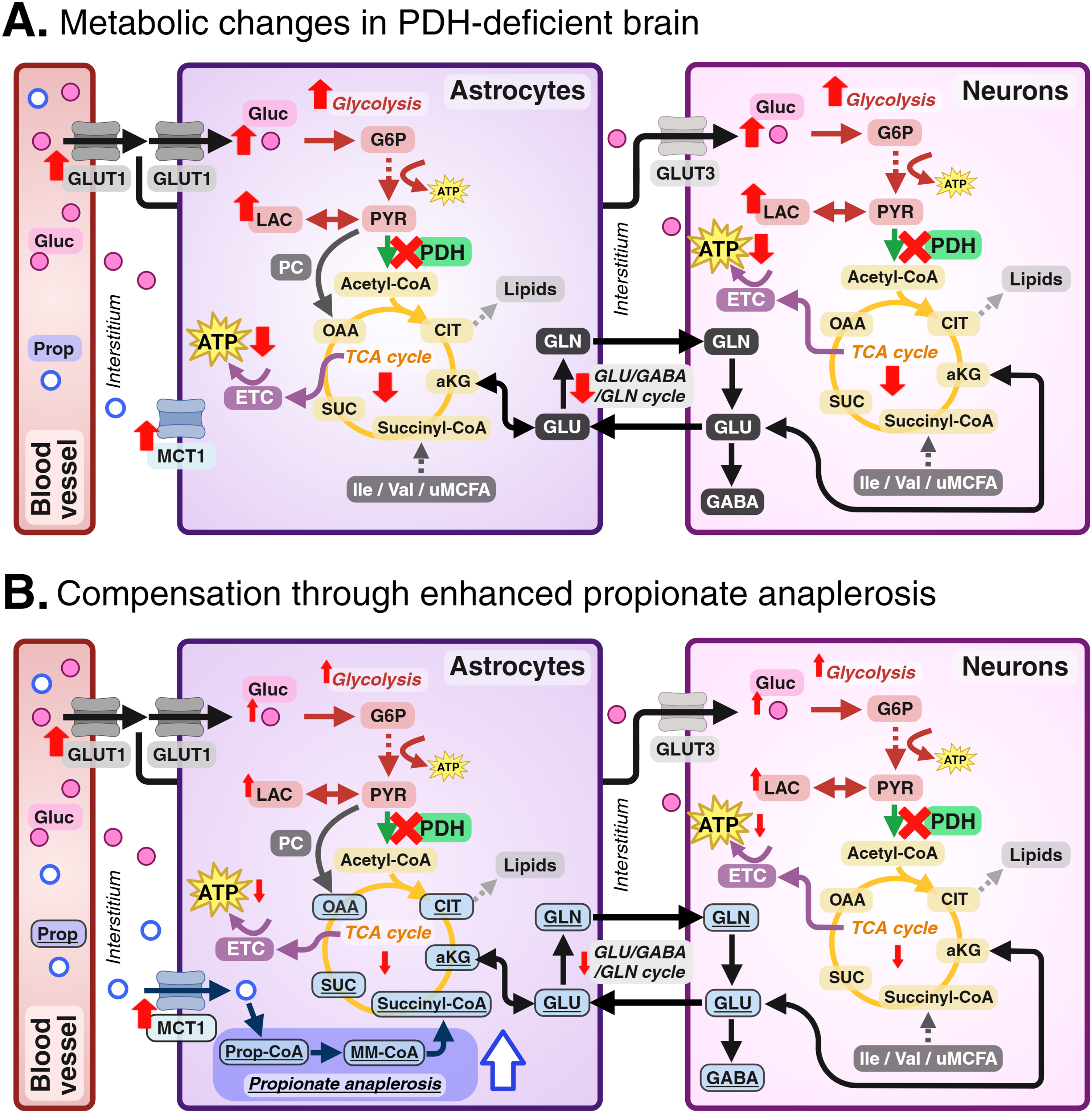
Simplified interpretation of the findings by Marin-Valencia et al. 2 (A) Metabolic deficits occurring in PDH-deficient mouse brain (red arrows). (B) PDH-deficient mice compensate for these changes through increased propionate anaplerosis (in blue, boxed and underlined), which is proposed to be a mechanism to overcome some of these metabolic deficits. Created in BioRender. Borges, K. (2024) BioRender.com/t72y168. Abbreviations: ATP, adenosine triphosphate; CIT, citrate; ETC, electron transport chain; Gluc, glucose; G6P, glucose-6-phosphate; GABA, γ-aminobutyric acid; GLU, glutamate; GLN, glutamine; GLUT1, glucose transporter 1; GLUT3, glucose transporter 3; Ile, isoleucine; LAC, lactate; MCT1, monocarboxylate transporter 1; MM-CoA, methylmalonyl-CoA; OAA, oxaloacetate; PC, pyruvate carboxylase; PDH, pyruvate dehydrogenase; Prop, propionate; Prop-CoA, propionyl-CoA; PYR, pyruvate; SUC, succinate; TCA, tricarboxylic acid; uMCFA, uneven medium chain fatty acid; Val, valine; α-KG, alpha ketoglutarate.
PDH-deficient mice were treated with (1) propionate (in drinking water), (2) propionate plus a ketogenic diet, or (3) a ketogenic diet alone, the current standard of treatment in people with PDH deficiency (see references by Marin-Valencia et al 2 ). Both ketogenic diet with and without propionate extended the lifespan of PDH-deficient mice. This coincided with reduced neuronal cell death and/or astrogliosis in several brain regions (except hippocampus), which also occurred in mice given propionate alone, despite no improvement in survival. Only mice receiving ketogenic diet plus propionate showed a statistical improvement in motor function.
This paper is important for 2 major reasons. Propionate was (a) anaplerotic and (b) therapeutic alone and when combined with ketogenic diet in PDH-deficient brains.
Propionate was a better source of anaplerotic succinate and glutamine in PDH-deficient compared to wild-type brains. When oxidizing glucose, carbons are lost from the Krebs cycle providing the backbones of lipids and amino acids. Anaplerotic substrates are important as they replenish carbons in the Krebs cycle, allowing it to run efficiently and ensure adequate ATP production. In brain, anaplerosis occurs in astrocytes mostly via carboxylation of pyruvate to the Krebs cycle intermediate oxaloacetate (Figure 1, PC, pyruvate carboxylase). The other anaplerotic pathway from valine, isoleucine, or rare uneven medium chain fatty acids via propionyl-CoA is less used. Metabolism of propionate was increased in PDH-deficient brains compared to wild-type, highlighting that during energy deficiency, brain can increase the use of propionate as substrate. Bolstering brain propionate supply in PDH deficiency is thus a potentially useful approach to ensure that propionate amounts available for metabolism and anaplerosis are nonlimiting. The ideal propionate amounts remain to be investigated.
In PDH deficiency, there is an obvious need for substrates other than pyruvate and potentially also anaplerosis, as the overall pool sizes of some, but not all, Krebs cycle intermediates and derived amino acids were low. On the other hand, it is difficult to evaluate to which extent propionate just serves as a substrate to provide extra fuel, as the activity of the main astrocytic anaplerotic enzyme, pyruvate carboxylase, should not be impaired in PDH deficiency.
A ketogenic diet with and without propionate extended lifespan and reduced neuropathological changes in PDH-deficient mice. Propionate monotherapy did not enhance survival or motor function, but was neuroprotective. These effects are consistent with earlier studies that found propionate to be anticonvulsant and confer anti-inflammatory effects in brain (see references by Marin-Valencia et al 2 ). Based on the findings presented here, 2 enhanced anaplerosis is an attractive explanation for the effects previously published. Anaplerotic therapies have been employed previously in other conditions associated with brain energy deficits, including deficiencies of Glut1 and pyruvate carboxylase.5,6 However, to be certain that enhanced anaplerosis is the mechanism responsible for the effects of exogenous propionate, future experiments that specifically inhibit propionate anaplerosis in PDH-deficient brains are required. As it is unlikely that propionate can provide enough fuel in PDH deficiency in the absence of a ketogenic diet (plasma concentrations of propionate are expected to be ∼50 µM, as opposed to ∼1-4 mM ketone body levels), the lack of effects of propionate alone on body weight, motor function, and survival is not surprising. We cannot comment on potential differences in the causes of mortality in these mice on different treatments, as no information was given.
Interestingly, the phenotype of PDH-deficient mice resembles that of mice with pilocarpine-status epilepticus-induced chronic epilepsy, which display low PDH activity and similar neuropathological changes including neuronal cell death, astrogliosis, and neuroinflammation (see McDonald et al 7 and references therein). This model of human temporal lobe epilepsy has responded well to triheptanoin, 7 which like propionate, is anaplerotic via metabolism of propionyl-CoA into succinyl-CoA. 8 Thus, propionate supplementation may be useful beyond PDH deficiency, in other epilepsies showing brain energy deficits. The doses of propionate used here 2 approximately doubled plasma propionate levels like what was seen in humans switching between low and high fiber diets. 9 Thus, modulating fiber intake could be one method of raising propionate levels in people.
An alternative explanation of the 13C-propionate experiments is that propionate can be gluconeogenic in liver, and can thus produce 13C-labeled glucose that could, via metabolism into 13C-pyruvate and subsequent carboxylation, be incorporated into the Krebs cycle and give rise to the reported changes. Comparison and modeling of 13C-glucose versus 13C-propionate metabolism showed that different labeling patterns in aspartate, glutamate, and glutamine arise from these 2 labeled substrates (see Figures S10 and S11 by Marin-Valencia et al 2 ). The authors claim that this indicates that the 13C-labeling in these amino acids was produced directly from 13C-propionate, especially in the PDH-deficient brains, where higher levels of this labeling were seen. Another limitation is that brains were not collected using methods that prevent postmortem changes, like head-focused microwave fixation. 10 Thus, lactate production from glucose is high, and the possibility of postmortem 13C-propionate incorporation cannot entirely be eliminated. Despite this, the difference in propionate metabolism between PDH-deficient and wild-type brains remains convincing.
There is some evidence in the paper that propionate may be anaplerotic in healthy brain also, although this was not discussed. In wild-type mice, there was 30% to 50% incorporation of 13C derived from 13C-propionate into the Krebs cycle-derived amino acids, glutamate, glutamine, and aspartate (Figure 6F by Marin-Valencia et al 2 ). Moreover, after propionate addition, lactate production from 13C-glucose was reduced by 30% (Figure S8D by Marin-Valencia et al 2 ). Hence, the effects of anaplerosis by propionate in healthy brain may be substantial, although gluconeogenesis from propionate in the liver may also at least partially explain the results.
In conclusion, Marin-Valencia et al (2024) discovered a new anaplerotic substrate for brain astrocytes, laying a foundation for new therapies based on propionate. It remains to be seen to which extent propionate is used in other conditions with energy deficits and in healthy brain.
Footnotes

Declaration of Conflicting Interests
The author(s) declared no potential conflicts of interest with respect to the research, authorship, and/or publication of this article.
Funding
The author(s) disclosed receipt of the following financial support for the research, authorship, and/or publication of this article: ESN was supported by an Australian Government Research Training Program (RTP) Scholarship.