
Abstract
This study aimed to design and develop flurbiprofen-loaded chitosan nanoparticles for ophthalmic drug delivery, with the objectives of enhancing formulation stability, sustaining drug release, and improving patient comfort. This study introduces an optimized chitosan-based nanoparticle system for ocular delivery of flurbiprofen, achieving high encapsulation efficiency, physiological compatibility, and sustained release for 20 h. The findings demonstrate a practical, patient-friendly approach that enhances drug bioavailability and stability compared with conventional eye drops. Flurbiprofen-loaded nanoparticles were prepared using gelation and optimized through a Box–Behnken design. The influence of formulation variables (0.1%–0.3% w/v chitosan concentration, 0.2–0.6 mL/min dropping rate, and 500–900 rpm mixing speed) was assessed. The optimized nanoparticles were evaluated for particle size, polydispersity index (PDI), zeta potential, pH, osmolarity, and entrapment efficiency (%). The optimized formulation was achieved at 0.2% (w/v) chitosan concentration, a dropping rate of 0.4 mL/min, and a mixing speed of 700 rpm. The nanoparticles exhibited a particle size of 110.0 ± 2.20 nm, PDI of 0.347 ± 0.03, and zeta potential of + 15.4 ± 3.8 mV. The entrapment efficiency was 80.89%. The formulation was adjusted to physiological conditions (pH 6.7, osmolarity 300 mOsm/kg). In vitro studies showed a controlled release of flurbiprofen over 20 h. The optimized chitosan-based nanoparticle system demonstrated favorable physicochemical stability, high encapsulation efficiency, and sustained release of flurbiprofen. Compared with conventional ocular formulations, this structured nanoparticle delivery system offers superior drug retention and extended therapeutic action.
INTRODUCTION
Topical ocular drug delivery faces significant challenges due to the complex anatomical and physiological barriers of the eye. Tear turnover, reflex blinking, and nasolacrimal drainage rapidly eliminate most of the administered drug, resulting in <5% reaching intraocular tissues. In addition, the low permeability of the corneal epithelium and enzymatic degradation mechanisms further restrict ocular bioavailability. Consequently, frequent instillation of high concentrations is often required, which may reduce patient compliance and increase the risk of ocular irritation.1,2
Non-steroidal anti-inflammatory drugs (NSAIDs) play a key role in controlling ocular inflammation by inhibiting prostaglandin synthesis. They are widely used in the management of postoperative inflammation, allergic conjunctivitis, and prevention of cystoid macular edema. However, conventional NSAID eye drops are hindered by their short precorneal residence time and limited corneal penetration, resulting in suboptimal therapeutic efficacy.3,4
To overcome these limitations, nanoparticle-based drug delivery systems have been developed. Owing to their small size, nanoparticles closely interact with the corneal and conjunctival epithelium, exhibit mucoadhesion, prolong precorneal retention, and provide controlled drug release, thereby reducing dosing frequency. They also protect the encapsulated drug from enzymatic degradation, enhancing therapeutic effectiveness. Among biocompatible and mucoadhesive polymers, chitosan stands out in ocular applications due to its ability to transiently open tight junctions and improve transcorneal transport. 5
Among nanoparticle preparation methods, the “dropping technique” has attracted attention because of its simplicity, reproducibility, and ability to generate particles with narrow size distribution. This method allows homogeneous nanoparticle formation under controlled addition and stirring conditions, avoids the use of harsh solvents, and ensures better safety for bioactive compounds. These advantages make it a suitable approach for both laboratory research and potential industrial translation.6,7
In this study, flurbiprofen-loaded chitosan nanoparticles were optimized using a Box–Behnken experimental design (BBED). The aim was to prolong ocular residence time, improve corneal penetration, and achieve controlled release to enhance bioavailability. Moreover, physiological pH and osmolality adjustments were considered to improve patient comfort. This study, therefore, provides a comprehensive and innovative approach to enhancing the ocular therapeutic efficacy of NSAIDs.
MATERIALS AND METHODS
Materials
Flurbiprofen, the active pharmaceutical ingredient employed in this study, was kindly gifted by Deva Pharmaceutical Industry Co. (İstanbul, Turkey). Chitosan, exhibiting a molecular weight between 50 and 150 kDa and a deacetylation degree of at least 75%, was obtained from Sigma-Aldrich. Tween® 80, sodium chloride, and glacial acetic acid (≥99%) were purchased from Merck KGaA (Darmstadt, Germany). Sodium hydroxide solution (1 N), used for pH adjustments, was also obtained from Sigma-Aldrich. A 0.1% (v/v) aqueous acetic acid solution was freshly prepared as the solvent medium for chitosan. All reagents employed in the formulation and analysis phases were of analytical grade. Throughout the study, freshly distilled water was utilized in all preparations.
Methods
Drug–excipient compatibility study
FT-IR spectral analysis
FT-IR analysis (PerkinElmer Spectrum 100, USA) was employed to evaluate the chemical integrity of the active pharmaceutical ingredient (flurbiprofen) and to investigate potential interactions between flurbiprofen and chitosan in the physical blends (1:1, w/w). Infrared spectra of pure flurbiprofen, pure chitosan, and their binary mixtures were recorded using potassium bromide (KBr) disk method within the spectral range of 4,000–550 cm−1. 8
Preparation of flurbiprofen-loaded chitosan nanoparticles
Flurbiprofen-loaded chitosan nanoparticles were prepared via a modified ionic gelation technique utilizing the dropping method. In this method, instead of using a commonly employed anionic crosslinker such as sodium tripolyphosphate (TPP), nanoparticle formation occurs through electrostatic interactions between the protonated amino groups of chitosan (–NH3+) and the deprotonated carboxylate groups of flurbiprofen (–COO−). Under mildly acidic conditions (pH ≈ 6.0–6.5), chitosan becomes partially protonated, while flurbiprofen, due to its weakly acidic nature, is ionized and forms negatively charged aggregates. The interaction of these opposite charges enables the self-assembly of chitosan–drug complexes, leading to the formation of stable nanostructures without the need for an external crosslinker. Moreover, Tween® 80 supports this process by reducing surface tension and preventing irregular aggregation. Therefore, the term modified ionic gelation refers to this specific mechanism in which the drug molecule itself acts as the anionic counter-ion responsible for nanoparticle formation. Initially, chitosan was dissolved in 0.1% (v/v) acetic acid solution under constant magnetic stirring at room temperature to obtain a clear polymeric solution. In a separate beaker, flurbiprofen (0.1% w/v) was dispersed in distilled water containing Tween® 80 (0.2% v/v) to improve its wetting and dispersion characteristics. The drug-containing phase was then added dropwise into the chitosan solution using a syringe pump at a controlled rate of 0.1–1.0 mL/min, while the mixture was stirred continuously at 500–1000 rpm.8,9
Nanoparticle formation occurred spontaneously because of ionic interactions between the positively charged chitosan and the negatively charged drug aggregates stabilized by the surfactant. The suspension was stirred for an additional 24 h to ensure uniform nanoparticle formation. The pH was adjusted to 6.5 using 1 N NaOH solution. Formulations were centrifuged at 15,000 rpm for 30 min and washed with distilled water to remove unbound materials before being stored at 4°C for further analysis. 10
Formulation optimization via design of experiments
To investigate the influence of formulation and process parameters on critical quality attributes, a Design of Experiments approach was employed. A three-factor, three-level Box-Behnken design was applied using Design-Expert® software (Stat-Ease Inc., Minneapolis, MN, USA). The independent variables included chitosan concentration (w/v %), dropping rate (mL/min), and stirring speed (rpm), while the dependent responses were particle size (nm) and entrapment efficiency (EE) (%).11,12 Experimental runs were generated, and the formulations were prepared according to the design matrix. The results were analyzed using response surface methodology to evaluate the main effects, interaction effects, and quadratic effects of the variables. Optimal formulation conditions were determined based on numerical desirability criteria. 13
Particle size, polydispersity index, and zeta potential
The mean particle size, polydispersity index (PDI), and zeta potential of the prepared nanoparticles were measured using dynamic light scattering analysis with a Zetasizer Nano ZS (Malvern Instruments, UK). Formulations were diluted appropriately with distilled water prior to measurement to prevent multiple scattering. Each sample was analyzed in triplicate at 25°C. The PDI values were used to evaluate the size distribution, and zeta potential provided information on the colloidal stability of the nanoparticles. 14
Entrapment efficiency (%).
To determine the EE, nanoparticle suspensions were centrifuged at 15,000 rpm for 30 min, and the free flurbiprofen in the supernatant was quantified using a UV–Vis spectrophotometer at 247 nm. 15
EE% was calculated using the formula:

Osmolality measurement
Osmolality measurement of the optimized flurbiprofen–chitosan nanoparticle formulations was performed using a freezing point depression osmometer (Advanced Instruments, Model 3250, USA). Approximately 50 µL of each formulation was placed in the sample holder, and osmolality was measured in triplicate at 25 ± 1°C. The osmometer was calibrated prior to analysis with standard solutions of 100, 290, and 1,000 mOsm/kg to ensure accuracy. The obtained values were directly compared with the physiological osmolality range of tear fluid (290–310 mOsm/kg).16,17
Determination of pH
The pH of the flurbiprofen-loaded chitosan nanoparticle formulations was measured using a calibrated digital pH meter (Ohaus, USA) at room temperature (25 ± 1°C).
Each sample was analyzed in triplicate, and the average value was recorded. The pH values were assessed to ensure compatibility with the physiological pH range of tear fluid (approximately 7.0–7.4), which is critical for minimizing ocular irritation upon topical administration.16,18
Sterilization
In ophthalmic formulations, sterilization is as important a parameter as pH and osmolarity. In this study, sterilization by filtration was preferred due to its practicality and the possibility that the formulation may be sensitive to heat. For this purpose, membrane filters with a pore size of 0.22 µm were used.
In vitro drug release study
The in vitro release profile of flurbiprofen from chitosan-based nanoparticles was evaluated using the dialysis bag diffusion technique, which simulates the controlled release environment relevant to ocular applications. A cellulose membrane dialysis bag with a molecular weight cut-off of 12–14 kDa was used as the semi-permeable barrier. Prior to use, the dialysis membrane was soaked in distilled water for 12 h to ensure flexibility and permeability. A measured volume (2 mL) of the nanoparticle suspension was carefully introduced into the dialysis bag and sealed tightly to prevent leakage. The bag was then immersed in 50 mL of phosphate-buffered saline (PBS, pH 7.4) maintained at 37 ± 0.5°C in a beaker placed on a magnetic stirrer operating at 100 rpm to mimic ocular surface dynamics. At predetermined time intervals (0.5, 1, 2, 4, 6, 8, 12, and 24 h), 5 mL samples were withdrawn from the external release medium and replaced immediately with an equal volume of fresh PBS to maintain sink conditions. The concentration of flurbiprofen in each sample was quantified using a UV–Vis spectrophotometer at a wavelength of 247 nm. Cumulative drug release was calculated and plotted as a function of time. This method provided insight into the sustained release behavior of the nanoparticle formulation and was used as a basis for subsequent kinetic modeling. 19
Stability studies
The stability of the optimized formulation was assessed over a 3-month period by storing it in a stability chamber at 40°C and 75% relative humidity (RH), in accordance with ICH guidelines. At predefined time intervals, the formulation was evaluated in terms of its physical appearance, pH value, and active pharmaceutical ingredient (API) content (n = 3). 20
Statistical evaluation
The formulation data were analyzed using regression modeling and one-way analysis of variance (ANOVA) with Design-Expert® software (Version 12.0.0). Additionally, experimental results obtained from various studies were statistically evaluated through one-way ANOVA. A p-value of <0.05 was considered to indicate a statistically significant difference between groups.
RESULTS
FT-IR Spectral Analysis
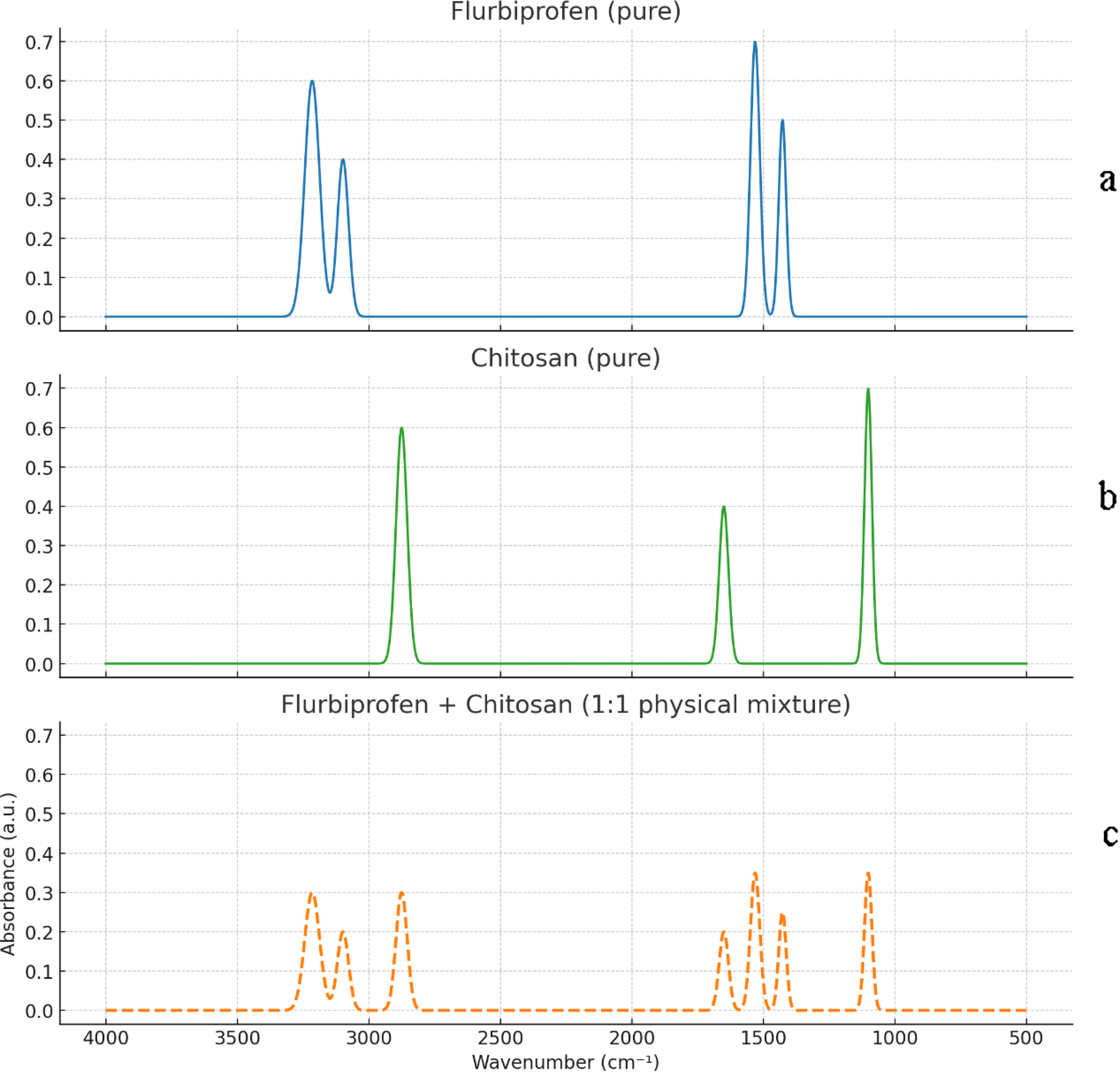
The FT-IR spectra illustrated in Figure 1 represent the pure active pharmaceutical ingredient (flurbiprofen), chitosan, and their physical mixture at a 1:1 (w/w) ratio. The spectral analysis concentrated on identifying the characteristic absorption bands of flurbiprofen, including CH2 stretching vibrations observed at 1427.6 cm−1, C = N stretching at 1,531 cm−1, C = CH stretching around 3098.4 cm−1, and C–H stretching near 3214.7 cm−1. Chitosan exhibited its own distinctive peaks, notably the C–O–C asymmetrical stretch at 1101.3 cm−1 and C–H stretching vibrations at 2875.4 cm−1. In the spectrum of the physical blend, a combination of signals corresponding to both components was detected.
FTIR spectra of
Box–Behnken Design: Data Evaluation and Optimization
Table 1 summarizes the 17 distinct formulations along with their respective response values, as designed within the BBED matrix using Design-Expert® software (Version 12.0.0, Stat-Ease Inc., USA).
Independent and Dependent Parameters Used in BBED, Along with Their Experimental Responses
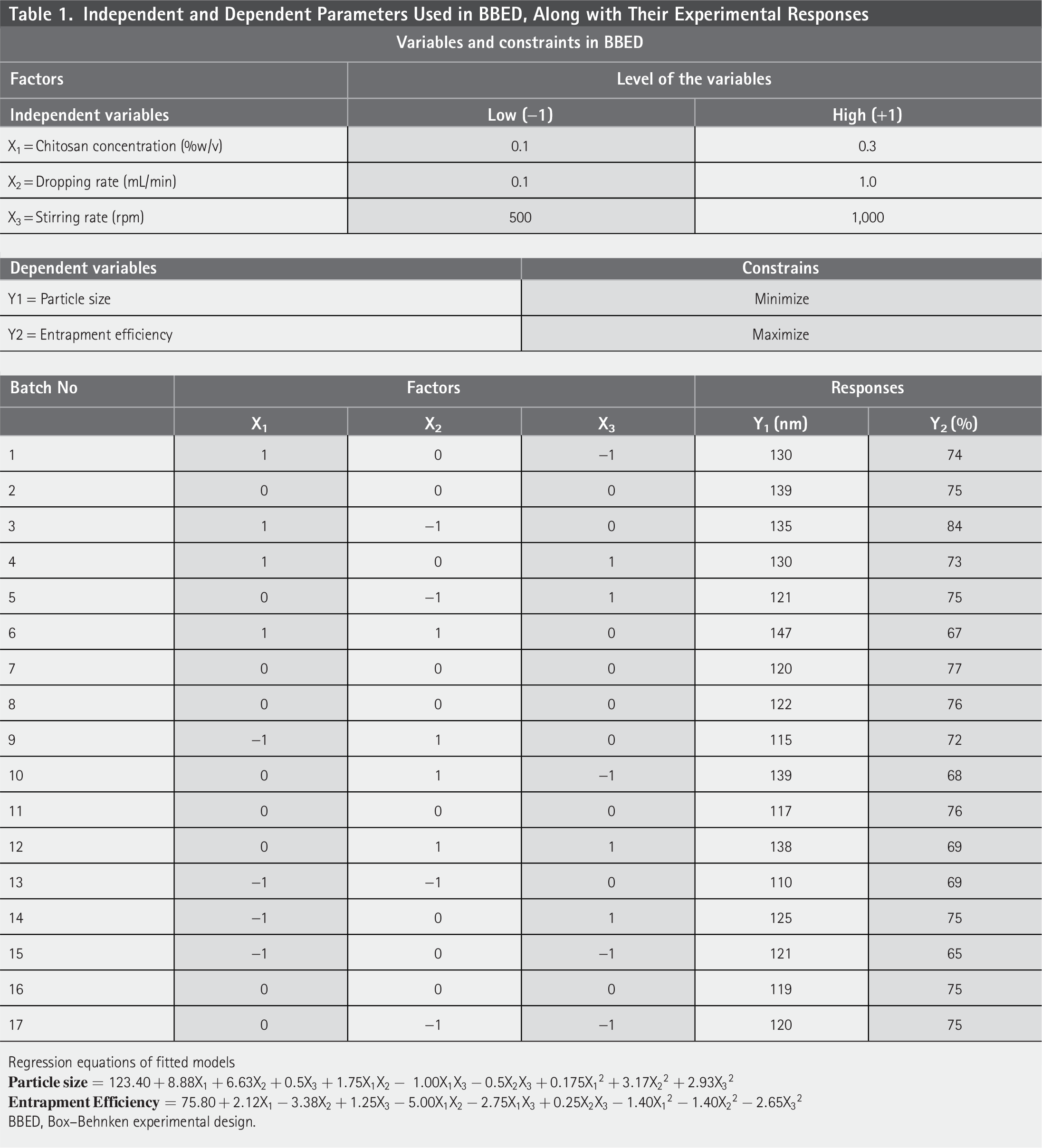
Regression equations of fitted models
BBED, Box–Behnken experimental design.
Based on the statistical parameters provided by the Design-Expert® software, including p-values with a significance threshold of p < 0.05, a suitable polynomial model was selected to describe the relationship between the critical formulation factors and their interactions. The coefficients of the resulting model reflected both primary effects and potential synergistic or antagonistic interactions influencing the response variables. The three-dimensional response surface plots illustrated in Figure 2 visually represent the influence of independent variables on the measured outcomes.

3D graphics demonstrating the effect of chitosan concentration, stirring rate, and dropping rate on particle size (micrometer) and entrapment efficiency (EE%).
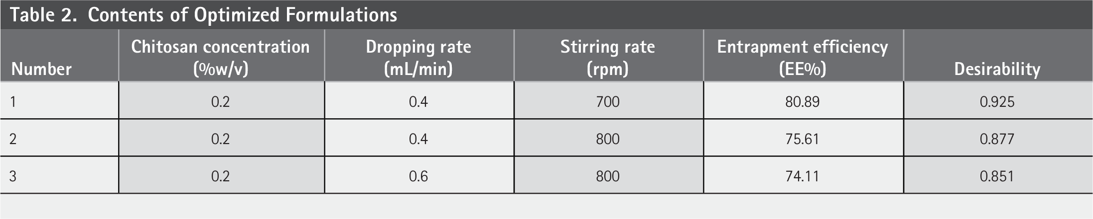
To determine the optimal formulation, the desirability function approach was applied by defining a specific design range. The desirability index varies between 0 and 1, where values closer to 1 reflect a higher degree of agreement with the targeted formulation criteria. 21 For validation experiments the software generated a total of 37 formulation solutions but the top 3 were shown. The optimized formulation identified through this method were subsequently prepared and subjected to experimental validation. As presented in Table 2, the experimental outcomes were in close agreement with the predicted values, falling within the 95% confidence interval, thereby confirming the reliability and predictive accuracy of the selected model.
Contents of Optimized Formulations
Evaluation of Optimized Formulation
Particle size, PDI, and zeta potential
The formulation containing 0.2% chitosan (w/v) and prepared with a dropping rate of 0.4 mL/min and a stirring rate of 700 rpm was accepted as the optimized formulation. The optimized nanoparticulate formulation demonstrated a mean particle size of 110.0 ± 2.20 nm, placing it well within the nanoscale range and suggesting its potential suitability for ocular drug delivery. The PDI was recorded as 0.347 ± 0.03, indicating a moderately narrow particle size distribution. Although the PDI slightly exceeds the ideal monodispersity threshold of 0.3, it still reflects an acceptable level of uniformity for pharmaceutical nanoparticle systems. The zeta potential of the formulation was found to be + 15.4 ± 3.8 mV, indicating moderate surface charge and electrostatic stability. While values above ± 30 mV are generally associated with strong colloidal stability, the positive charge observed here may still contribute to muco-adhesion in ocular tissues, particularly due to interactions with the negatively charged ocular surface.
Entrapment efficiency (%)
The % active substance (flurbiprofen) loading capacity of the optimized flurbiprofen-containing ocular nanoparticle formulation was determined as 80.89%.
Osmolarity Measurement
The osmolality of the optimized formulation was measured using a freezing point depression osmometer, yielding a mean value of 302 ± 3 mOsm/kg (n = 3). This value falls within the physiological range of tear fluid (290–310 mOsm/kg), confirming the isotonicity of the developed formulation. Maintaining isotonicity is a critical factor in ophthalmic drug delivery, as deviations can cause irritation or reflex tearing, ultimately reducing drug bioavailability. The osmometer results therefore, support the ocular compatibility and patient comfort of the optimized nanoparticle formulation.
pH Value
The pH value of the nano formulation optimized by the experimental design was determined as 6.7. The optimum pH value of formulations used for ophthalmic purposes is between 6.5 and 7.5. The risk of irritation in formulations in this pH range is quite low. In addition, this pH value provides a favorable environment to maintain the chemical stability of flurbiprofen and chitosan, supporting the overall integrity of the formulation and its compatibility with the eye.
In Vitro Release Study
The in vitro drug release profile of the formulation was evaluated using the dialysis bag method in phosphate-buffered saline solution (pH 7.4), which simulates the tear environment. The nanoparticle suspension was studied in a continuously shaking environment at 37 ± 0.5°C and the amount of flurbiprofen in the samples taken at specified time intervals was analyzed by UV-spectrophotometric method (λmax = 247 nm). According to the release data obtained, the formulation showed a limited initial release within the first 2 h, followed by a controlled and sustained release profile. In total, the drug release rate was recorded as 77.14% ± 2.01 over a 20 h period. This release behavior is attributed to the gradual drug release property of the chitosan matrix and the contribution of the particle structure to the controlled release. Figure 3 shows the plot of the release of the active substance against time in a decreasing manner. 22 Additionally, the log cumulative drug release graph was drawn according to first-order kinetic values and added as Figure 4.
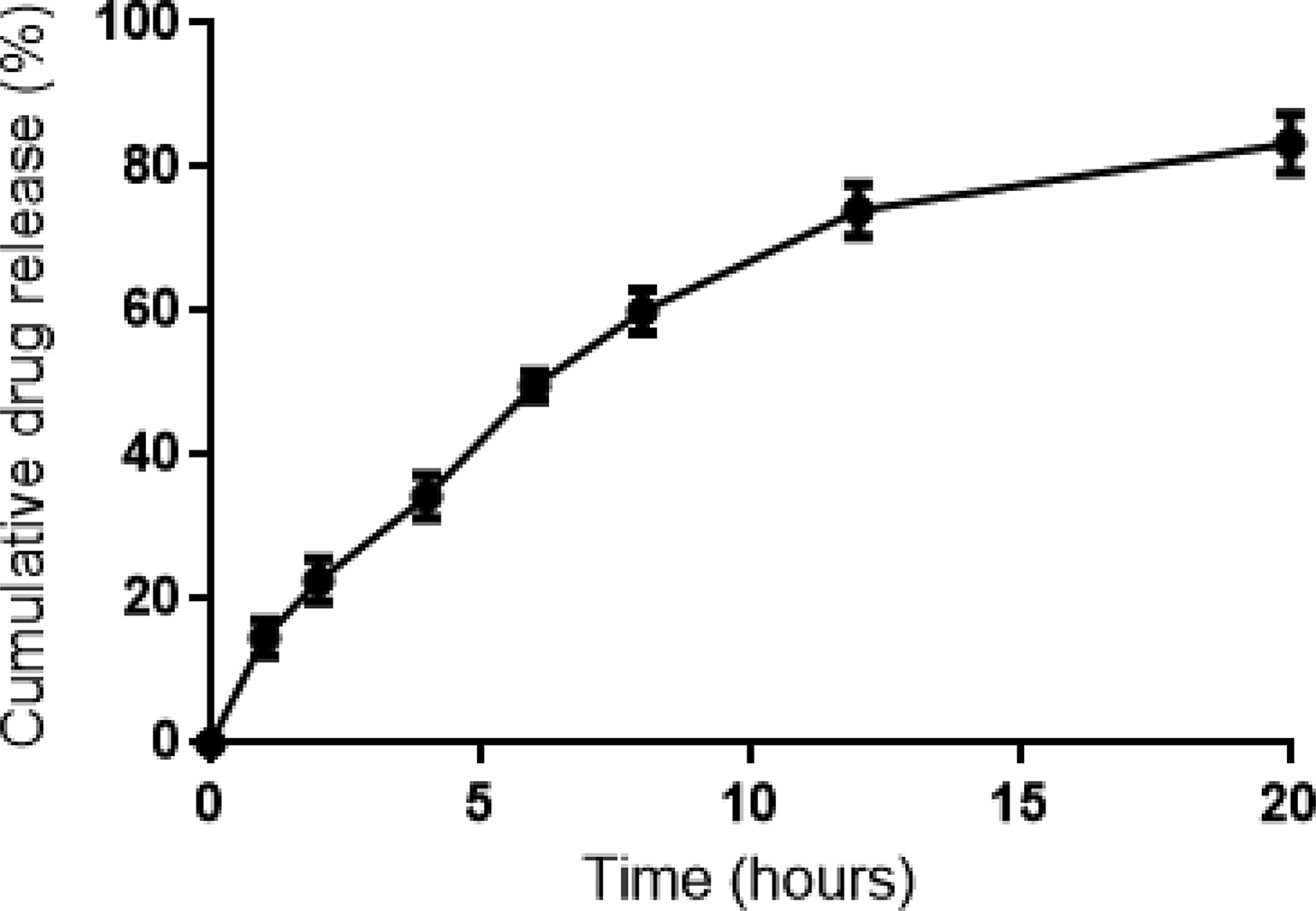
Cumulative release of flurbiprofen from the optimized formulation.
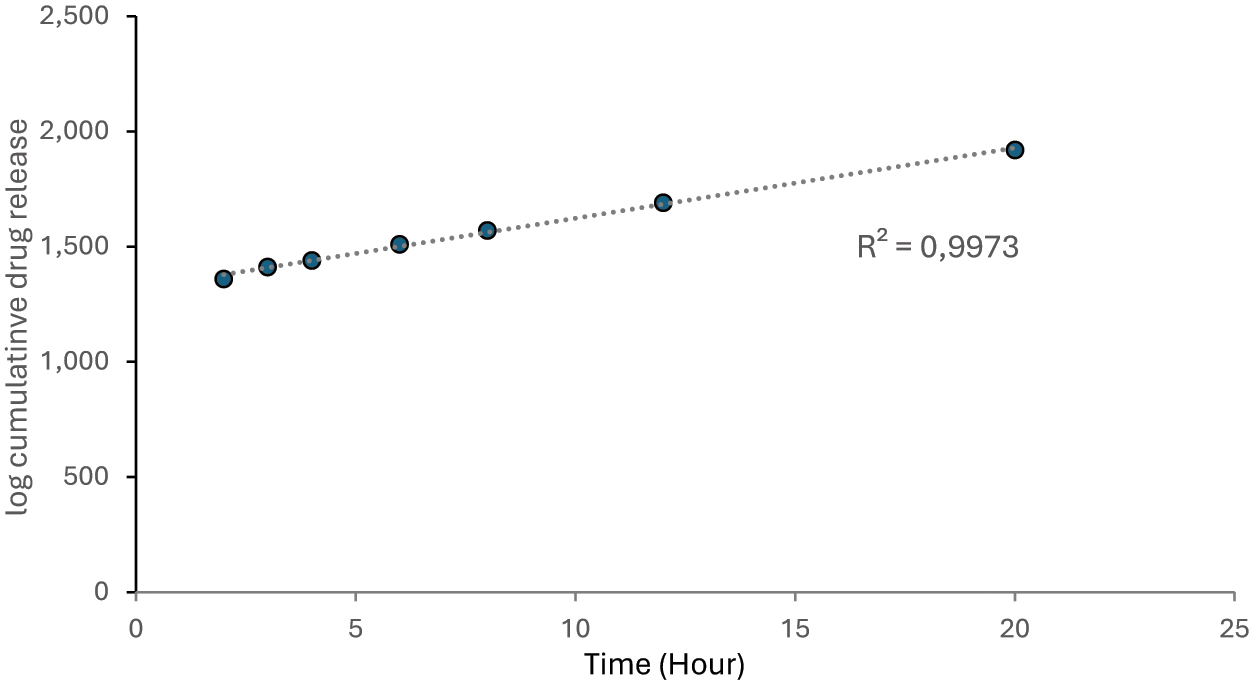
Log of cumulative drug release versus time.
The fit of the in vitro release data to different kinetic models was examined and the best-fitting model was determined according to the R2 values obtained. The first-order model showed the highest correlation with a value of R2 = 0.9973, indicating that the release rate of the drug depends on the remaining drug concentration. Such kinetic behavior is in agreement with the diffusion-controlled release mechanism observed mostly in nanoparticle systems. In contrast, the Higuchi (R2 = 0.9737) and Korsmeyer-Peppas (R2 = 0.9746) models also showed high agreement. These results suggest that matrix diffusion is effective in the release mechanism and the chitosan structure releases the drug in a controlled manner. The zero-order model had the lowest R2 value (0.7938), indicating that the release profile does not occur at constant rate. Overall, the data obtained reveal that the optimized flurbiprofen-loaded nanoparticle formulation has a controlled and concentration-dependent release mechanism and therefore offers a suitable system for ophthalmic applications.
Stability of the Formulation
Stability testing of optimized flurbiprofen–chitosan nanoparticulate formulation under acceleration storage condition (40°C/75% RH) was performed for a predetermined period as per ICH guidelines. During the study, no significant differences are observed in the visual appearance, pH (6.5), particle size (115.4 nm), PDI (0.415), and zeta potential (+13.4 ± 1.7 mV) and the drug content was found to be within limits without showing any evidence of degradation. These results are indicative of formulating stability.
DISCUSSION
The FT-IR spectrum of a 1:1 physical mixture of flurbiprofen and chitosan was compared with the spectra of pure flurbiprofen and pure chitosan (1:1) to check for interactions. C = CH and C–H stretching bands near the characteristic peaks of flurbiprofen were clearly visible in both the pure drug and the physical mixture. Similarly, peaks specific to chitosan were also present in the mixture spectrum. No significant shift, disappearance, or broadening of the peaks was observed in the physical mixture, indicating that no chemical interaction occurred between flurbiprofen and chitosan at the molecular level. These results indicate that the physical structure of the mixture and the structural identity of each component in the formulation were preserved.23,24 According to the Box-Behnken experimental design used in the optimization of the formulation, the parameters of chitosan concentration, dropping rate and stirring speed, which were selected as independent variables, were examined at three levels and their effects on the dependent variables, particle size and active substance % loading capacity, were statistically evaluated according to ANOVA results. Accordingly, it was statistically determined that particle size was affected by chitosan concentration. Since the tendency of cross-linking or agglomeration between chitosan chains may increase as the chitosan concentration increases, more chitosan in the mixture may increase the particle diameter by increasing the surface coating (p < 0.01). On the other hand, slow dropping rate provides more diffusion and controlled nucleation in the mixing medium and may lead to the formation of smaller and homogeneous particles. Fast dropping speed can be explained by the fact that sudden local concentration increase may lead to sudden precipitation and irregular nucleation and larger and heterogeneous particles (p = 0.02). When the relationship between stirring speed and particle size is analyzed, it can be concluded that low stirring speed may cause insufficient energy in the mixture and diffusion between phases will be slow, nucleation may decrease and large, heterogeneous particles may form. On the other hand, in case of mixing at excessively high speed, there is a risk of structural deterioration due to turbulence and foaming, and an increase in particle size as a result of agglomeration (p = 0.04).25–27 Another dependent variable is active ingredient loading capacity (EE%). The active ingredient loading capacity is affected by all the independent variables. As chitosan concentrations increased, the loading capacity was also enhanced, though at very high concentrations the active ingredient loading efficiency decreased. This can be due to the fact that the active drug might remain outside and may not be encapsulated within the polymer matrix, and thus might reduce the loading efficiency (p = 0.01). The same independent variable, slow dripping, increased. The reason for this can be that there can be more time of interaction between chitosan and flurbiprofen, and thus the loading capacity can increase (p < 0.01). It was observed in the study that active substance loading capacity and stirring rate are inversely related to each other (p = 0.03). This may be attributed to the reason that at an elevated stirring rate, flurbiprofen in the system is more likely to escape without diffusion or get trapped in foam.28–30
The optimized formulation, which contains 0.2% chitosan and was prepared at a stirring rate of 700 rpm with a dropping rate of 0.4 mL/min, produced nanoparticles with suitable properties for eye applications. The average particle size of 110.0 ± 2.20 nm supports the use of this system within the eye. Specifically, particle sizes below 200 nm help increase penetration into eye tissues and lower the risk of irritation. The measured PDI value was 0.347 ± 0.03, indicating a uniform distribution that is acceptable for pharmaceutical nanoparticle systems. Additionally, the zeta potential of the formulation was + 15.4 ± 3.8 mV, showing moderate electrostatic stability. While this value does not exceed the ± 30 mV threshold, the positive surface charge may boost the mucoadhesive effect by interacting with the negatively charged structures of the eye surface. This interaction might prolong the time the formulation stays on the eye surface and improve the drug’s effect. Overall, the particle size, distribution uniformity, and surface charge values suggest that the optimized nanoparticle system is a stable, effective, and well-tolerated method for delivering drugs in ocular applications.31–33 The improved ophthalmic nanoparticle formulation with flurbiprofen had a substance loading capacity of 80.89%. This high rate shows that flurbiprofen was well-encapsulated in the chitosan matrix and that the formulation process was efficient. In ophthalmic uses, high loading capacity is crucial. It ensures there is enough medicine in the target tissue and can reduce how often patients need to take the drug, which may lead to better compliance. Moreover, electrostatic interactions and possible hydrogen bonds between flurbiprofen and chitosan might have helped keep the active substance in place. Such a high loading rate means that the formulation can provide effective drug doses without needing too many extra ingredients. Overall, these results suggest that this system could be a good option for delivering medications in eye treatments.34–37 Following sterilization by 0.22 µm filtration, no significant alterations were observed in particle size, PDI, zeta potential, or pH, indicating that the optimized formulation maintained its stability and integrity.
In the optimized nanostructured formulation, the role of excipients other than NaCl in the total osmotic effect was found to be quite low. Therefore, NaCl was chosen as the main osmotic agent to control the osmolarity in the formulation. As a result of the measurement, it was determined that the osmotic pressure of the formulation was at an acceptable level. Keeping osmolarity close to tear fluid is essential to avoid issues such as discomfort, irritation, or reflex tear production during drop administration. The ability to accurately adjust the osmolarity using only NaCl meant that there was no need to add other ions to the formulation, which could affect the stability of the nanoparticle system. This method not only improves patient comfort but also supports the overall stability and compatibility of the formulation with the eye. Furthermore, the physiological pH 6.7 further confirmed the suitability of the developed system for ocular administration. 38
In terms of drug release, the formulation exhibited a controlled sustained release pattern (for over 20 h) after a relatively small initial release in the first 2 h. Such release is believed to be due to the chitosan matrix forming a diffusion barrier that allows a slow delivery of the drug. Among different kinetic models employed, the best-fit was obtained with the first-order kinetic model with R2 = 0.9985, indicating that the rate of release is proportional to the amount of drug remaining within the system. Higuchi and Korsmeyer–Peppas models were equally highly correlated, supporting the dominance of matrix diffusion during the release process. Overall, pH and drug release data revealed by the optimized nanoparticle formulation are that they possess a controlled drug release mechanism based on concentration, apart from providing an eye-physiologically compatible system. These findings strongly validate that the developed system can be utilized as a stable and efficient ophthalmic drug carrier. 39
Compared with previous flurbiprofen or chitosan-based ocular formulations, the system developed in this study achieved higher encapsulation efficiency (80.89%; typically 60%–70%) and longer release time (20 h; 8–12 h in the literature). 23 Furthermore, osmolarity and pH were carefully adjusted to the physiological range; this parameter has often been neglected in previous studies. These unique features highlight the innovative nature of the study and demonstrate that it combines patient comfort, formulation stability, and therapeutic efficacy in a single, optimized platform. After accelerated stability testing, a slight increase in PDI was observed, from 0.347 to 0.415. This indicates a minor broadening in particle size distribution but may not be considered as significant aggregation. Since the PDI value remained below 0.5, it is regarded as acceptable in terms of colloidal stability for ocular nanoparticle formulations. Therefore, this variation was interpreted as a normal fluctuation during storage rather than a sign of deterioration. The poststorage drug content was analyzed by dissolving the nanoparticles in 0.1% acetic acid and measuring the absorbance at 247 nm using UV–Vis spectrophotometry. As a result, 97.8 ± 1.6% of the initial drug content was retained, confirming the maintenance of chemical stability throughout the storage period.
CONCLUSIONS
In the current research, flurbiprofen-loaded chitosan nanoparticle formulation was formulated and optimized for ophthalmic drug delivery using BBED. Optimized formulation displayed desired physicochemical attributes such as nanoscale particle size of 110.0 ± 2.20 nm, acceptable homogeneity with PDI value of 0.347 ± 0.03 and zeta potential of + 15.4 ± 3.8 mV. These values support the colloidal stability and mucoadhesive potential of the formulation. Besides, 80.89% entrapment efficiency was obtained in the formulation, indicating that flurbiprofen was encapsulated in the chitosan matrix successfully. Osmolarity was precisely adjusted to approximately 300 mOsm/kg by the addition of NaCl alone and pH was determined to be 6.7. Both are within the physiologically acceptable range for ophthalmic preparations. In vitro drug release studies demonstrated controlled and prolonged release for up to 20 h. The data indicated that the release was optimally described by a first-order kinetic model (R2 = 0.9985), indicating that the release rate is concentration-dependent on the drug remaining. Accelerated stability testing confirmed that the formulation was stable with no degradation over time. The optimized flurbiprofen–chitosan nanoparticle system can be considered an innovative ocular drug delivery approach that combines high encapsulation efficiency, controlled drug release, and physiological compatibility. The integration of statistical optimization and stability verification makes this system a strong candidate not only for formulation but also for future preclinical and clinical applications.
AUTHORS’ CONTRIBUTIONS
In this article, hypothesis development, literature research, experimentation, data analysis, and preparations and correction of the article were carried out by EDÖ.
Footnotes
DISCLOSURE STATEMENT
Since the article is single-authored, there is no conflict of interest to declare.
FUNDING INFORMATION
The study was not supported by any person or organization.