
Abstract
Ischemic stroke (IS) is an important disease leading to high disability and mortality, and the current clinical treatment is limited. Tongnao Decoction (TND) is a traditional Chinese herbal formula for treating IS, but its pharmacological mechanism remains unclear. This study aims to elucidate the molecular mechanism through network pharmacology, molecular docking, and related experimental verification. First, the bioactive components of TND, along with their potential targets and IS-related gene targets, were identified through multiple databases. Subsequently, an “herb-active component-disease gene target” network and a protein–protein interaction (PPI) network were constructed. Combined with enrichment analysis, key biological processes and signaling pathways were identified. Following this, molecular docking experiments were conducted to preliminarily validate drug–target interactions. Finally, the efficacy of the relevant pathway targets was validated in a photochemically induced mouse cerebral ischemia model. A total of 90 active compounds and 615 target genes were screened. PPI network analyses suggested that TP53, EGFR, STAT3, AKT1, and IL-6 were the hub targets. TND significantly modulates inflammatory biological processes and the PI3K-Akt signaling pathway during IS treatment. Molecular docking analysis indicated that the primary components of TND may exhibit favorable binding affinity to multiple hub target proteins, including TP53, EGFR, STAT3, AKT1, and IL-6. Further in vivo experiments showed that TND dramatically improved neurological function, reduced neuronal damage, and decreased IL-6 expression in brain tissue. In addition, TND stimulated the PI3K/Akt/GSK-3β pathway. These findings imply that TND and its key bioactive components, coryincine, dihydrocapsaicin, and 4 (4′-hydroxybenzyloxy)benzyl methylether exert therapeutic effects on IS through the IL-6/PI3K/Akt/GSK-3β pathway.
Keywords
INTRODUCTION
Ischemic stroke (IS) is a sudden-onset cerebrovascular event with symptoms such as coma, verbal difficulties, and movement abnormalities. It threatens patient survival and imposes a significant socioeconomic burden due to neurological disabilities. Globally, 12.2 million new strokes occur annually (total number of patients: 101 million), with 87% being ischemic.1,2 IS pathogenesis involves dysregulation of energy metabolism, oxidative stress, and neuroinflammation. 3 Currently, intravenous tissue plasminogen activator and endovascular thrombolysis for major vessel blockage are the principal therapies for IS. However, these treatments are limited by a short clinical window, numerous contraindications, and the danger of adverse effects, such as cerebral edema and bleeding, leaving many patients without benefit. 4 These limitations necessitate the development of alternative therapeutic strategies. Chinese herbal formulas, with fewer side effects and multi-component, multi-target characteristics, present a unique promise for the treatment of IS. Tongnao Decoction (TND) is a traditional Chinese medicine (TCM) formula approved as a hospital preparation by the Jiangsu Provincial Hospital of TCM for the treatment of acute IS for over a decade. Annually, approximately 500 patients with acute IS are treated with TND, and its clinical efficacy has been well-substantiated. 5 TND consists of Chuanxiong Rhizoma (Chuanxiong), Gastrodia elata Blume (Tianma), Uncariae Ramulus Cumuncis (Gouteng), Rhodiola rosea L. (Hongjingtian), Arisaema erubescens (Wall.) Schott (Zhinanxing), Anemone altaica Fisch. Ex C.A.Mey. (Jiujiechangpu), Hirudo nipponica Whitman (Shuizhi), and Bombyx batryticatus (Jiangcan). Plant identities were recognized using the Plant List database (http://www.theplantlist.org), and animal names were cross-checked with the Chinese Pharmacopoeia (2015). TCM theory holds that “wind, phlegm, and blood stasis” are the primary pathological factors in IS. In TND, Chuanxiong and Tianma serve as the king medicinals, promoting blood circulation and unblocking meridians. Modern pharmacology has also discovered that they possess neuroprotective effects with anti-thrombotic and anti-inflammatory properties.6,7 The supporting components Zhinanxing, Gouteng, and Jiujiechangpu dispel wind and resolve phlegm. Hongjingtian, Shuizhi, and Jiangcan act as adjuvant agents, collectively achieving phlegm-resolving, meridian-unblocking, and blood-activating effects. Through multicomponent, multi-target synergistic actions, these eight herbs jointly address the complex pathological state of intertwined wind, phlegm, and stasis following IS. Our previous studies demonstrated that TND promotes angiogenesis in ischemic cerebral microvessels and significantly prevents atherosclerosis by enhancing endothelial cell survival and lowering oxidative stress and inflammation.5,8 Despite these findings, the integrative pharmacological mechanisms of TND in IS remain largely unknown.
Network pharmacology integrates systems biology and computational prediction to analyze regulatory networks in TCM formulations. The molecular docking approach, by modeling the ligand–receptor binding conformation and measuring the binding free energy, offers a reliable identification of the interaction mechanisms among the therapeutic components and primary targets of TCM. 9 It enhances the multi-target drug discovery efficiency and is widely applied in drug mechanism research and molecular docking. Although TCM has a centuries-old tradition in stroke treatment, its mechanisms remain complex and poorly understood. Integrating TCM research with network pharmacology can help clarify the unique ways in which TCM addresses various ailments.
In this study, we applied network pharmacology approaches to investigate the potential pharmacological mechanisms of TND treating IS, based on our prior Ultra-Performance Liquid Chromatography–Quadrupole Time-of-Flight Mass Spectrometry (UPLC-QTOF-MS) results. 5 We constructed a herb-compound-target network for TND target/pathway identification. Subsequently, molecular docking and a photothrombotic stroke model were used to validate the key target proteins involved in the proposed pathways. These results provided important experimental data for the further development and utilization of TND (Fig. 1).
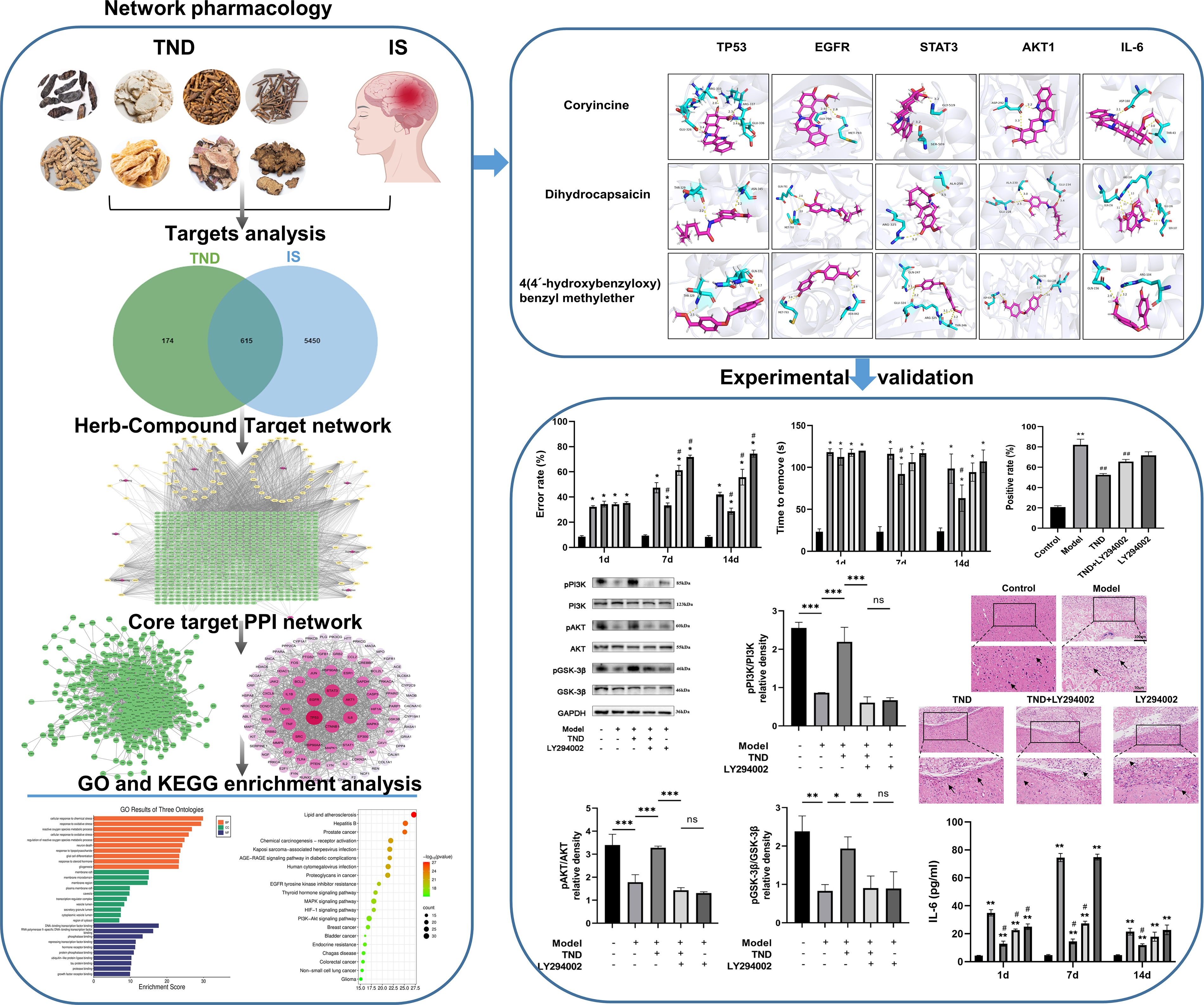
Workflow chart of the research. AKT1, AKT serine/threonine kinase 1; EGFR, epidermal growth factor receptor; GO, Gene Ontology; IL-6, interleukin-6; IS, ischemic stroke; KEGG, Kyoto Encyclopedia of Genes and Genomes; PPI, protein-protein interaction; STAT3, signal transducer and activator of transcription 3; TND, Tongnao Decoction; TP53, tumor protein p53.
MATERIALS AND METHODS
Identification of TND Active Ingredients
Pharmacokinetic assessment of TND began with compound acquisition from the TCM Systems Pharmacology Database (TCMSP; https://tcmspw.com). 10 The selection criteria were oral bioavailability (OB) ≥ 30% and drug-likeness (DL) ≥ 0.18. Compounds meeting these criteria are considered to possess broad pharmacological activity and are typical constituents of herbal medicines. 11 For components unavailable in TCMSP, the Encyclopedia of Traditional Chinese Medicine (ETCM) 2.0 platform (http://www.tcmip.cn/ETCM2) was employed to identify bioactive constituents using a Quantitative Estimate of Drug-likeness threshold (Compounds with QED ≥ 0.5 were considered to possess favorable drug-like characteristics). 12 The active constituents of TND’s component herbs have been previously characterized using UPLC-QTOF-MS. 5
Identification of Target Genes Associated with TND and IS
Target genes related to TND were identified using the TCMSP database, and protein target gene normalization was conducted using the UniProt protein database (https://www.uniprot.org/). 13 The GeneCards database (https://www.genecards.org/), OMIM database (https://www.omim.org/), 14 TTD database, and DrugBank database (https://go.drugbank.com/) were used to predict the targets associated with IS. A Venn diagram was constructed to display the overlapping goals.
Establishment of Protein–Protein Interaction (PPI) Network
Intersecting TND-IS target genes were examined using the STRING database (https://cn.string-db.org/), 15 with Homo sapiens as the species, with an acceptable interaction score of 0.7. Protein–protein interactions were evaluated using the default settings. The resulting network was displayed with Cytoscape 3.10.1, with topological features assessed using the Centiscape 2.2 plug-in. Core therapeutic targets for TND against IS were determined by evaluating degree, betweenness, and closeness centrality metrics, where nodes exceeding mean values across these indices were designated as significant.
Functional Enrichment Analysis (Gene Ontology/ Kyoto Encyclopedia of Genes and Genomes)
Enrichment analysis of STRING-derived data was performed using Metascape (https://metascape.org/). Gene ontology (GO) terms were stratified into cellular component (CC), biological process (BP), and molecular function (MF). The top 10 entries for each GO category (CC, BP, and MF) and the top 20 Kyoto Encyclopedia of genes and genomes (KEGG) pathways are shown and analyzed. Statistical significance was set at p < 0.05.
Molecular Docking
Molecular docking validated the expected connections between TND’s key active components and core target genes. The active chemical structures were collected from PubChem (https://pubchem.ncbi.nlm.nih.gov/), whereas the crystallographic data of the protein of choice originated from the PDB (http://www.rcsb.org/). Receptor proteins undergo preprocessing: removal of water molecules and native ligands, followed by the insertion of nonpolar hydrogens and charge assignment. Docking simulations were performed using AutoDock Vina 1.2.2, with binding results displayed using PyMOL 2.6.0.
Animal Models and Therapeutic Regimens
Thirty-five 6–8 week-old male C57BL/6J mice were received at SiPeiFu Biotechnology (Suzhou, China). Animals resided in uniform conditions (22–26°C, 30%–55% moisture, and a 12-h light/dark phase). All procedures received ethical approval (No. 2024DW-028-01), in compliance with China’s State Council Laboratory Animal Administration Regulations. The mice were acclimatized for at least 1 week before the study. The mice were randomly assigned to five groups (n = 6): control, model, model + TND (TND), model + TND + LY294002 (TND + LY294002), and model + LY294002 (LY294002). A photothrombotic stroke model was induced by injecting Bengal rose (Sigma, USA) into the tail vein for 5 min, followed by irradiation with a 5-mm-diameter fiber optic for 15 min. TND (2 g/(kg⋅d) via gavage for 2 weeks) and LY294002 (Selleck Biotechnology Inc., USA), a selective phosphatidylinositol 3-kinase (PI3K) inhibitor that competitively binds to the ATP-binding site of PI3K catalytic subunits and blocks downstream Akt phosphorylation, was administered at 7.5 mg/kg/d for 2 weeks to the TND and LY294002 groups, respectively. The dose and treatment duration for LY294002 were established based on the relevant literature.16,17 All animal experiments were conducted in accordance with the animal research: reporting of In vivo experiments guidelines 2.0. At the experimental endpoint, mice were euthanized by intraperitoneal injection of an overdose of sodium pentobarbital (150 mg/kg), followed by cervical dislocation to ensure death before tissue collection. TND was prepared by the Pharmacy Department of Jiangsu Provincial Hospital of Traditional Chinese Medicine (Nanjing, China) according to the standardized formula. The herbal components were authenticated by Professor Minghua Wu, Department of Traditional Chinese Medicine, and voucher specimens were deposited at the hospital pharmacy. The TND dosage and production procedure were similar to those used in our previous findings. 5 The composition ratio of the herbal ingredients was 1:1:3:1.5:1:1:0.5:1 (Chuanxiong Rhizoma: Gastrodia elata Blume: Uncariae Ramulus Cumuncis: Rhodiola rosea L.: Arisaema erubescens (Wall.) Schott: Anemone altaica Fisch. ex C.A. Mey.: Hirudo nipponica Whitman: Bombyx batryticatus).
Irregular Ladder Walking Test
Photothrombotic stroke surgery was performed as described in the “Animal Models and Therapeutic Regimens” section. For the irregular ladder walking test, mice were trained for 1 week before surgery on a horizontal ladder (100 cm in length, 19 cm in height, and 10 cm in width) with irregularly spaced rungs (1 to 2 cm spacing) to prevent task memorization. 18 Testing was conducted on days 1, 7, and 14 post-surgery (corresponding to during and after TND and/or LY294002 treatment). Each mouse performed three trials per session, and the average was recorded. Video recordings were analyzed by a blinded observer who quantified the percentage of contralateral forelimb foot slips (defined as misses or slips through the rungs) relative to total steps taken.
Adhesive-Removal Test
The adhesive-removal test was performed to evaluate sensorimotor function and motor asymmetry. Mice underwent a 3-day training period before surgery to familiarize them with the procedure. Testing was conducted on days 1, 7, and 14 post-surgery (corresponding to the pre-treatment, treatment period, and post-treatment stages of TND and/or LY294002 treatment). A 3 × 3 mm2 adhesive tape was applied to the center of the glabrous skin of each forepaw. The time required for the mouse to sense (indicated by shaking the paw) and remove the tape was recorded by a blinded observer, with a maximum cutoff time of 120 s. Three trials were performed per session, with a 5-min intertrial interval, and results were averaged. 19
H and E Staining
Brain tissues were fixed in 4% paraformaldehyde for 24 h, dehydrated through a graded ethanol series, and embedded in paraffin. Sections (5 µm thickness) were cut using a cryostat (Leica RM2235, Germany). After deparaffinization in xylene (2 × 10 min) and rehydration through graded ethanol, sections were stained with hematoxylin solution (Beyotime, China; cat. no. C0105S) for 5 min, rinsed in running tap water, differentiated in 1% acid alcohol for 30 s, and counterstained with eosin solution (Beyotime, China; cat. no. C0105S) for 2 min. Sections were then dehydrated, cleared in xylene (2 × 5 min), and mounted with neutral resin. For each animal, three nonconsecutive sections from the peri-infarct region were imaged using an Olympus CKX53 light microscope at 100× magnification. Images were analyzed using ImageJ software (NIH, USA) with background correction applied uniformly across all images.
Enzyme-Linked Immunosorbent Assay
Plasma IL-6 levels were quantified using a commercially available sandwich Enzyme-Linked Immunosorbent Assay (ELISA) kit (Proteintech, Wuhan, China; cat. no. KE10007). Blood samples were collected via cardiac puncture and centrifuged at 1000×g for 15 min at 4°C to obtain plasma. According to the manufacturer’s protocol, 100 µL of plasma or standard was added per well and incubated for 2 h at 37°C. After washing, 100 µL of biotinylated detection antibody (1:100 dilution) was added and incubated for 1 h at 37°C. Following additional washes, 100 µL of HRP-conjugated streptavidin (1:100 dilution) was added and incubated for 40 min at 37°C. After final washes, 100 µL of TMB substrate solution was added and incubated for 20 min at 37°C in the dark. The reaction was terminated with 100 µL of stop solution, and absorbance was measured at 450 nm using a microplate reader (BioTek Synergy H1, USA). IL-6 concentrations were calculated from a standard curve.
Western Blot Analysis
Ischemic hemisphere brain tissues were homogenized in radioimmunoprecipitation assay lysis buffer (Beyotime, China) supplemented with protease inhibitor cocktail (1:100) and phosphatase inhibitors (1:100). Protein concentrations were determined using a bicinchoninic acid protein assay kit (Beyotime, China). Equal amounts of protein (20 µg per lane) were separated by 10% sodium dodecyl sulfate polyacrylamide gel electrophoresis and transferred to polyvinylidene fluoride membranes (0.45 µm; Millipore, USA) using wet transfer (100 V, 90 min, 4°C). Membranes were blocked with 5% nonfat milk in TBST for 1 h at room temperature and incubated overnight at 4°C with primary antibodies: PI3K (1:1,000; Proteintech, 20584-1-AP), p-PI3K (1:1,000; Cell Signaling Technology, #17366), Akt (1:2,000; Proteintech, 10176-2-AP), p-Akt (1:2,000; Cell Signaling Technology, #4060), GSK-3β (1:1,000; Affinity Biosciences, AF5016), p-GSK-3β (1:1,000; Cell Signaling Technology, #5558), and β-actin (1:10,000; Proteintech, 66009-1-Ig). After washing with tris-buffered saline tween (3 × 10 min), membranes were incubated with HRP-conjugated secondary antibodies (1:5,000; goat anti-rabbit or goat anti-mouse; Proteintech) for 1 h at room temperature. Protein bands were visualized using an enhanced chemiluminescence detection kit (Millipore, USA) and imaged using a ChemiDoc XRS + imaging system (Bio-Rad, USA). Band intensities were quantified using ImageJ software (NIH, USA) without image smoothening, and phosphorylated protein levels were normalized to their respective total protein levels.
Immunofluorescence Analyses
Paraffin-embedded brain sections (3 µm) were deparaffinized, rehydrated, and subjected to antigen retrieval in citrate buffer (pH 6.0) at 95°C for 20 min. After cooling, sections were blocked with 10% normal goat serum for 1 h at room temperature. Sections were then incubated overnight at 4°C with anti-IL-6 primary antibody (1:200; Proteintech, 21865-1-AP). After washing with PBS (3 × 5 min), sections were incubated with Alexa Fluor 488-conjugated secondary antibody (1:500; Invitrogen, USA) for 1 h at room temperature in the dark. Nuclei were counterstained with DAPI (1 µg/mL; Beyotime, China) for 10 min. Sections were mounted with anti-fade mounting medium and imaged using a P250FLASH scanning microscope (3DHISTECH, Hungary) at 400 × magnification. For each animal, three sections were analyzed, and five random fields per section were captured. Background correction was applied uniformly using Image-Pro Plus 6.0 software (Media Cybernetics, USA), and IL-6-positive cells were quantified by a blinded observer.
Statistical Analysis
All in vivo data were tested for outliers using Grubb’s test (α = 0.05), and no significant outliers were identified. Normality was assessed using the Shapiro–Wilk test, and homogeneity of variance was evaluated using Levene’s test before ANOVA. All datasets met the assumptions of normality and homogeneity of variance. One-way analysis of variance followed by Tukey’s post hoc test was used for multiple group comparisons. Group sizes (n = 6 per group) were determined based on previous studies using similar stroke models and outcome measures, which demonstrated adequate statistical power (>80%) to detect meaningful differences in behavioral and molecular endpoints. 20 A formal power analysis using G*Power software (effect size f = 0.5, α = 0.05, power = 0.8) confirmed that n = 6 per group was sufficient for our experimental design.
RESULTS
Herb-Compound-Target Network Construction
By searching the TCMSP and ETCM databases and meeting the screening criteria (OB ≥ 30%, DL ≥ 0.18; QED ≥ 0.5), a total of 90 active components of TND were identified. (Supplementary Table S1). The PubChem database was then leveraged to check the PubChem IDs of these components; those missing a valid PubChem ID were excluded. Gene normalization was performed using the Uniprot protein database (https://www.uniprot.org/) to convert protein names to standardized gene symbols, ensuring consistency across different databases and eliminating redundancy arising from synonymous nomenclature. After removing duplicates, 789 compound-related targets were identified. Using the keywords “Ischemic Stroke” and “Cerebral Infarction,” 6,065 disease-related targets were collected from the GeneCard, OMIM, TTD, and DrugBank databases. A Venn chart was generated to identify the overlap of TND-related targets and IS-related targets, suggesting 615 common target genes (Fig. 2A), which were linked to the 90 active compounds. Using Cytoscape software, a network of herb-compound-target was constructed to describe the interactions between herbs, their constituents, and potential targets with diseases. The network included 711 nodes and 2,729 edges (Fig. 2B). Nodes with a higher number of edges have larger degree values, and the size of each node represents its relative importance. By performing topology analysis, the top three active components ranked by degree value were coryincine, dihydrocapsaicin, and 4 (4′-hydroxybenzyloxy)benzyl methyl ether, indicating their central positions in the network and potentially crucial roles in TND’s therapeutic mechanism (Supplementary Table S2).

Herb-Compound-Target Network Construction.
PPI Network Analysis
To identify key proteins mediating TND’s actions of TND against IS, 615 candidate targets were screened using the STRING database. This generated a PPI network (579 nodes; 4,957 edges; Fig. 3A). Topological analysis via Centiscape 2.2 calculated degree, betweenness, and closeness centrality metrics. The PPI network was then refined based on these topological parameters, with targets with degree, betweenness, and closeness values above the average considered as core targets. The refined network contained 92 nodes and 1,129 edges. The node size and color intensity were positively correlated with the degree value. The five primary target genes, ranked by degree value, were tumor protein p53 (TP53), epidermal growth factor receptor (EGFR), signal transducer and activator of transcription 3 (STAT3), AKT serine/threonine kinase 1 (AKT1), and interleukin-6 (IL-6), which are regarded as crucial targets in the pharmacological mechanism of TND for IS (Fig. 3B).
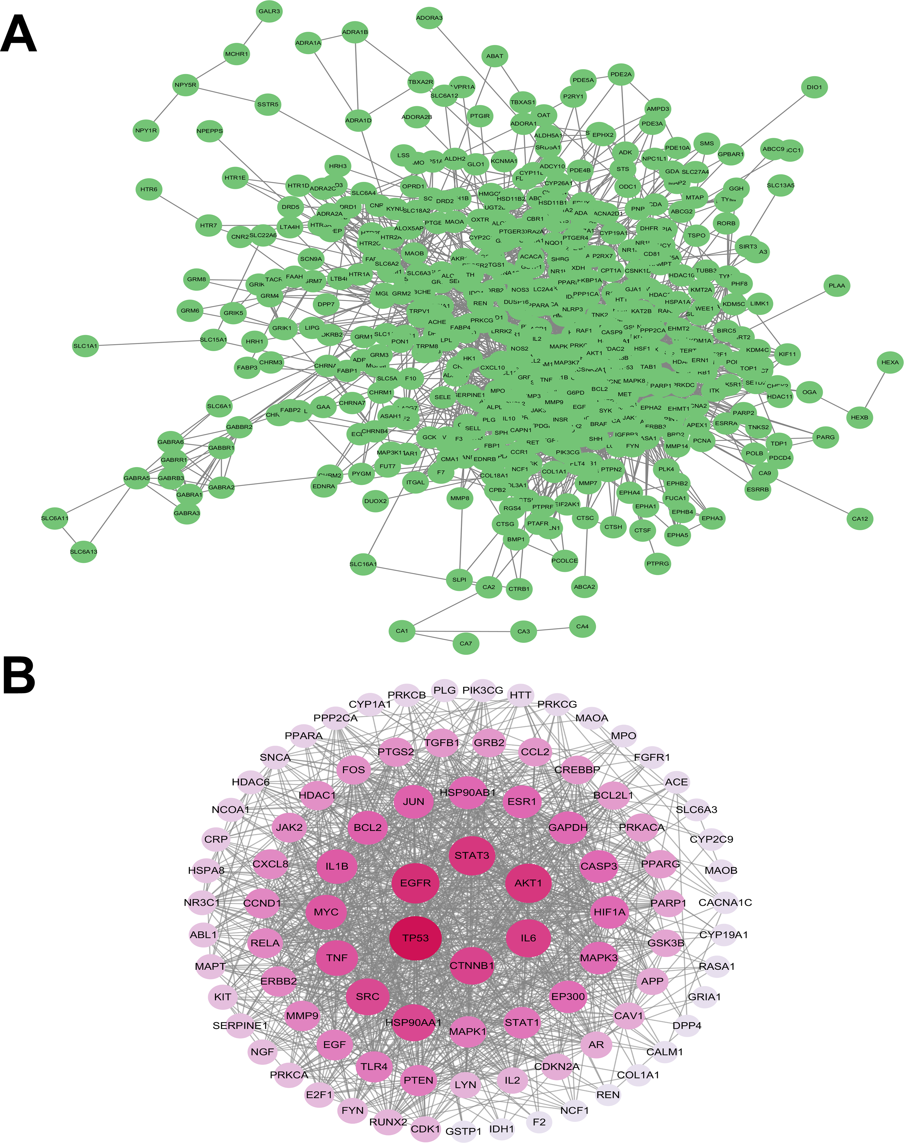
PPI Network Analysis.
Functional Enrichment (GO/KEGG) Analysis
GO enrichment analysis revealed the important BPs underlying the benefits of TND against IS. We obtained 2,994 significant terms (p < 0.05), comprising 117 CC terms describing subcellular localization, 2,655 BP terms describing physiological functions, and 222 MF terms describing biochemical activities. The highest point, 10 entries per category, was visualized (Fig. 4A), including key BPs related to neuroinflammation such as the response to oxidative stress, cellular response to chemical stress, and response to lipopolysaccharide (LPS). KEGG pathway sequencing identified 193 enriched pathways (p < 0.05), with lipids and atherosclerosis, mitogen-activated protein kinase (MAPK) signaling, and phosphatidylinositol 3-kinase/protein kinase B (PI3K-AKT) signaling exhibiting special IS significance (Fig. 4B). These findings imply TND’s beneficial actions may involve these pathways. Notably, the inflammatory and PI3K-Akt signaling pathways demonstrated the strongest association.

Functional enrichment of core TND targets in IS.
Molecular Docking of Major Components to Key Targets
According to the network pharmacology results, the three main active ingredients in TND—Coryncine, dihydrocapsaicin, and 4 (4′-hydroxybenzyloxy)benzyl methylether—were chosen to perform molecular docking with the important targets TP53, EGFR, STAT3, AKT1, and IL-6. It is generally accepted that a more negative binding free energy value suggests greater stability of the drug–target interaction. 21 Molecular docking research demonstrated that the free binding energies of the three principal agents of action with the key target proteins were all below −4.9 kJ/mol (Table 1, Fig. 5). These results suggest that the active compounds may possess strong affinity for their respective target proteins, supporting the hypothesis that TND’s active ingredients could possibly treat or mitigate IS through these targets.
The Bonding Energy of Three Active Ingredients and Five Target Genes (kcal/mol)

AKT1, AKT serine/threonine kinase 1; EGFR, epidermal growth factor receptor; IL-6, interleukin-6; STAT3, signal transducer and activator of transcription 3; TP53, tumor protein p53.
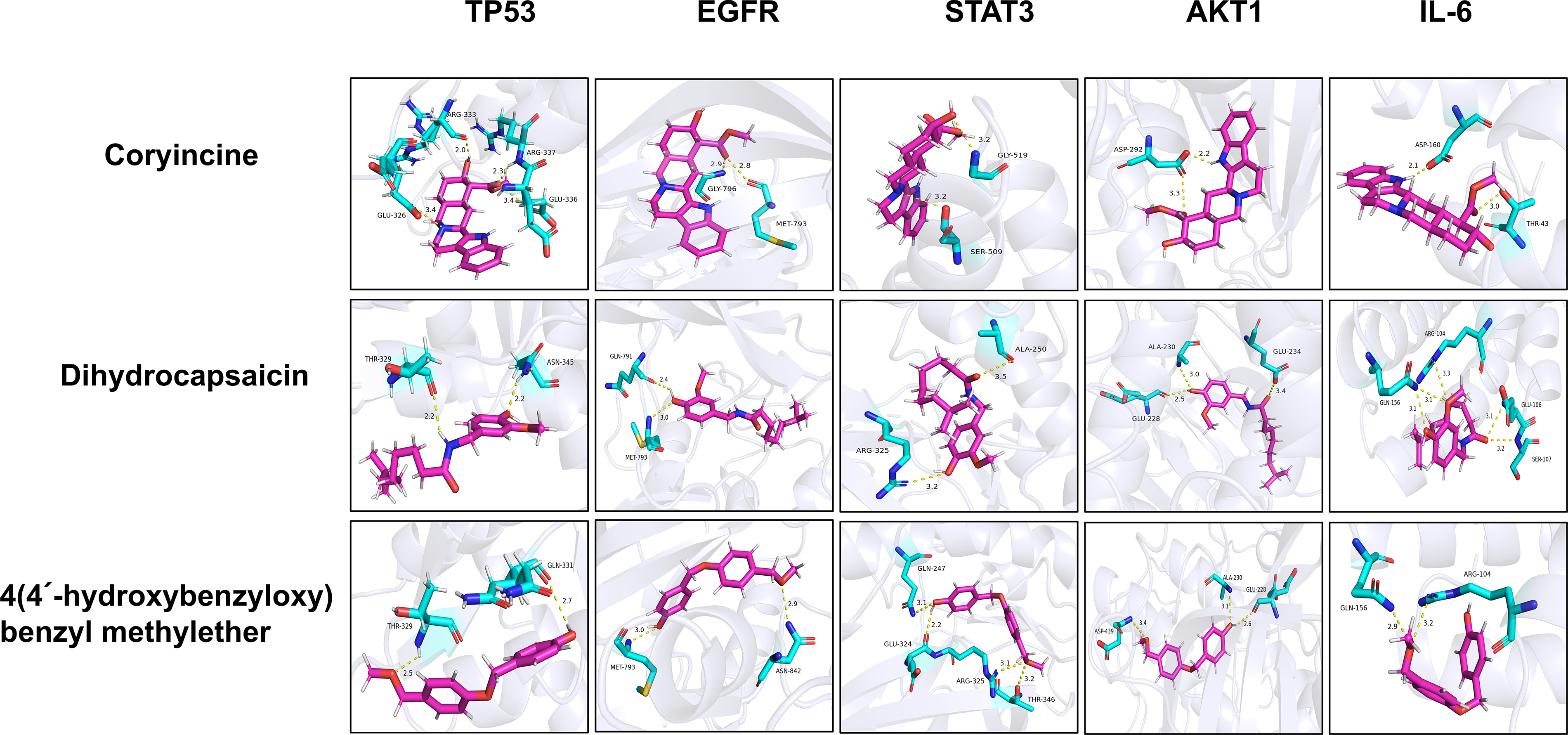
Molecular docking validation of TND bioactives against core targets. TND, Tongnao Decoction.
TND Alleviated the Neurological Damage of IS Mice
To further validate the mechanism of action of TND in IS, in vivo animal experiments were performed. Figure 6 depicts the results of the irregular ladder walking test and adhesive removal test in each group of mice on days 1, 7, and 14 after photothrombotic stroke. As opposed to the control group, after photothrombotic stroke, mice had a considerably greater mistake rate (p < 0.05) and an increasingly longer duration (p < 0.05) for tape removal from the injured forelimb, and the treatment of TND antagonized these changes. This suggests that treatment with TND could reduce neurological impairment in mice after photothrombotic stroke.
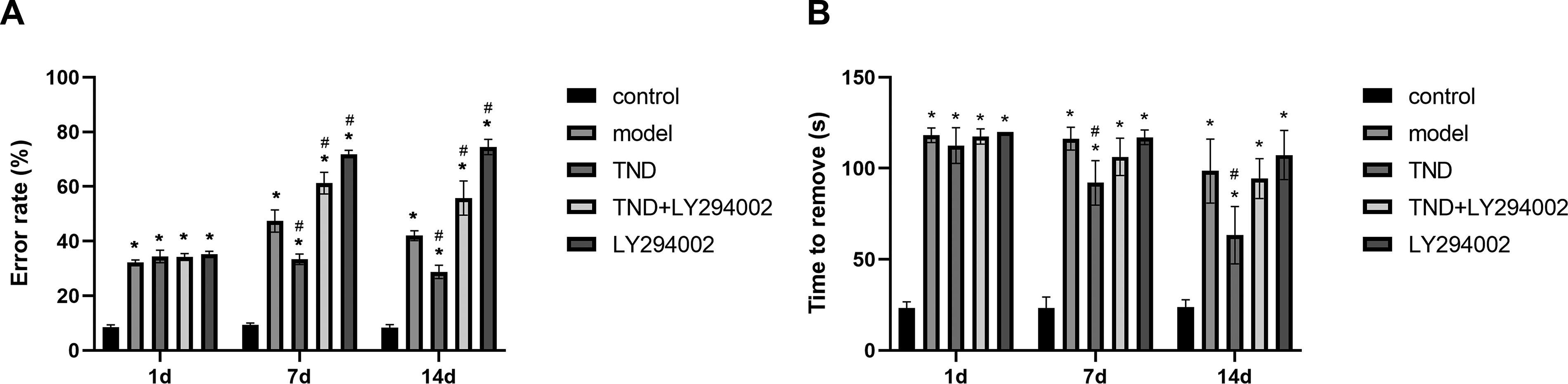
Contribution of TND on neurological function in mice with photothrombotic stroke.
TND Inhibited Inflammation after Photothrombotic Stroke
IL-6, a known pro-inflammatory cytokine, activates immune cells, increases the generation of additional inflammatory mediators, and amplifies the local inflammatory response. It is positively correlated with the severity of neurological deficits. 22 ELISA results demonstrated that blood levels of IL-6 in mice with photothrombotic stroke were considerably increased, whereas IL-6 expression levels decreased following treatment with TND (p < 0.05) (Fig. 7A). The immunofluorescence results further confirmed TND’s inhibitory effect on IL-6 in the brain (Fig. 7C and D). Pathological staining of brain tissues showed that neurons in the cortex of the brains of control mice exhibited a regular layout with a maintained cellular structure and rounded nuclei. The model group exhibited cortical neuron depletion featuring shrunken somata, disrupted cytoarchitecture, and inconspicuous nuclei. Some cells exhibited vacuolar degeneration, whereas the nuclei were either atrophied or necrotic. In addition, the proportion of infiltrating inflammatory cells was significantly higher than control group. After treatment with TND, the cerebral cortex cells of mice in the TND group had a better morphology, the cell structure was reasonably preserved, and inflammation of the brain tissue was obviously reduced (Fig. 7B). As observed in Figure 7B and C, TND attenuates the inflammatory response and possibly confers neuroprotection after photothrombotic stroke. However, all of these changes can be partially reversed by adding LY294002.
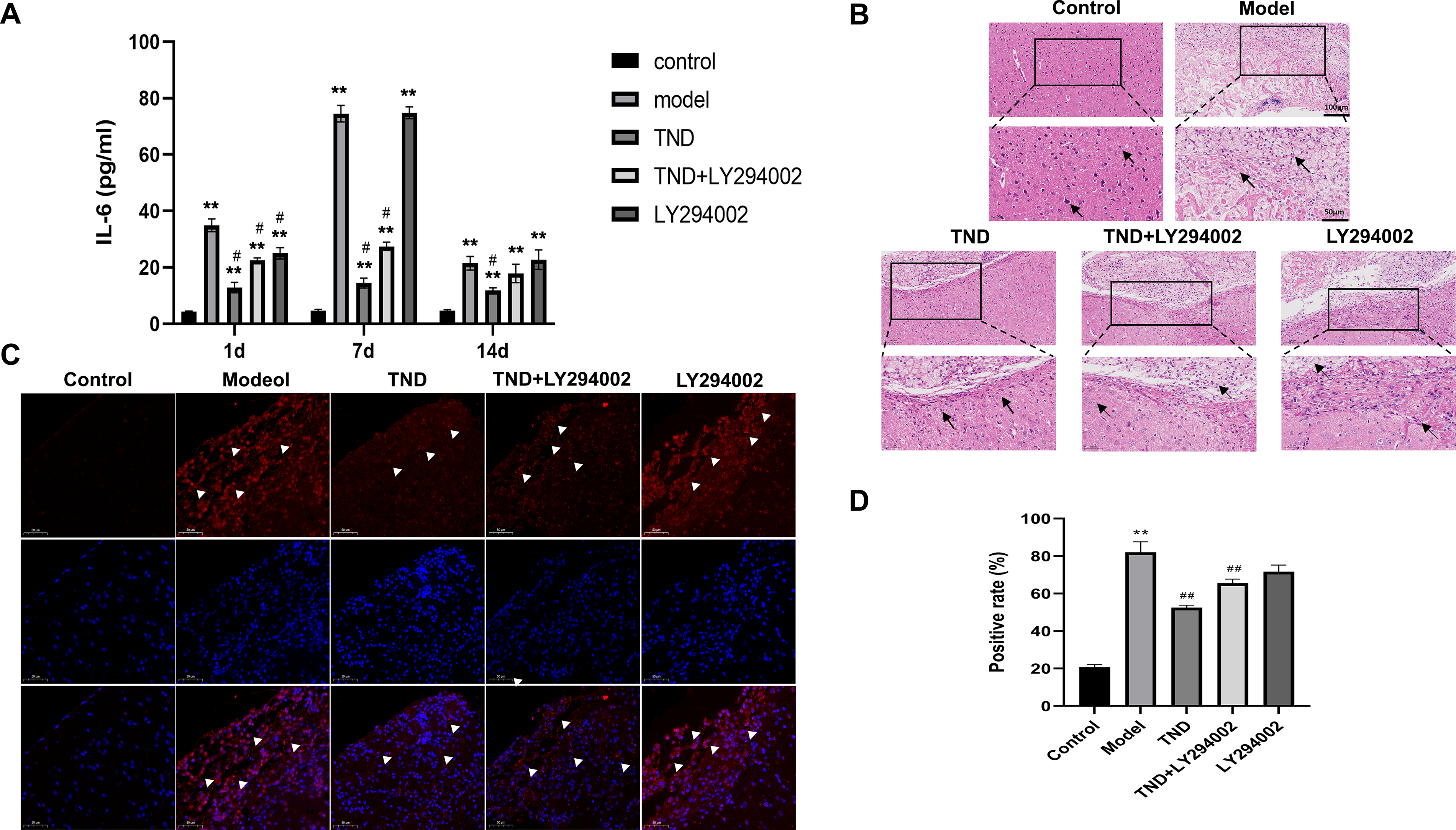
TND attenuates neuroinflammation post-photothrombotic stroke.
TND Activates the PI3K-Akt Signaling Pathway in Photochemically Induced Stroke Mice
To elucidate TND’s molecular mechanism, we analyzed PI3K/Akt/GSK-3β pathway protein expression. Figure 8 shows significantly reduced phosphorylation of PI3K, AKT, and GSK-3β in photothrombotic stroke mouse brains. TND treatment markedly reversed these deficits. Critically, validation experiments using LY294002 (a PI3K-specific inhibitor) confirmed pathway involvement; the compound blocked TND-induced PI3K/Akt activation in post-stroke brain tissue.
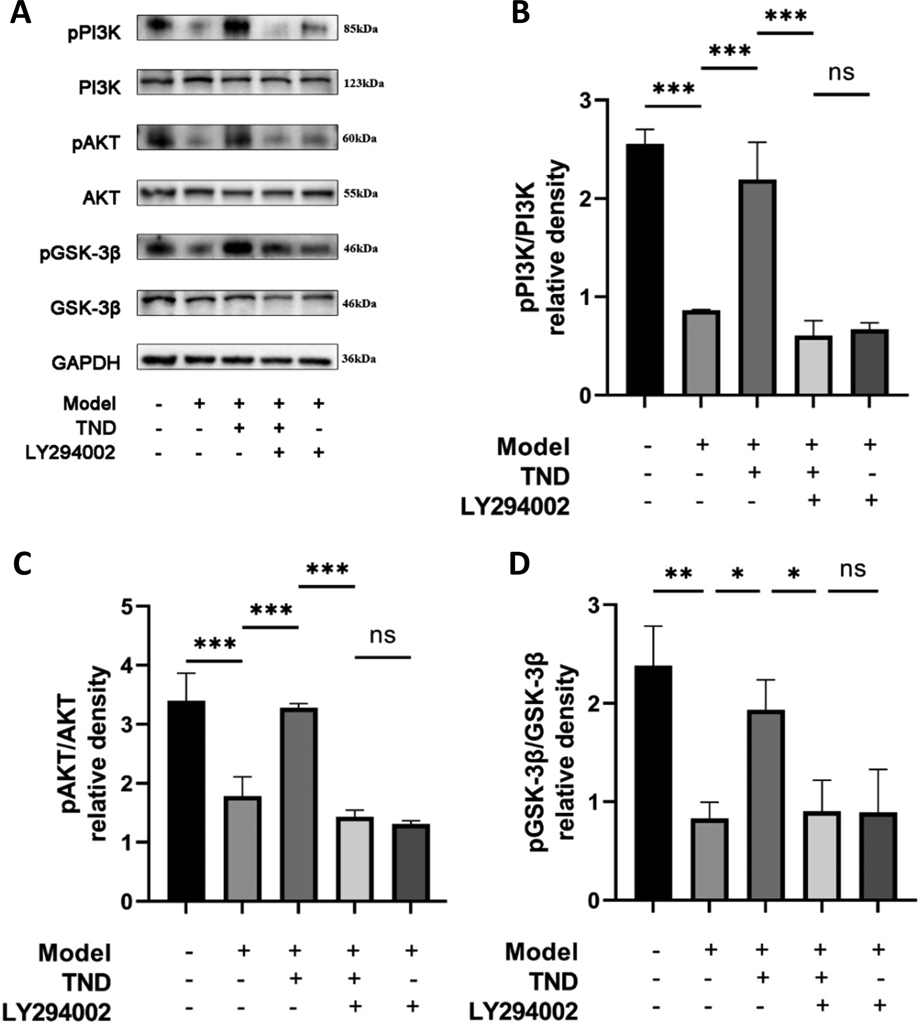
Effects of TND on PI3K/AKT/GSK-3β pathway activity in the brain tissue.
DISCUSSION
Summary and Interpretation of Findings
Insufficient cerebral blood flow initiates complex signaling cascades, causing irreversible brain injury and neurological deficits. Neuroinflammation critically drives stroke pathophysiology and influences penumbra evolution and ischemic injury progression. Consequently, targeting neuroinflammatory pathways represents a key therapeutic strategy for mitigating cerebral damage and enhancing functional recovery post-stroke. 23 TND is a novel TCM formulation derived from established Chinese medicinal recipes, utilized for the clinical management of several neurological disorders, and has demonstrated efficacy as a therapeutic agent for the treatment of IS.24,25 However, the complexity of its components complicates our understanding of the pharmacological mechanisms underlying its anti-IS effects, which significantly hinders its broader clinical adoption. Network pharmacology, an emerging interdisciplinary field, is particularly equipped to shed light on the molecular basis of TCM for diseases characterized by various components, targets, and pathways. This study extensively examined the bioactive constituents of TND and their mechanism of action against IS using network pharmacology analysis, molecular docking, and animal experimental validation. We discovered that one way by which TND could exert its anti-IS effects could plausibly be through the IL-6/PI3K/Akt/GSK-3β signaling pathway.
The current investigation identified multiple overlapping targets among the various chemicals in TND, indicating that TND exerts its anti-inflammatory effects via the synergistic interaction of its constituents. The principal active component of TND is coryncine, followed by dihydrocapsaicin and 4 (4′-hydroxybenzyloxy)benzyl methyl ether. Coryncine is an alkaloid extracted from the poppy, Corydalis bungeana Turcz. It exhibits anti-inflammatory effects by blocking the activation of the nuclear factor κB (NF-κB) signaling pathway. 26 Research indicates that dihydrocapsaicin alleviates pathological changes induced by cerebral ischemia through anti-apoptotic and antioxidant mechanisms while also blocking the aggregation of NLR family pyrin domain-containing three inflammasomes and the expression of NF-κB, thereby exerting anti-inflammatory effects.27,28 4 (4′-hydroxybenzyloxy)benzyl methylether, a natural phenolic chemical obtained from the traditional Chinese medicine Tianma, 29 also displays a broad spectrum of anti-inflammatory and antioxidant capabilities. 30 Collectively, the above results show that multiple components of TND have favorable therapeutic effects on IS.
To examine the possible mechanism of TND in the treatment of IS, we identified IS-related targets of the core components of TND through a PPI network. Our study found that the key targets of TND for treating IS were TP53, EGFR, STAT3, AKT1, and IL6, all of which are intimately associated with inflammatory reactions. The strong binding of the core compounds to these targets was further confirmed by molecular docking. The TP53 maintains genomic stability through cell cycle and apoptosis regulation. Critically implicated in oxidative stress and mitochondrial dysfunction, p53 inhibition modulates neuroinflammatory, autophagic, and mitophagic pathways to attenuate neuronal injury in ischemia-reperfusion.31,32 As a transmembrane ErbB kinase, EGFR mediates proliferative signaling triggered by EGF ligand engagement. Ligand binding (e.g., EGF and tumor necrosis factor-alpha [TGF-α]) activates downstream PI3K-Akt and MAPK cascades, promoting post-ischemic angiogenesis. 33 STAT3 operates as a critical regulator of the post-ischemic inflammatory response, triggered by ischemic stimuli and strongly manifested in peri-infarct neurons and neuronal cells. It is also a critical component of the acute stage response factor complex that interacts with IL-6. 34 Furthermore, STAT3 in neutrophils/macrophages induces neuroprotection by modulating microglia/macrophage polarization. 35 A genome-wide association study of 200,000 participants demonstrated that attenuated IL-6 signaling activity is related to an 11% reduction in IS risk. Furthermore, elevated circulating IL-6 levels are independently associated with the incidence of stroke in individuals without an established vascular disease. Elevated IL-6 concentrations also predict increased severity, instability, and progression of carotid atherosclerotic plaques. 22 As a key inflammatory mediator following stroke, IL-6 amplifies local inflammation by activating and attracting neutrophils and monocytes and by promoting vascular endothelial cells to generate adhesion molecules and other inflammatory mediators. Clinically, higher circulating IL-6 levels correlate with elevated body temperature, early neurological deterioration, larger infarct volumes, and poorer long-term functional outcomes. 36 AKT1 is a key effector molecule in the PI3K/Akt pathway that regulates cell survival, metabolism, and apoptosis by phosphorylating downstream target proteins. In IS, AKT1 activation attenuates oxidative stress, prevents neuronal apoptosis, stimulates neovascularization, and curtails pro-inflammatory factor secretion (e.g., TNF-α and IL-6) by inhibiting NF-κB nuclear translocation, thereby tempering neuroinflammation. 37 Cumulatively, these results identify TP53, EGFR, STAT3, AKT1, and IL6 as core molecular determinants of acute IS pathogenesis.
GO enrichment indicated that TND was associated with primary BPs, including oxidative stress, reactive oxygen species (ROS) metabolic processes, and response to LPS. Meanwhile, KEGG pathway analysis indicated TND mitigates acute ischemic stroke by modulating lipid metabolism, atherosclerosis, MAPK signaling, and PI3K/Akt signaling. This aligns with prior evidence that TND regulates lipid metabolism and atherosclerosis. 8 Oxidative stress, characterized by an imbalance between ROS production and antioxidant defense mechanisms, plays a crucial role in the pathophysiology of cerebral ischemia. These ROS act as vital signaling mediators that drive microglial M1 polarization, enhance peripheral leukocyte infiltration, and promote cytokine production by inflammatory cells. The consequent oxidative stress and neuroinflammation critically exacerbate ischemia-reperfusion injury pathology. 38 LPS is a component of the outer membrane of gram-negative bacteria and can activate the innate immune system through the Toll-like receptor 4 signaling pathway, leading to the upregulation of proinflammatory cytokines (such as TNF-α, IL-1β, and IL-6) and recruitment of microglia and peripheral immune cells to the ischemic site, thereby promoting oxidative stress, cell death, and disruption of the blood-brain barrier. 39 Both MAPK and PI3K/AKT cascades critically regulate post-ischemic inflammation. The current findings indicate that the activated MAPK signaling pathway contributes to the stimulation of microglia and production of pro-inflammatory factors in brain tissue after IS. 40 Conversely, PI3K/AKT signaling confers essential neuroprotection during ischemia/reperfusion injury. Activation of the PI3K/AKT pathway enhances the activity of antioxidant enzymes, reduces ROS and oxidative stress, and attenuates brain tissue injury. In addition, PI3K/AKT protects neuronal cell function by reducing inflammatory responses and apoptosis. 41 The PI3K/Akt/GSK-3β signaling cascade operates through sequential phosphorylation events. Upon activation by upstream signals, PI3K phosphorylates phosphatidylinositol-4, 5-bisphosphate (PIP2) to generate phosphatidylinositol-3, 4, 5-trisphosphate (PIP3), which recruits Akt to the plasma membrane. Akt is then phosphorylated at Thr308 by phosphoinositide-dependent kinase 1 (PDK1) and at Ser473 by mTOR complex 2, resulting in full activation. Activated Akt subsequently phosphorylates Glycogen synthase kinase-3β (GSK-3β) at Ser9, thereby inhibiting its kinase activity. 37 Research has found that GSK-3β is activated following cerebral ischemia-reperfusion, exacerbating stroke outcomes by inducing inflammation and apoptosis. 42 Inhibiting GSK-3β alleviates neuroinflammation in middle cerebral artery occlusion models. 43 IL-6 signaling intersects with the PI3K/Akt pathway through multiple mechanisms. Upon binding to its receptor complex (Interleukin-6 Receptor/glycoprotein 130), IL-6 can activate downstream signaling pathways (e.g., Janus kinase/signal transducer and activator of transcription 3 [JAK/STAT3]), which subsequently phosphorylate and activate PI3K either directly or through adaptor proteins such as Insulin Receptor Substrate-1 and Grb2-associated binder 1. This IL-6-induced PI3K activation leads to downstream Akt phosphorylation. 44 In addition, IL-6 signaling modulates the expression of various PI3K pathway components. 45 Taken together, these interconnected signaling networks position IL-6 as a central hub linking neuroinflammation to the PI3K/Akt/GSK-3β neuroprotective pathway, providing a mechanistic framework for understanding TND’s therapeutic effects against IS.
This study utilized TND to intervene in photothrombotic stroke mice to understand its mechanism of action and validate network pharmacology findings. Behavioral assessments suggested that the model group displayed substantial neurological deficits, while the TND-treated group showed notable improvement. Pathological examination revealed that the brain tissue of the control group displayed normal cellular morphology with no significant infiltration of inflammatory cells. In contrast, neuronal damage was more evident in the model group and was accompanied by considerable inflammatory cell infiltration. After treatment with TND, the cellular architecture was substantially intact, and the total number of cells associated with inflammation was reduced. Compared with the model group, TND treatment significantly elevated PI3K expression and enhanced the phosphorylation of AKT (P-AKT) and GSK-3β (P-GSK-3β) in the brains of photothrombotic stroke mice. Concurrently, TND reduced IL-6 expression, both systemically (serum) and centrally (brain tissue). These findings collectively indicate that TND alleviates neuroinflammation in this stroke model, likely mediated through PI3K/Akt/GSK-3β signaling activation. Previous studies have demonstrated that the PI3K/Akt/GSK-3β pathway is significantly impaired following IS and that its restoration confers neuroprotection.37,46 Several antioxidant and anti-inflammatory agents have been shown to activate this pathway in IS models. For example, resveratrol attenuates cerebral ischemia-reperfusion injury by activating the PI3K/Akt pathway and inhibiting GSK-3β. 47 Similarly, Scutellarin protects against ischemic brain damage through PI3K/Akt-mediated inhibition of apoptosis. 48 Among TCM formulations, Buyang Huanwu Decoction has been reported to promote neurological recovery after stroke via PI3K/Akt activation, 49 and Da Qin Jiu decoction exerts neuroprotection through similar mechanisms. 50 These findings collectively support the therapeutic potential of targeting the PI3K/Akt/GSK-3β pathway for IS treatment and align with our observations regarding TND’s mechanism of action.
Strengths and Limitations
This study presents several notable strengths: first, we employed a comprehensive multi-methodological approach that integrated network pharmacology, molecular docking, and experimental validation, providing robust evidence for TND’s therapeutic mechanisms. The systematic identification of bioactive compounds and target genes through multiple authoritative databases (TCMSP, GeneCards, OMIM, and STRING) ensures comprehensive coverage of potential therapeutic targets. Second, the molecular docking analysis demonstrated strong binding affinities between key active compounds and target proteins, supporting the biological plausibility of the proposed mechanisms. Third, our in vivo validation employed multiple complementary assessment methods, including behavioral tests, histopathological analysis, ELISA, Western blotting, and immunofluorescence, providing multilevel evidence for TND’s neuroprotective effects. Fourth, the mechanistic validation using the PI3K inhibitor LY294002 provided crucial evidence for pathway specificity, strengthening our conclusions about the PI3K/Akt/GSK-3β signaling mechanism. Despite these strengths, several limitations should be acknowledged. First, the relatively short follow-up period (14 days) limits our understanding of long-term therapeutic effects and functional recovery, which are crucial for clinical translation. Second, we did not conduct dose-response studies to determine optimal TND dosing, which would be essential for clinical application. Third, while we observed concurrent changes in IL-6 expression and PI3K/Akt/GSK-3β pathway activation following TND treatment, our study did not directly demonstrate the mechanistic involvement of IL-6 in regulating this signaling pathway. Future studies employing IL-6 knockdown/knockout models or IL-6 neutralizing antibodies are warranted to establish the direct relationship between IL-6 and PI3K/Akt/GSK-3β pathway modulation. Fourth, among the five key targets identified through network pharmacology (TP53, EGFR, STAT3, AKT1, and IL-6), we only experimentally validated IL-6 in the present study. The possible roles of TP53, EGFR, STAT3, and AKT1 in the pharmacological efficacy of TND were not investigated and need to be carefully evaluated in future studies to provide a comprehensive understanding of TND’s multi-target therapeutic mechanisms. In addition, while excluding compounds without PubChem identifiers is essential for computational methods and data standardization to ensure research reproducibility, this approach may inadvertently overlook phytochemicals with pharmacological relevance. Such database-dependent filtering could systematically favor well-characterized compounds over novel bioactive constituents. Future studies employing alternative structural databases, direct compound isolation, or advanced analytical techniques may provide a more comprehensive characterization of bioactive features in TND. These limitations highlight important directions for future research to advance the clinical translation of TND for the treatment of IS.
CONCLUSIONS
This study employed a combination of network pharmacology and molecular docking methods alongside animal experiments to preliminarily elucidate the potential pharmacological mechanisms of TND in treating IS, identifying its active components, core genes, and possible signaling pathways. Animal validation demonstrated that TND improved neurobehavioral and histopathological deficits in mice, the mechanism possibly through reducing IL-6 levels while activating the PI3K-Akt signaling pathway. Molecular docking analysis indicated that the primary active components of TND (Coryncine, dihydrocapsaicin, and 4 (4′-hydroxybenzyloxy)benzyl methylether) exhibited favorable binding affinity with their targets (TP53, EGFR, STAT3, AKT1, and IL-6). However, the pathways and compound-target interactions identified in this study are predictive and require further experimental validation before inferring clinical relevance. Taken together, this study provides an optimized approach for elucidating the pharmacological mechanisms of TND and supplying novel targets for the treatment of IS.
DATA AVAILABILITY STATEMENT
The data supporting the findings of this study are available from the corresponding author upon reasonable request.
AUTHORS’ CONTRIBUTIONS
J.G. and Y.C.: Visualization, writing—original draft, and software. L.L. and F.M.: Software, methodology. X.Y. and Y.D.: Software, methodology, conceptualization. Y.L. and Y.W.: Supervision. H.J. and Z.C.: Supervision and funding acquisition. W.L.: Formal analysis. Y.Z. and M.W.: Conceptualization and validation. All authors have read and agreed to the published version of the article.
Footnotes
DISCLOSURE STATEMENT
The authors declare that they have no competing interests.
FUNDING INFORMATION
This work was supported by National Natural Science Foundation of China (82274428, 82474435), Natural Science Foundation of Jiangsu Province (BK20241996), Jiangsu Provincial Administration of Traditional Chinese Medicine (ZT202102), Key Medical Research Project of the Jiangsu Provincial Health Commission (K2023009), and Weihai Traditional Chinese Medicine Science and Technology Project (2024 N-17).
Supplemental Material
Supplemental Material
Abbreviations
References
Supplementary Material
Please find the following supplemental material available below.
For Open Access articles published under a Creative Commons License, all supplemental material carries the same license as the article it is associated with.
For non-Open Access articles published, all supplemental material carries a non-exclusive license, and permission requests for re-use of supplemental material or any part of supplemental material shall be sent directly to the copyright owner as specified in the copyright notice associated with the article.