
Abstract
The therapeutic efficacy of ellagic acid could be improved by developing suitable carrier system. The present research was to develop ellagic acid-loaded microsponges impregnated with chitosan-guar gum hydrogel and evaluate its potential as novel carrier system for wound therapy. Ellagic acid-loaded microsponges were fabricated using ethyl cellulose by the quasi-emulsion solvent diffusion method and incorporated into chitosan and guar gum hydrogel. Prepared microsponges were found to be spherical, non-aggregated particles ranging in size from 100 to 300 µm. The hydrogel formulation was assessed for pharmaceutical characteristics, rheology, drug release, antioxidant and antimicrobial activities. FTIR confirmed that the bioactive and the excipients used are chemically compatible. Spreadability data confirm precise and easy dosage application of microsponge hydrogel in the skin. Results of rheological studies revealed that microsponge-loading considerably increased elastic strength, while frequency and temperature sweep tests demonstrated viscoelastic stability at 25°C. A sustained ellagic acid release for 12 h following the Higuchi kinetics model (R2 = 0.987) was noticed with microsponge loaded chitosan-guar gum hydrogel. The good antioxidant activity exhibited at doses (50–100 µg/mL) by the developed hydrogel implies that the formulation successfully preserves bioactive antioxidant properties. Developed hydrogel retained antimicrobial activity against the test organisms and showed notable inhibition against Pseudomonas aeruginosa at low concentration (12.5 µg/mL). This study establishes an effective topical formulation of ellagic acid-loaded microsponges with potential antioxidant and antimicrobial efficacy, highlighting its possibility to be used for wound healing.
INTRODUCTION
Skin acts as an intricate barrier to environmental challenges such as penetration of UV rays, moisture evaporation, and invasion of microorganisms. As a protective shield from these challenges, this layer of the body is continuously exposed to injuries, and hence, wound healing is a remarkable process for the survival of living beings. 1 Major cascades involved in the healing process of wounds include the initial inflammatory phase, followed by the proliferation and remodeling phase. Key players involved in these phases include skin stem cells, immune cells, growth factors, cytokines, and extracellular matrix. 2 Hence, any therapeutic agent promoting these cascades may prove worthy for the promotion of wound healing. In recent years, plant-derived natural moieties have been frequently explored and used as an alternative to synthetic options. 3 Owing to increasing chronic wound conditions, researchers are investigating the medicinal properties of bioactives of natural plants to improve wound healing. 4 Various secondary metabolites of plants, including polyphenols, possess biological activities that play a key role in wound healing. 5
Ellagic acid (EA) is a polyphenolic, thermostable phytonutrient found in strawberries, pomegranates, and almonds. 6 This bioactive possesses diverse activities, including hepatoprotective, antiulcerogenic, neuroprotective, gastroprotective, anti-inflammatory, antimicrobial, and antihemorrhoid.7–9 Antioxidant, anti-inflammation, and anticoagulant properties of EA help in promoting it as a vital therapeutic agent for wound healing.8–10 In addition, this moiety promotes collagen synthesis, ROS scavenging, modulation of inflammatory mediators and inhibits lipid peroxidation, 11 which are also useful in wound healing. Furthermore, EA promotes angiogenesis, which is essential for delivering oxygen and nutrients to the wound site during repair. 12 According to reported studies, this phytoconstituent promotes fibroblast proliferation and migration, which is required for wound contraction and new tissue formation. 8 It also inhibits melanogenesis, which may aid in addressing scar formation issues during wound healing. 13 However, its therapeutic efficacy is limited by the low water solubility (owing to high crystallinity and strong hydrogen bonding) and photosensitivity. 14 As a result, achieving the full potential of this bioactive in wound healing requires encapsulating it in an appropriate carrier system.
Microparticles and nanoparticle-based approaches have emerged as the most promising technology for delivering active compounds. In this context, microsponges have recently gained popularity due to their highly porous, cross-linked polymeric construction, which allows for high drug loading, sustained release, and the preservation of labile phytochemicals from degradation. 15 Compared with other carrier systems, microsponge-based formulations offer several merits, including better formulation flexibility, decreased side effects, improved elegance, and superior stability, thereby addressing inherent challenges associated with the encapsulated moiety. 16 Therefore, selecting this porous carrier system could improve EA stability, reduce local irritation, and maintain steady drug levels at the site of application. In addition, formulations based on microsponges can improve the solubility and bioavailability of hydrophobic phytochemicals. Typically, they have a size range of 5–300 µm, which ensures drug localization within the epidermis, and are useful in reducing systemic and local cutaneous complications, making them a commendable solution for delivering natural products in topical therapy. 17 The potential of microsponges as an ideal carrier capable of encasing various moieties such as anti-acne agents, anti-inflammatory and immunomodulatory agents, sunscreens, and essential oils has been documented.17,18 Indeed, microsponge technology has reached commercialization for topical applications, with Retin-A Micro® (tretinoin) already in the market, which demonstrates this method has translated from laboratory research to clinical use. 19
Microsponges are generally integrated into some suitable vehicle such as ointments, lotion, creams, emulgel, or gels for improving their topical delivery. 17 In this context, the incorporation of drug-loaded microsponges into a hydrogel matrix will increase the efficacy of topical therapy by providing adequate hydration and bioadhesion that prolong residence time on the skin or wound surface. The incorporation of microsponges within gel matrices for various applications, including wound healing, has been reported in the literature. 20 Drugs such as resveratrol, metronidazole, nebivolol, and silver sulfadiazine have been successfully encapsulated into microsponges based gel and evaluated for wound healing applications.21,22 Thus, it is hypothesized that the incorporation of EA into a microsponge-loaded hydrogel system could be a promising strategy for improving wound healing. However, selecting the right gelling agent is critical for successful wound healing therapy. In this context, chitosan is a well-known polysaccharide polymer prepared by deacytylation of chitin, which is nontoxic, biodegradable, and biocompatible. 23 Chitosan has been used to make gel and has antibacterial properties, which could be useful in the formulation of a wound-healing product. 24 Its physicochemical properties can be improved with appropriate cross-linkers such as glutaraldehyde. 25 Chitosan gels are used to treat a variety of wounds, including burns, surgical wounds, and infected or diabetic wounds, by promoting the various stages of healing, from hemostasis to tissue regeneration. 26 In addition, chitosan can reduce inflammation and oxidative stress in wounds by blocking pro-inflammatory cytokines, such as TNF-α. 27 Furthermore, chitosan-based hydrogels are known to aid in wound healing by maintaining moist environment, promoting cell proliferation, and possessing inherent antibacterial activity. 28 The porous, hydrated structure of chitosan hydrogels serves as a scaffold for new tissue formation and can be loaded with growth hormones, stem cells, or antibacterial agents to further expedite healing. 29
Guar gum is a nonionic compound of galactose and mannose with good swelling properties. Although this polymer is insoluble in major organic solvents, it produces viscous solution at low temperature and is stable at pH 5–7. 30 Besides its antimicrobial, antioxidant, and antihemorrhagic activity, it may play synergistic role in wound healing potential. 31 Hence, these two polymers were chosen to develop gel system for EA-loaded microsponge in the current study. A literature search has revealed that no study has been conducted using EA microsponges and chitosan-guar gum (CGG) hydrogel to manage wound healing. Thus, the objective of the present investigation was to develop EA-loaded microsponge-impregnated CGG (EAMS-CGG) hydrogel as novel carrier system for wound therapy. In addition, the antioxidant and antimicrobial potential of the EAMS-CGG was evaluated, as oxidative stress and microbial infection are two main factors that delay wound healing. These studies can provide an early biological indicator of the developed EAMS-CGG’s suitability for topical wound management. Microsponges were prepared by the quasi-emulsion solvent diffusion method. Preliminary studies were carried out to select the concentrations of chitosan, guar gum, and glutaraldehyde for hydrogel preparation. The developed formulation was subsequently evaluated for its general characteristics, rheological behavior, antioxidant, and antimicrobial properties.
MATERIALS AND METHODS
Chemicals and Reagents
EA was procured from Sigma-Aldrich (Bangalore, India). Ethyl cellulose, polyvinyl alcohol (PVA), and sodium hydroxide were procured from Thomas Baker (chemicals), Mumbai, India. Ethanol, sodium chloride, and disodium hydrogen orthophosphate were supplied by CDH (New Delhi, India). Chitosan and guar gum were purchased from High Purity Laboratory Chemicals, Mumbai, India. Dichloromethane (DCM) and potassium dihydrogen phosphate were obtained from Sisco Research Laboratories (Mumbai, India). 2,2-Diphenyl-1-picrylhydrazyl (DPPH) was obtained from Hi-Media (Mumbai, India). All chemicals and reagents used in this study were of analytical grade and utilized without further purification.
Fabrication of Ellagic Acid Loaded Microsponges (EAMS)
EAMS were prepared using the quasi-emulsion solvent diffusion method reported earlier with slight modifications.32,33 Briefly, ethyl cellulose (700 mg) was dissolved in 20 mL DCM, and EA (100 mg) was added to obtain the internal phase. This organic phase was introduced dropwise into 100 mL of an aqueous solution containing PVA (100 mg) under continuous mechanical stirring (Remi Elektrotechnik Ltd., Mumbai, India) at 12298 × g for 3 h at room temperature. During emulsification, DCM diffused and evaporated, resulting in the formation of microsponges. The obtained microsponges were collected by filtration using Whatman filter paper (0.45 µm), washed repeatedly with double Milli Q water to remove residual PVA and solvent traces, and dried at 25°C for 24 h. The product (Supplementary Fig. S1) was stored in desiccator for further characterization and evaluations. EA content was measured by UV spectrophotometer at λ max (255 nm), and the calibration curve was prepared in methanol and phosphate buffer (Supplementary Figures S2, S3, and S4). The production yield 34 and the % encapsulation efficiency were determined according to the standard protocols. 35
Scanning Electron Microscopy
Scanning electron microscopy images were obtained to analyze the morphology and surface topography of EAMS. The samples were gold coated using a sputter coater under an argon atmosphere to improve conductivity. 36 The images were examined using SEM instrument (Model 7610 F Plus, JEOL, Peabody, MA, USA) with a 3 kV accelerating voltage and at a magnification of 50×.
Preformulation Study of Hydrogel
Preformulation studies were carried out using two cross-linkers (PVA and glutaraldehyde). Briefly, four batches (PVP1–PVP4) of CGGH were prepared by varying the amount of chitosan (0.75%–1.5%), while keeping guar gum (0.25%) and PVA (0.25%) amounts constant (Supplementary Table S1). Then, another four batches (GA1–GA4) of gels were prepared by varying the amount of chitosan (1 and 1.25%) and guar gum (0.25% and 0.40%), while keeping the glutaraldehyde (0.1%) amount constant (Supplementary Table S2). All formulations were checked visually for consistency and physical appearance.
Fabrication of EAMS-CGG Hydrogel
Hydrogel was prepared according to the previously reported method with minor modifications. 37 Briefly, chitosan (1% w/v) and guar gum (0.25% w/v) were dispersed in 50 mL of 1% v/v acetic acid with constant stirring (300 rpm) for 2–4 h using mechanical stirrer. Then the required quantity of microsponges was weighed (containing 20 mg EA) and added slowly to the preformed gel and mixed at low speed (200–300 rpm) for 15–30 min. Subsequently, 0.1% v/v glutaraldehyde solution was added dropwise with continuous stirring to provide crosslinking within the gel matrix. The remaining 50 mL of 1% acetic acid was then added dropwise, and the dispersion was stirred for 30 min. The gel was kept covered overnight to remove entrapped air. The pH of the developed gel was adjusted to 5.5–6.0 with 0.1M NaOH. Similarly, blank MS-CGG hydrogel and blank CGG hydrogel were also prepared.
Characterization of EAMS-CGG Hydrogel
pH
The pH of the hydrogel samples was measured by a pH meter (RG-101-02, Rescholar Equipment, Ambala, India).
FTIR analysis
Samples of pure EA, EAMS, and EAMS-CGG hydrogel were subjected to spectral analysis using FTIR spectrometer (Model BX-II, PerkinElmer, Waltham, MA, USA). Sample spectra were determined in the range of 4,000–400 cm−1 by the KBr technique.
Spreadability assessment
The spreadability of EAMS-CGG hydrogel and the corresponding blank formulation (CGG hydrogel) were evaluated according to the method reported earlier. 38 Briefly, 0.5 g of the gel was placed at the center of a clean glass plate. Another glass plate was carefully placed on top of the gel to ensure uniform contact. Subsequently, varying weights (5, 10, 20, 50, 100, and 200 g) were placed on the upper plate and allowed to rest for 1 min. After the removal of the weights, the diameter of the spread gel was measured, and the spreadability (S) was calculated using the equation reported earlier. 38
Rheology Study
Rheological characteristics of the CGG hydrogel and EAMS-CGG hydrogel were measured using rheometer (MCR 102, Anton Paar, Graz, Austria) with Peltier temperature controller. 39 Sample (∼1 mL) was loaded onto the lower plate, and the upper plate was lowered to a gap of 1.00 mm, and the system was equilibrated at 25°C for 5 min before starting the test. Linear viscoelastic region (LVER) was established in order to maximize the strain and comprehend the elastic to viscous characteristics of the hydrogel. Oscillatory frequency sweep measurements were performed over a frequency sweep of 0.1–100 rad/s, and data were recorded. From the resultant force/torque and phase data, the RheoCompass software determined the loss modulus (G″), storage module (G′), complex viscosity, torque (τ), and other rheological behavior of samples. Similarly, temperature-dependent rheological properties (temperature-sweep) of both formulations were also determined in the LVER region using the same rheometer. Temperature sweeps were performed from 25°C to 50°C at 0.5°C·min−1 with constant strain (0.1%) and constant frequency (10 rad·s−1). Various rheological characteristics of the formulations were recorded by the rheometer software at each temperature point after thermal equilibration.
In Vitro Ellagic Acid Release
In vitro release studies were carried out using vertical Franz diffusion cells recommended for topical gel formulation 40 with 5 mL reservoir capacity and diffusion area of 0.74 cm2. Cellulose nitrate membrane (molecular weight cutoff at 12–14 kDa) was mounted between donor and receptor compartments. The receptor medium used was phosphate-buffered saline (pH 7.4): ethanol (70:30, v/v) maintained at 32 ± 0.1°C and stirred at 500 rpm with magnetic bar. 41 Each formulation weighing 50 mg equivalent to EA was placed on the donor side. At predefined time intervals, 4 mL of the sample was removed from the receiver fluid and replenished with the same volume of fresh receptor fluid. Samples were analyzed by UV-Vis spectrophotometer following appropriate dilution. The release data were analyzed using different kinetic models 42 to explore the pattern of EA release from the tested products.
Evaluation of Antioxidant Activity
Free radical scavenging activity was evaluated using the DPPH assay. Test samples (pure EA, EAMS, and microsponge-loaded hydrogel) were prepared from the stock solutions to get various concentrations (2, 4, 6, 8, and 10 µg/mL) in methanol. DPPH stock (0.1 mM) was prepared in methanol. Samples were mixed with 1.5 mL of DPPH solution, vortexed, and incubated in the dark at room temperature for 30 min. Absorbance was recorded at 517 nm using Genesys UV-Visible spectrophotometer (Thermo Fisher Scientific, Madison, USA). Methanol with DPPH (no sample) served as the control and methanol alone as the blank. Percent radical scavenging was calculated according to the equation described earlier. 43
In Vitro Antimicrobial Activity
The minimum inhibitory concentration (MIC) of EA, EAMS, and EAMS-CGG hydrogel was determined using the standard broth dilution method. Gram-positive (Staphylococcus aureus, ATCC 25923) and gram-negative strains (Escherichia coli, ATCC 25922, and Pseudomonas aeruginosa, ATCC 27853) were used. Nutrient broth was prepared by following the standard protocol. Bacterial cultures were activated by inoculating 1 mL of sterile nutrient broth with each test strain and incubated for 24 h at 37°C in a BOD incubator. The activated cultures were then diluted in 0.9% NaCl solution (100 mL) to achieve uniform bacterial suspensions. Test samples were prepared to have final concentrations of 100, 50, 25, 12.5, 6.25, and 3.125 µg/mL. For each bacterial strain and concentration, 0.1 mL of the standardized bacterial suspension was added to the test tubes. Negative controls (nutrient broth without test samples) and positive controls (broth with bacteria but without the test compound) were included in parallel. All tubes were incubated for 24 h at 37°C in a BOD incubator. After incubation, bacterial growth was evaluated visually by observing turbidity. The MIC was defined as the lowest concentration of the test sample that completely inhibited visible growth.
Stability Study
The stability of EAMS-CGG hydrogel was carried out for four weeks under refrigerated conditions (2–8°C) by storing it in amber-colored containers. 44 Parameters such as visual appearance, drug content, pH, and viscosity were examined.
RESULTS AND DISCUSSION
Preformulation Study of Hydrogel
Preformulation studies were carried out to select the cross-linker (PVA/glutaraldehyde) for preparing gel formulations and were observed visually for consistency and physical appearance. PVA-cross-linked batches (PVA1–PVA4) showed decrease in consistency with decrease in chitosan amount, as well as pale-yellow discoloration within 2 to 3 days (at room temperature), indicating that PVA was not appropriate for the formulation (Supplementary Table S1). However, GA1–GA4 gels prepared using glutaraldehyde demonstrated variable consistency, with batch GA2 (chitosan 1.0%, guar gum 0.25%, glutaraldehyde 0.1%) having a semi-solid gel composition with no color change in the formulation after one week of testing (Supplementary Table S2). It has been reported that glutaraldehyde stabilizes CGG hydrogels and forms covalent imine (Schiff-base) bonds with chitosan’s main amine groups, resulting in a strong three-dimensional polymer network. 45 In addition, through hydrogen bonding or physical entanglement, glutaraldehyde bifunctional aldehyde groups can interact with guar gum to generate a porous, mesh-like structure that is required for controlling the drug release. 46 Hence, the formulation GA2 was selected for EA loading. Preparation of EAMS was carried out by the quasi-emulsion solvent diffusion method. The production yield and % entrapment efficiency of EAMS were 84.06 ± 1.8% and 87.32 ± 1.6%, respectively.
Scanning Electron Microscopy
The morphology and particle size of the prepared microsponges have been regularly examined using scanning electron microscopy. 17 The observed micrograph of EAMS is displayed in Figure 1 . The prepared microsponges showed that the particles, which ranged in size from 100 to 300 µm, were spherical, non-agglomerated, and had few surface pores (pores might have been filled due to EA encapsulation). The absence of aggregation of microsponges could be due to the inclusion of PVA in the formulation as reported earlier. 47 In addition, the rapid evaporation of organic solvent (DCM) from the microsponges surface would have resulted in the pore formation. 48

Scanning electron microscopy image of ellagic acid-loaded microsponges. The scale bar represents 100 µm.
Characterization of EAMS-CGG Hydrogel
pH
The pH of the prepared EAMS-CGG, blank MS-CGG, and blank CGG hydrogels were 5.3 ± 0.46, 5.5 ± 0.38, and 5.4 ± 0.42, respectively. The observed pH here with all the hydrogels is comparable with human skin pH (4.5–6.5) and is safe for the topical applications in chronic wounds. Various studies have reported that formulations with slightly acidic pH are best suited for topical wound application as they enhance epithelialization, wound closure, and collagen deposition, supporting fibroblast activity, collagen formation, DNA synthesis, and oxygenation.49,50
FTIR analysis
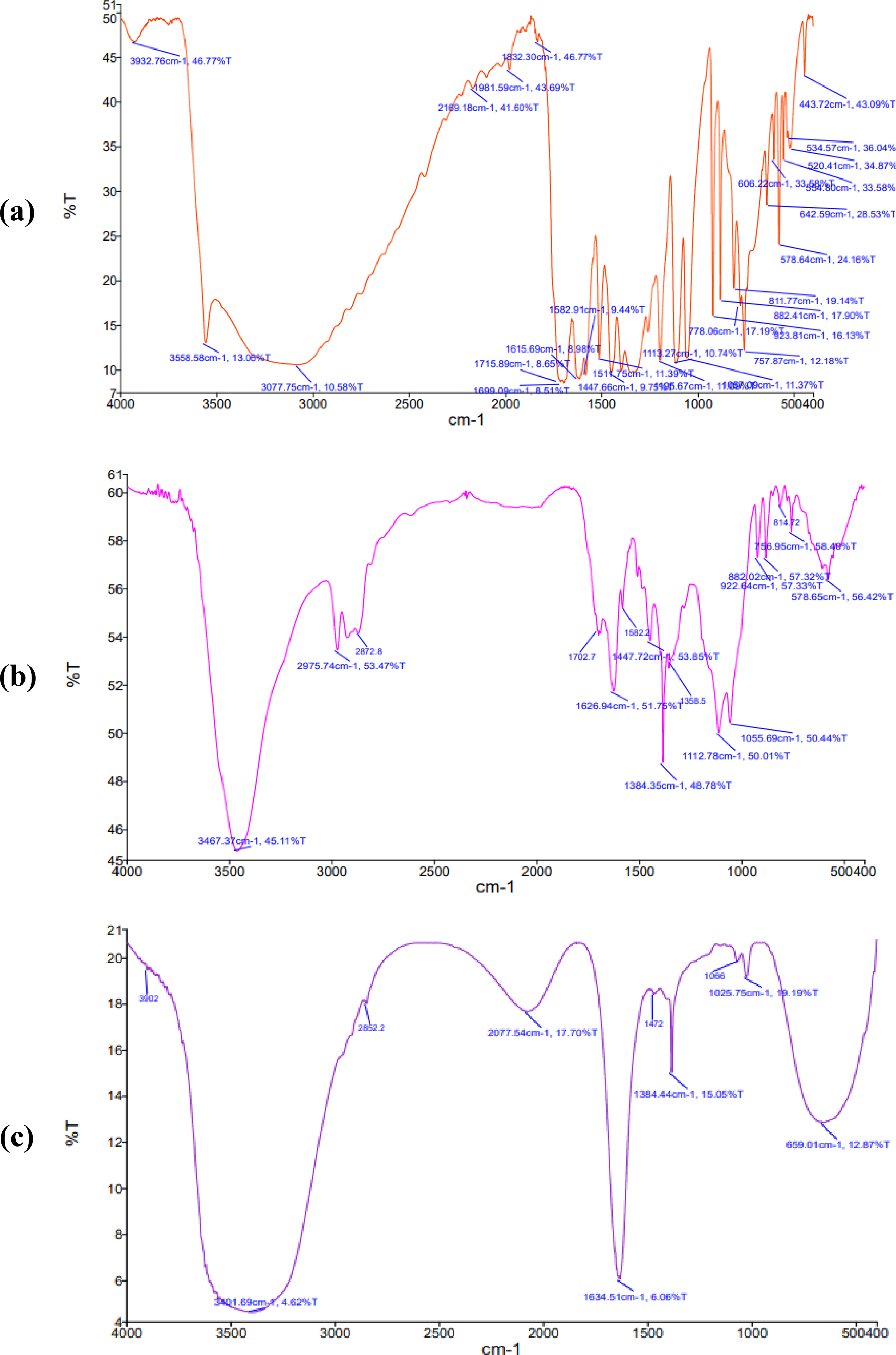
When developing formulations, FTIR spectroscopy is typically used to detect potential chemical interactions between the drug and excipients during formulation development. Functional groups in excipients can occasionally interact with drug molecules and cause drug degradation, which influences the stability as well as bioavailability of the drug. FTIR spectra of pure EA, EAMS, and EAMS-CGG hydrogel are presented in
Figure 2
. The presence of EA is confirmed by characteristic peaks corresponding to their functional groups at 3,558.58 cm−1 (-OH group), 3,077.75 cm−1 (symmetric C-H stretching), 1,715 cm−1 (C = O), 1,615/1,517 cm−1 (C = C vibration), 1,447 cm−1
FTIR spectra of
Spreadability assessment
The efficacy of topical therapy can be enhanced by applying a uniform layer of formulation that delivers the required dose to the infected skin surface. 53 It has been reported that the selection of an appropriate formulation is crucial for improving drug therapeutic efficacy in topical and transdermal therapy. 54 In clinical applications, the gel systems are more favorable than other products because they can match the contours of the skin and minimize local skin responses. 55 For topical applications, the hydrogel should exhibit properties such as ease of spreading and removing from containers with minimal shear force. Therefore, spreadability is a key component of gel formulations for precise dosage delivery to the application location. Usually, a larger spreadability coefficient value indicates better spreadability on the skin. Spreadability is determined by the formulation’s viscosity and the physical properties of the polymers employed to produce it. Figure 3 shows the spreadability of EAMS-CGG hydrogel and the corresponding blank formulation under different applied weights (5–200 g). It is apparent from the figure that both formulations showed typical viscoelastic behavior, with the spread area increasing as the weight increased. In addition, the spreading area with applied load was higher for blank gel (3–5.6 cm) than the microsponge-loaded gel (2.7–4.5 cm); however, both values remain within an acceptable range, indicating their suitability for topical wound therapy. 56 The observed spreading area is also in agreement with an earlier study. 57 The decrease in spreading area with microsponge-loaded gel observed here could be attributed to the integration of microsponges, which may have increased the gel’s overall consistency.
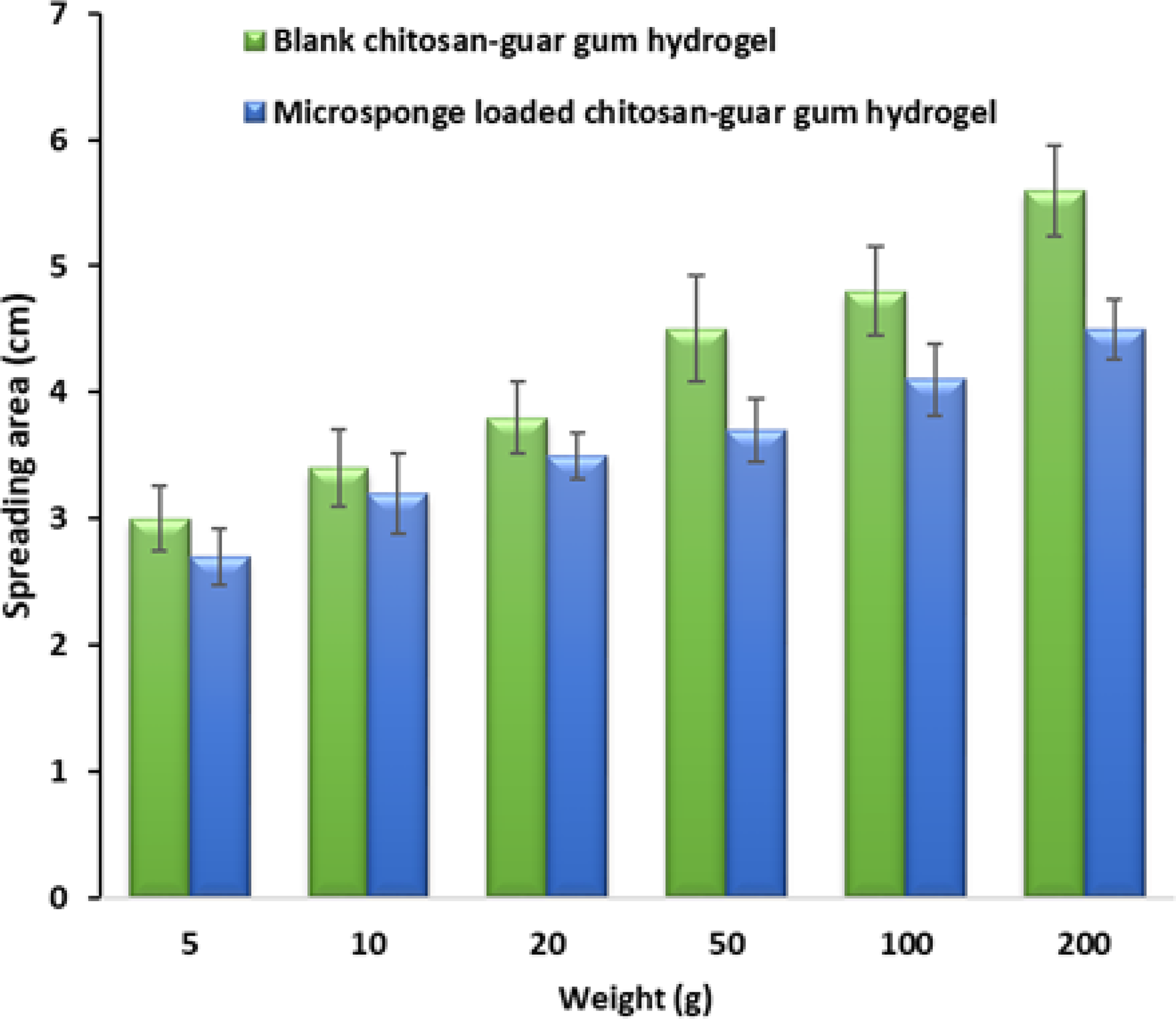
Observed spreading area of blank chitosan-guar gum hydrogel and microsponge-loaded chitosan-guar gum hydrogel with different applied weights.
Rheology Study
The prepared hydrogels viscoelastic behavior under oscillatory shear was examined using the frequency sweep method. The applied strain was kept within the LVER region while the experiment was conducted in the frequency range of 0.1–100 rad/s at 25°C. The LVER provides reliable and accurate data for interpretation by ensuring that the material reaction remains linear and unaffected by structural changes. In this case, the storage modulus (G′) depicts elastic or solid-like behavior, but the loss modulus (G″) exhibits viscous or liquid-like reactivity. 39 For well-structured, solid-like systems, G′ is often frequency-independent, but, in fluid-like materials, it becomes frequency-dependent. 58 Therefore, comparing the frequency-dependent changes in G′ and G″ between EAMS-CGG hydrogel and blank CGG hydrogels provides information about their viscoelastic stability and internal structural organization.
Results of the frequency sweep test of the blank CGG and EAMS-CGG hydrogels are presented in Figures 4 and 5, respectively. The profile in Figure 4 showed that both G′ and G″ increased with frequency, but G′ was greater than G″. Supplementary Table S3 shows that G′ and G″ values were low, ranging from 0.0004 to 57 Pa and 1.99 to 35.6 Pa, respectively, over the observed frequencies. The corresponding loss factor values, which were higher (0.2–20,000), revealed a weaker structural network, indicating that viscous behavior dominated elasticity, particularly at lower frequencies. Figure 5 demonstrates a significantly stronger elastic response than the blank formulation. The data in Supplementary Table S4 indicated a significant increase in G′ (113–237 Pa) and G″ (2.35–29.2 Pa), with consistently lower loss factor values (0.016–0.171) than the CGG blank, demonstrating a strong elastic and solid character of the EAMS-CGG. The possible reasons for the increase in G′ after microsponge incorporation could be linked to the improved cross-linking density and stronger intermolecular interactions within the hydrogel matrix, which in turn would have restricted polymer chain mobility. A change from primarily viscous to elastic behavior is confirmed by the decreased loss factor values seen here, indicating the critical function that microsponges play in maintaining the hydrogel network. From the clinical application perspective, the filled gel will probably resist creep and maintain its form better than the blank, which will spread more readily due to its higher relative viscous contribution and lower G′.
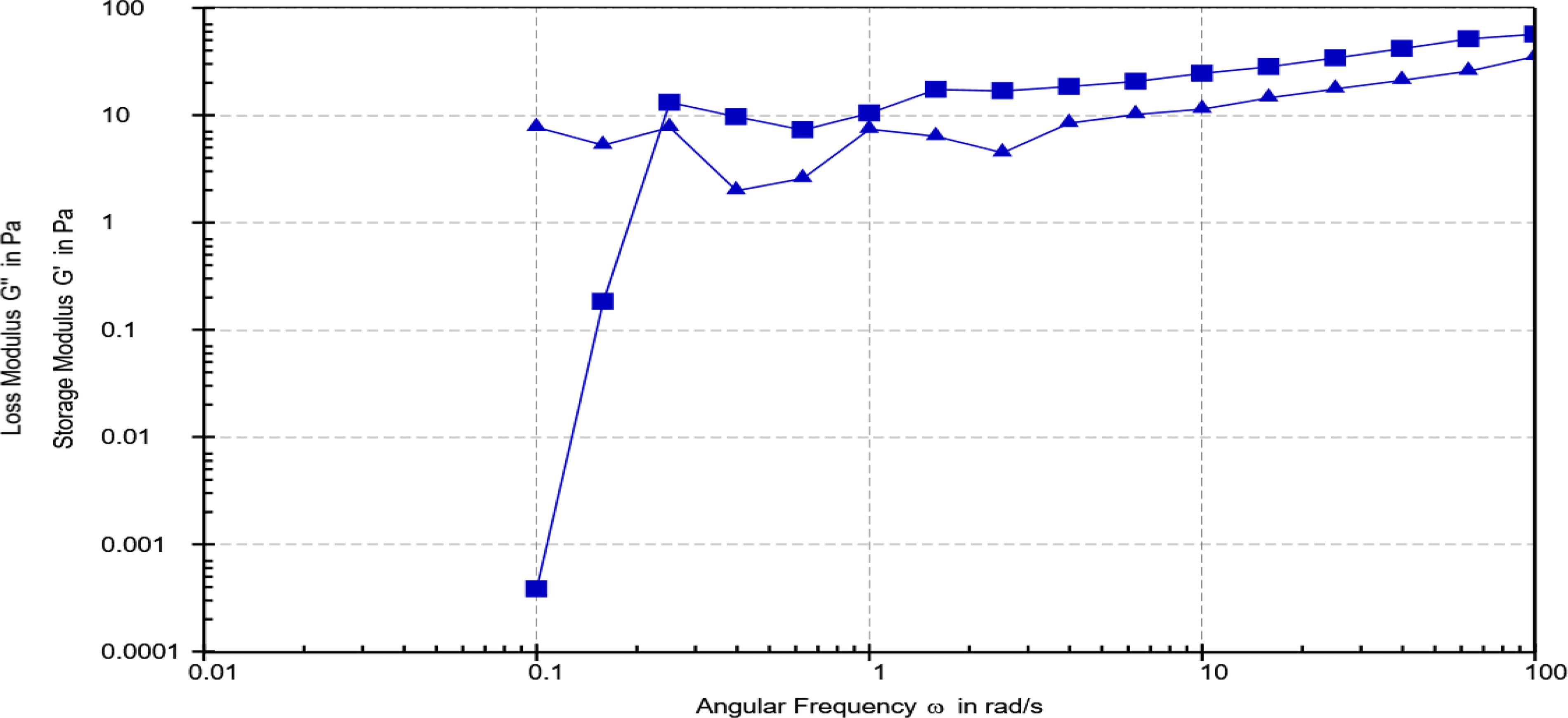
Frequency sweep test of blank chitosan-guar gum hydrogel showing storage modulus (G′, ■) and loss modulus (G″, ▲) values measured over the angular frequency range of 0.01–100 rad/s at 25°C.
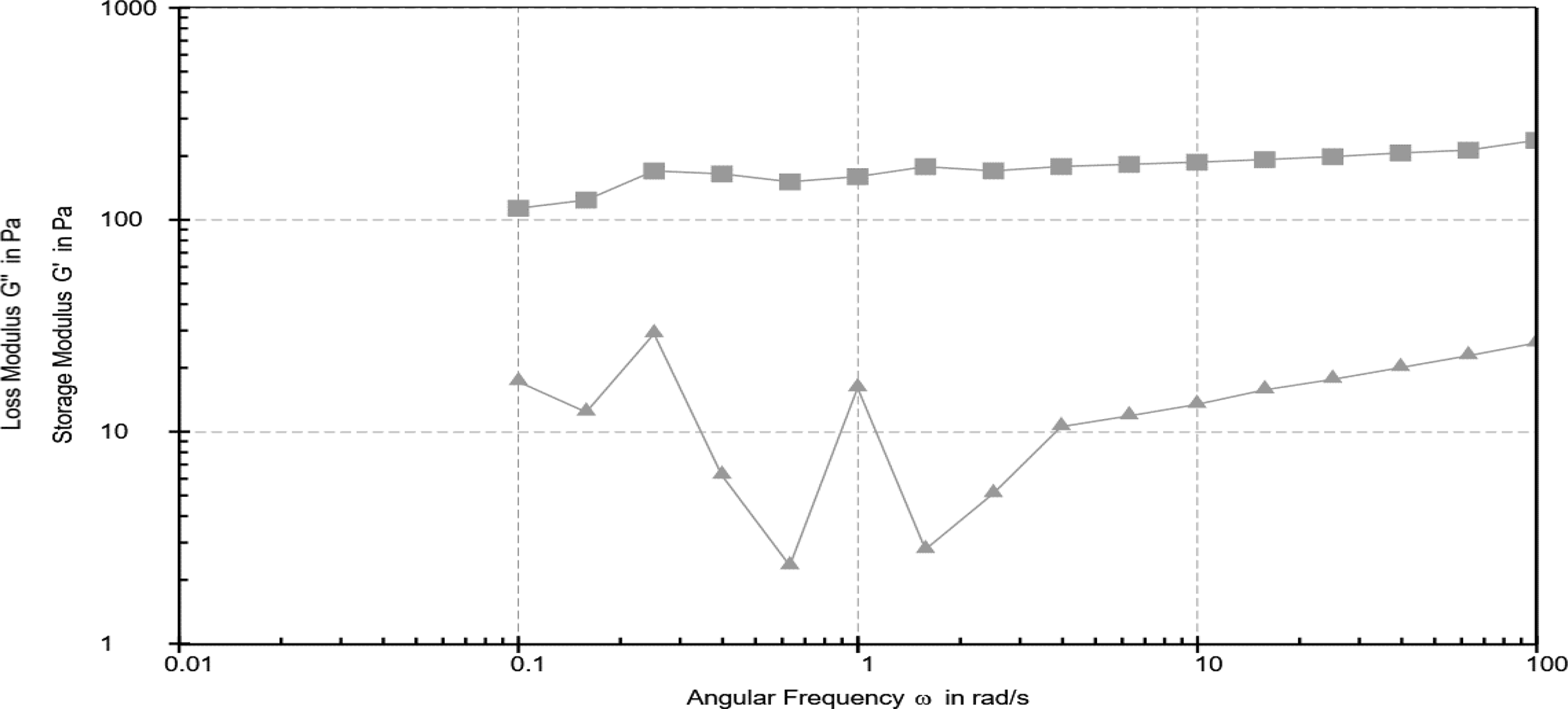
Frequency sweep test of microsponge-loaded chitosan-guar gum hydrogel showing storage modulus (G′, ■) and loss modulus (G″, ▲) values measured over the angular frequency range of 0.01–100 rad/s at 25°C.
In addition to the frequency sweep measurement, the thermal stability of the blank and EAMS-CGG hydrogels was evaluated using the rheological temperature sweep study. Temperature sweeps reveal the hydrogel network’s resistance to thermal softening, offering insight into its durability at physiologically relevant and higher temperatures, whereas frequency sweeps measure the viscoelastic response to oscillatory shear at a constant temperature. 59 Results of the temperature sweep test of the blank CGG and EAMS-CGG hydrogels are presented in Figures 6 and 7 , and Supplementary Tables S5 and S6, respectively. The profile of blank hydrogel in Figure 6 showed changes in G″ values along with a relatively low G′ (20–25 Pa at 25°C), which gradually reduced with increasing temperature. At high temperatures, the loss factor (tan δ) often surpassed 0.4 and in some cases neared 0.7, suggesting a primarily viscous reaction and insufficient structural integrity (Supplementary Table S4). These results imply that the blank hydrogel matrix is susceptible to heat and cannot sustain a stable elastic network when heated. On the other hand, the G′ of the EAMS-CGG hydrogel was almost an order of magnitude greater (G′ = 200 Pa at 25°C) and very stable throughout the studied temperature range (25–50°C) ( Figure 7 ). A robust cross-linked network and a dominant elastic character were confirmed by the consistently low tan δ values (0.04–0.07), underscoring the reinforcing function of microsponges inside the CGG matrix. Furthermore, the complex viscosity of the EAMS-CGG system remained significantly greater than that of the blank, indicating increased structural robustness. This significant difference is consistent with the frequency sweep results, which showed that the formulation is mechanically and thermally resilient due to the microsponge inclusion, as observed with higher G′ values across frequencies tested. The combined results of the two sets of rheological tests support that the hydrogels loaded with microsponges have higher viscoelastic stability. Similar results have been shown for other polysaccharide hydrogels reinforced with microparticles, where the addition of rigid microstructures enhanced the G′ and decreased temperature sensitivity by limiting the mobility of polymer chains and forming extra physical crosslinks.60,61 In addition to stabilizing the hydrogel network, this reinforcement improves its resistance to environmental stress during application, handling, and storage. Overall, the potential of the EAMS-CGG hydrogel to retain high viscosity and elastic strength observed here is beneficial for the topical drug delivery. Stable rheological behavior indicates that the system may be able to maintain its structural integrity while being applied to the skin surface, which could lead to better patient compliance and prolonged drug release. However, the rheological behavior after drug release was not studied in order to check the mechanical and viscoelastic stability during application. It should be noted that in the developed formulation, the EA was encapsulated within polymeric microsponges and dispersed within the crosslinked CGG hydrogel matrix. Therefore, the structural integrity and viscoelastic behavior of the gel are likely governed by the polymer network rather than by the drug itself. Therefore, it is not anticipated that the diffusion of EA from the microsponges will significantly change the hydrogel’s rheological properties. However, further research is recommended to confirm this.
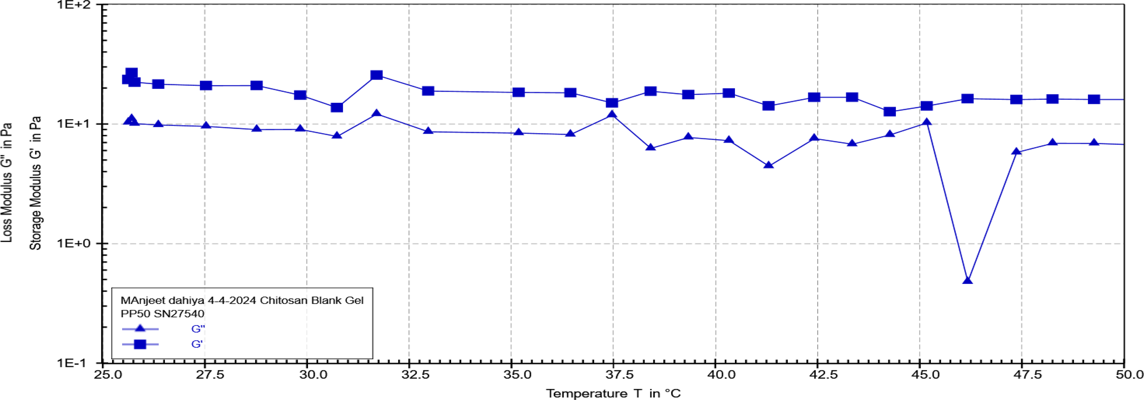
Temperature sweep test of blank chitosan-guar gum hydrogel showing storage modulus (G′, ■) and loss modulus (G″, ▲) values measured over the range of 25–50°C at a constant angular frequency (10 rad/s).
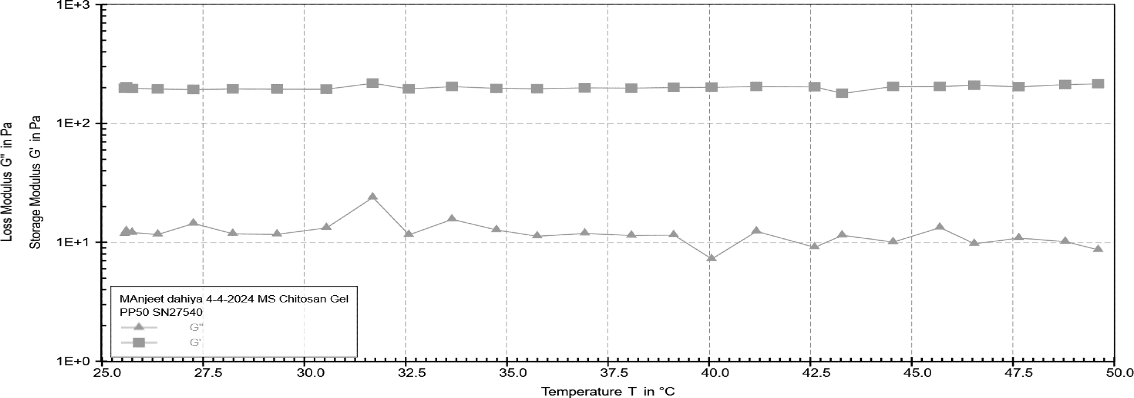
Temperature sweep test of microsponge-loaded chitosan-guar gum hydrogel showing storage modulus (G′, ■) and loss modulus (G″, ▲) values measured over the range of 25–50°C at a constant angular frequency (10 rad/s).
In Vitro Ellagic Acid Release
In vitro drug release studies are essential to predict the in vivo performance because they provide insights into the developed formulations. 62 The cumulative drug release profiles of formulations are presented in Figure 8 . It is evident that the release profile of pure EA differs from the other two microsponge formulations, which are quite comparable. The profile shows burst release of EA in the first hour, with 42% from pure form and ∼25% from EAMS, indicating a reduction in drug release from developed formulations. This slow initial EA release from the microsponge formulation is advantageous for topical therapy as it reduces the risk of higher drug levels in the skin surface. The drug release from EAMS and EAMS-CGG hydrogel continued to release drug for 12 h, while the pure drug release was completed in 6 h. The percentage of drug released in 6 h was 97.68%, 70.78% and 64.37% with EA, EAMS, and EAMS-CGG hydrogel, respectively. The sustained release observed here with microsponges is advantageous, as it can provide constant drug level at the wound site, thereby enhancing wound healing. Similar findings have been reported in the literature, where ∼52% of EA was released after 6 h when formulated into a topical gel. 63 Common kinetic models were fitted to release data, and the R2 value was determined to identify the best fit. The results indicate higher R2 values (0.981, 0.989, and 0.987 for EA, EAMS, and EAMS-CGG hydrogel, respectively) with the Higuchi model, consistent with diffusion-controlled release. The high R2 values for the Higuchi model suggest that the release is predominantly by diffusion through the porous matrix and/or surrounding vehicle under the experimental conditions. Similar release kinetics were observed for glabridin from the microsponge-loaded gel. 47
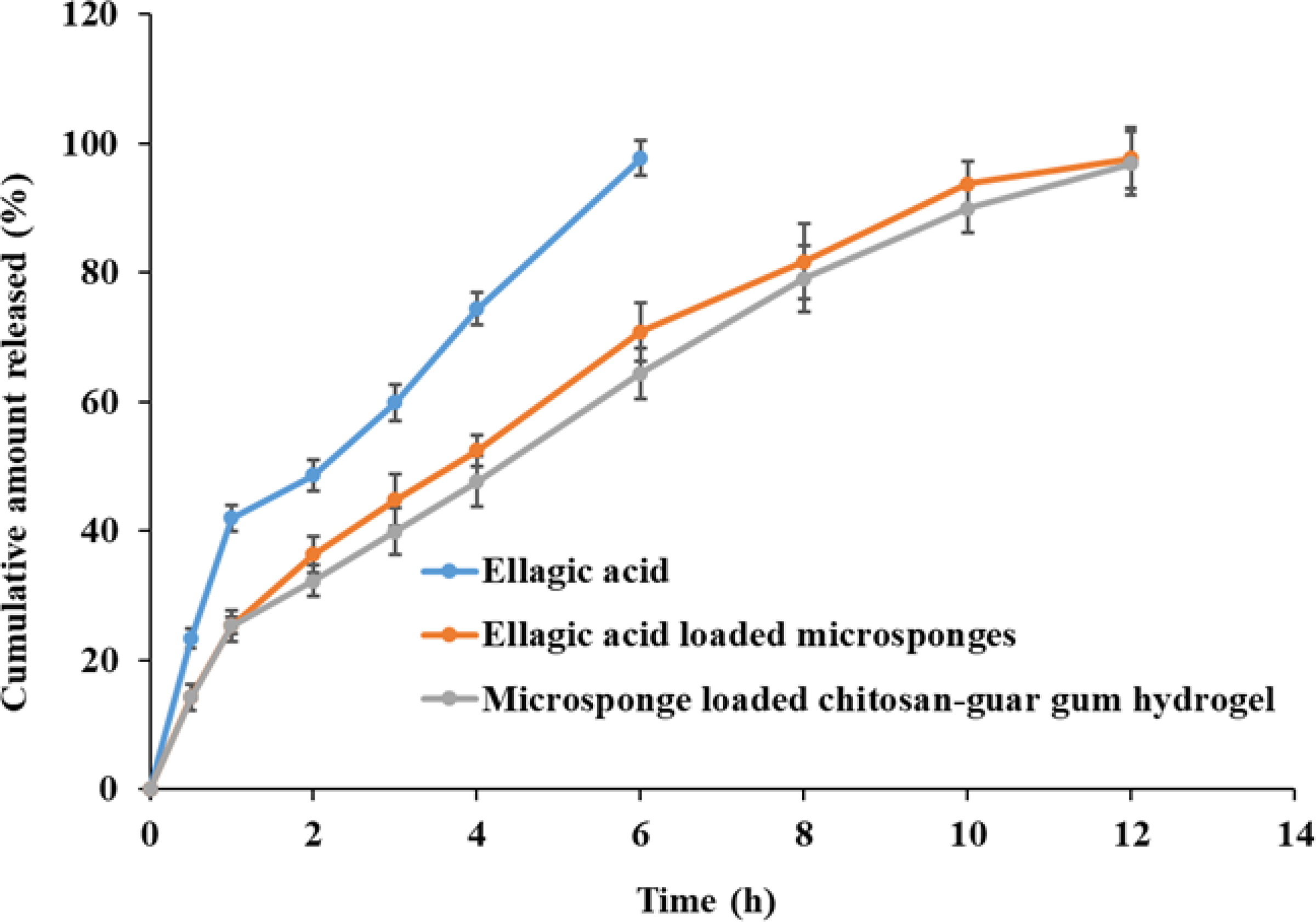
Cumulative amount of drug released from pure ellagic acid, ellagic acid-loaded microsponges, and microsponge-loaded hydrogel.
Evaluation of Antioxidant Activity
The antioxidant potential of pure EA, EAMS, and EAMS-CGG hydrogel was evaluated using the DPPH radical scavenging assay. The DPPH method is a popular and reliable tool to assess the free radical scavenging efficiency of bioactives. 64 EA is known to inhibit neutrophilic reactive oxygen species production in a dose-dependent manner, which contributes to its high antioxidant capabilities and possible therapeutic advantages. Its capacity to neutralize free radicals such as superoxide (O2−) and hydroxyl (OH−) ions is widely recognized. 65 In this experiment, better antioxidant activity is indicated by a greater decrease of DPPH radicals, which is correlated with greater discoloration of the DPPH solution. Figure 9 shows that all three formulations had dose-dependent free radical scavenging activity. EA had the highest inhibitory effect (91.32 ± 2.46% at 100 µg/mL), followed by EAMS (86.96 ± 2.24%) and EAMS-CGG (87.15 ± 0.98%) among the formulations tested. The differences between EA acid and its formulations were more noticeable at lower doses (5–20 µg/mL), indicating that the encapsulation of EA in hydrogels and microsponges may have decreased its immediate antioxidant ability. The antioxidant activity of formulations, however, approached that of EA at higher doses (50–100 µg/mL), suggesting effective release of the active ingredient from the microsponge and hydrogel matrices. Consistent with the observed scavenging activity, the gradual disappearance of the violet color of the DPPH solution confirmed the decrease of DPPH radicals. Comparable patterns have been noted for natural antioxidants and encapsulated polyphenols, where incorporation of hydrogel or microparticles moderately delayed radical scavenging at low concentrations but retains antioxidant activity. These results imply that the hydrogel and microsponge systems successfully preserve EA’s antioxidant properties while providing the possible advantages of increased stability and constant release.
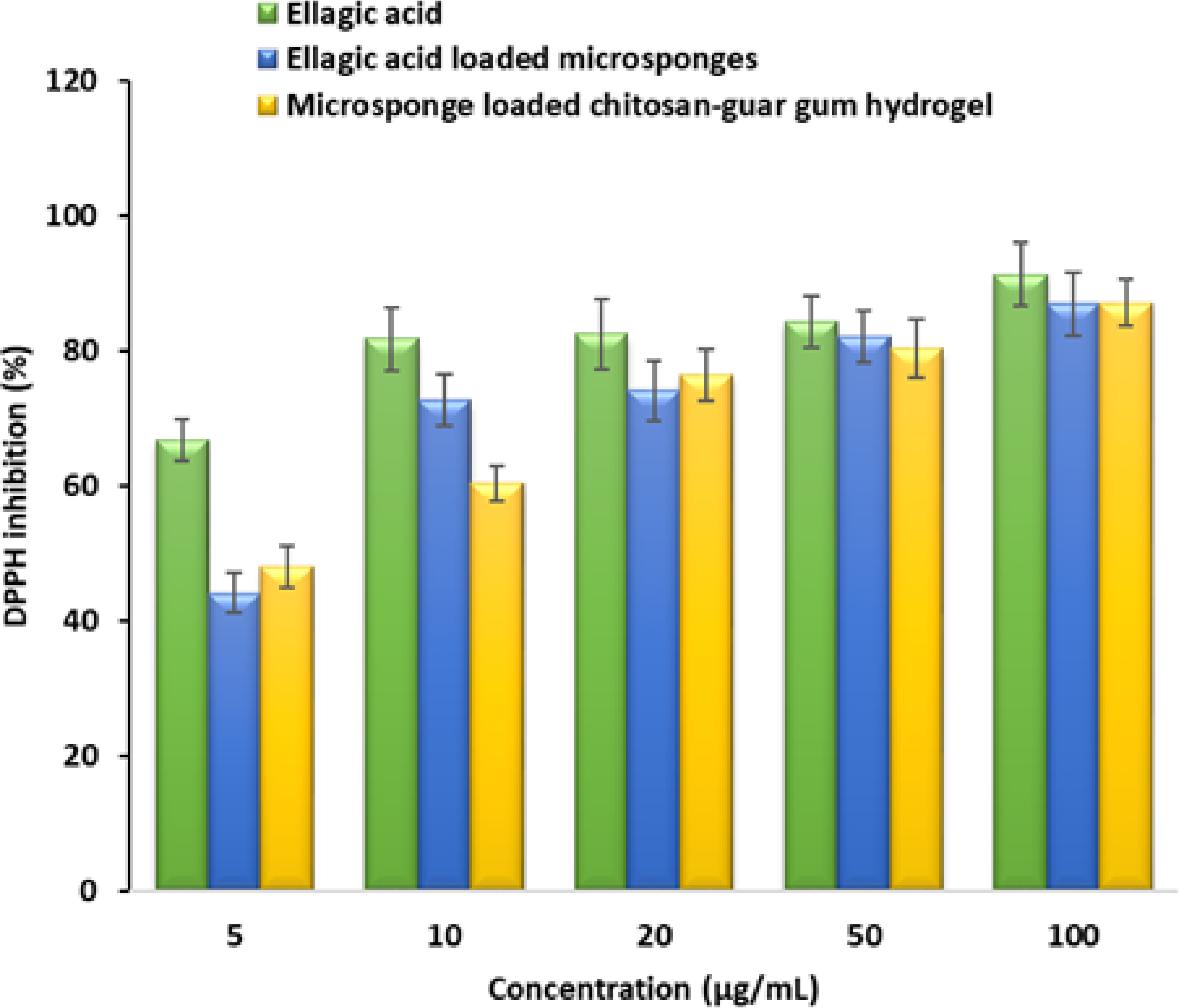
DPPH radical scavenging activity of pure ellagic acid, ellagic acid-loaded microsponges, and microsponge-loaded hydrogel.
In Vitro Antimicrobial Activity
Antimicrobial activity of pure EA, EAMS, and EAMS-CGG hydrogel was evaluated against Gram-positive (S. aureus) and Gram-negative bacteria (E. coli and P. aeruginosa) to assess the efficacy of developed formulations. Literature indicates that both Gram-positive bacteria and Gram-negative bacteria are frequently associated with infections, with P. aeruginosa and S. aureus being among the most prevalent pathogens responsible for chronic infections.51,66 The results presented in Figures 10 – 12 of broth-dilution assays demonstrated clear antimicrobial activity of all the tested products against three bacterial strains at 50 µg/mL. In addition, it also revealed that the EA maintains a high level of antibacterial activity upon encapsulation in microsponges. The visual observation of the lowest concentration showing no turbidity indicated that EA was most effective against E. coli (12.5 µg/mL), while EAMS and EAMS-CGG hydrogel needed 25 µg/mL. Interestingly, EAMS-CGG achieved inhibition against P. aeruginosa at 12.5 µg/mL, outperforming the other two that showed effect at 25 µg/mL. Given that Pseudomonas is a frequent opportunistic pathogen in chronic wounds, this observation is of utmost importance, and the possible reason for this excellent activity could be due to the synergistic effects of chitosan or better bacterial contact. Moreover, a comparison of the inhibition by EAMS-CGG hydrogel against the three strains tested shows that this formulation is more effective against P. aeruginosa, followed by E. coli and S. aureus at the same concentration. Notably, turbidity grading demonstrated that S. aureus continuously exhibited greater resistance than E. coli and P. aeruginosa, with visible growth persisting at intermediate concentrations. At 25 µg/mL, EA showed suppression of S. aureus growth, but EAMS and EAMS-CGG hydrogel showed inhibition of these microbes at 50 µg/mL. Overall, the results of this study demonstrate the importance of the developed microsponge-based formulation as a potential topical therapy for wound management. Indeed, the antioxidant and antibacterial activities demonstrated in this study provide preliminary biological evidence for the prepared formulation’s possible use in wound management. However, further mechanistic research and in vivo evaluation would provide more insight into the therapeutic significance and translational potential of this hydrogel system.
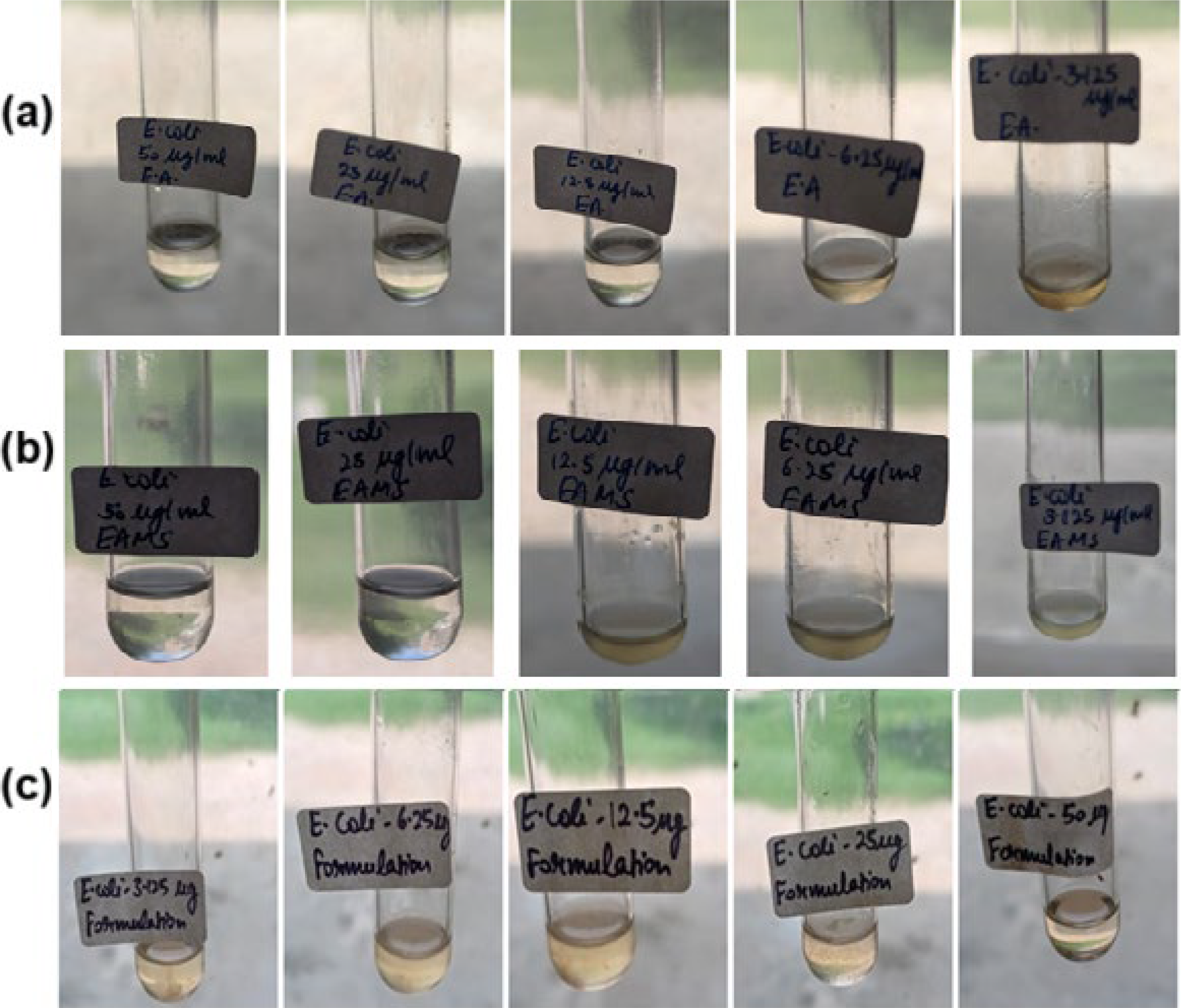
Photographic images showing the antimicrobial activity of pure ellagic acid
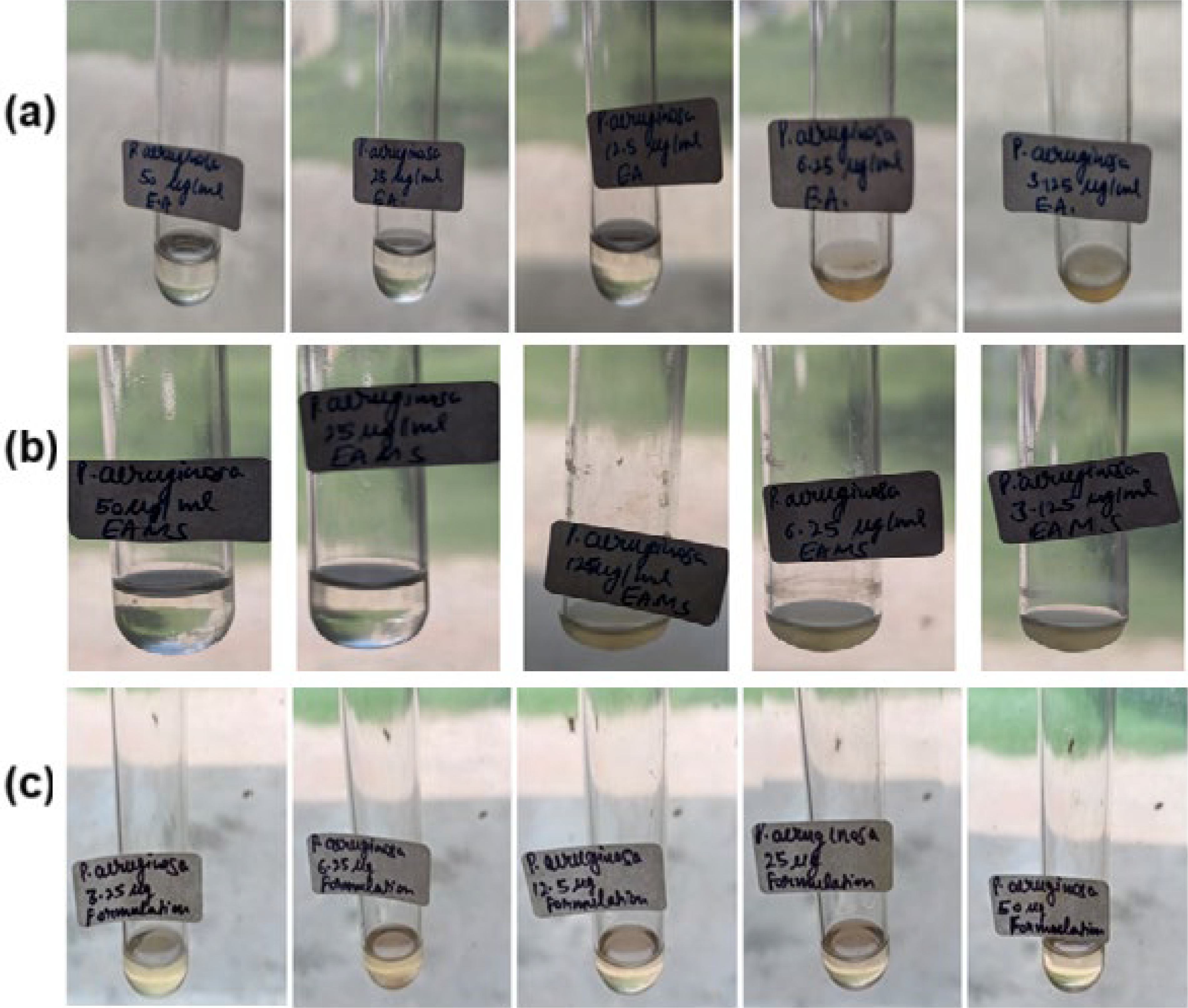
Photographic images showing the antimicrobial activity of pure ellagic acid
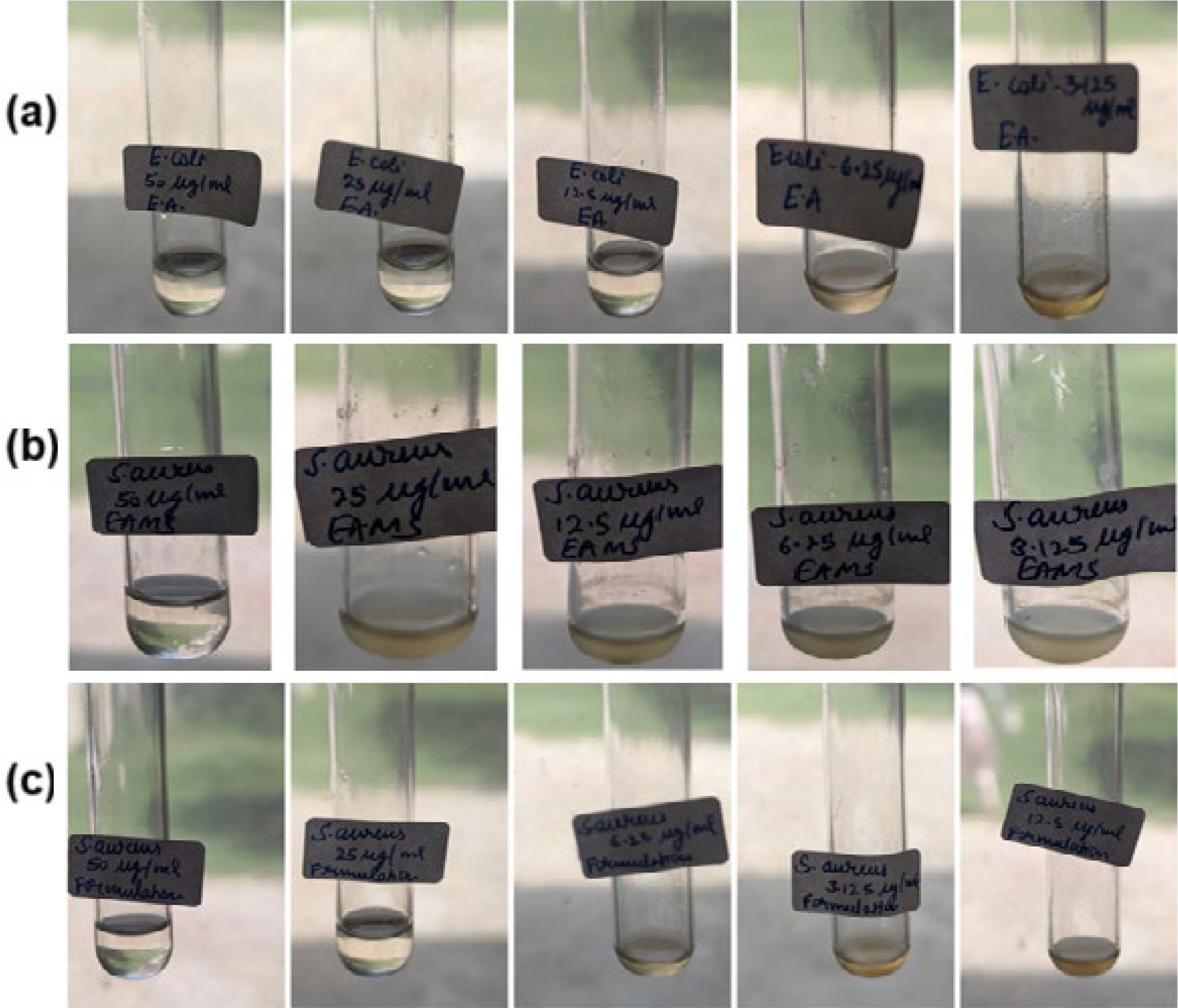
Photographic images showing the antimicrobial activity of pure ellagic acid
Stability Study
Visual analysis of the formulation reveals that the off-white color was stable for four weeks. There was no significant change in the drug content (98.51 ± 1.22%), pH (5.4 ± 0.31), or viscosity (32,500 ± 452 mPa·s) of the tested formulation during the study period at 2–8°C. Overall, the results of stability studies of EAMS-CGG hydrogel show adequate stability throughout the study period, indicating the potential for its clinical use.
CONCLUSIONS
This study assesses the feasibility of developing EAMS-CGG hydrogel and its prospective for topical therapy in wound healing. Microsponges were prepared with ethyl cellulose, DCM, and PVA and were incorporated into a selected hydrogel (GA2) prepared using chitosan (1.0%), guar gum (0.25%), and glutaraldehyde (0.1%). FTIR investigations demonstrated that the bioactive remains intact and chemically compatible within the developed EAMS-CGG hydrogel. The good spreadability noticed with the developed formulation suggests its suitability for topical wound therapy. The rheological investigation indicates that microsponge loading significantly improves the structural integrity and mechanical stability of the hydrogel, which is advantageous for topical applications requiring controlled drug delivery and prolonged retention at the site of application. In vitro drug release studies showed slow and sustained drug release from microsponges, which is advantageous and can provide steady drug level at the wound site, thereby enhancing wound healing. The antioxidant effect exhibited by the microsponge systems indicates successful preservation of EA’s antioxidant activity. Moreover, the developed formulation showed greater antimicrobial efficacy against both Gram-positive and Gram-negative bacterial strains tested. Overall, the results here demonstrate that the developed microsponge hydrogel is a suitable candidate for the management of wound healing, and further in vivo studies are recommended to assess its clinical efficacy.
AUTHORS’ CONTRIBUTIONS
Conceptualization: A.B.N. and R.R.; Data curation: M.D., A.B.N., and R.R.; Formal analysis: M.D., A.B.N., V.K., A.S.A., S.J., B.A., R.M.A., and R.R.; Investigation: M.D., A.B.N., V.K., A.S.A., S.J., B.A., R.M.A., and R.R.; Writing—original draft: M.D., A.S.A., S.J., B.A., R.M.A., and R.R.; Writing—review and editing: A.B.N., V.K., and R.R. All authors read and approved the final article.
DATA AVAILABILITY STATEMENT
The data presented in this study are contained within the article.
Footnotes
ACKNOWLEDGMENTS
The authors acknowledge the Deanship of Scientific Research, Vice-Presidency for Graduate Studies and Scientific Research, King Faisal University for the support.
DISCLOSURE STATEMENT
The authors declare no conflict of interest.
FUNDING INFORMATION
This work was supported through the Ambitious Researcher track-Research Articles by the Deanship of Scientific Research, Vice Presidency for Graduate Studies and Scientific Research, King Faisal University, Al-Ahsa, Saudi Arabia [Grant Number KFU261885].
Supplemental Material
Abbreviations used
References
Supplementary Material
Please find the following supplemental material available below.
For Open Access articles published under a Creative Commons License, all supplemental material carries the same license as the article it is associated with.
For non-Open Access articles published, all supplemental material carries a non-exclusive license, and permission requests for re-use of supplemental material or any part of supplemental material shall be sent directly to the copyright owner as specified in the copyright notice associated with the article.