
Abstract
Objective(s):
Sentinel lymph node (SLN) mapping commonly involves presurgical administration of a radioactive colloid and intraoperative injection of a blue dye near the tumor. Combining gamma scintigraphy and visual inspection could reduce false-negative rates. This study introduces novel imaging agents—radioactive nanoliposomes (NLs) encapsulating patent blue dye—for lymph node scintigraphy.
Materials and Methods:
PEGylated (PEG-NLs) and non-PEGylated (non-PEG-NLs) NLs were prepared using the thin-film hydration method with patent blue dye and labeled with 99mTc-hexamethylpropylene-amine-oxime (99mTc-HMPAO). Lymphatic drainage of the radiolabeled liposomes was assessed in BALB/c mice following subcutaneous footpad injections. Planar imaging was performed at 0.5 and 1 hour postinjection.
Results:
The mean diameter, zeta potential, and polydispersity index of the PEG-NLs were 130.7 ± 0.348 nm (n = 3), −22.4 ± 0.54 mV, and 0.118 ± 0.12, respectively. These values for the non-PEG-NLs were 120.46 ± 0.506 nm (n = 3), 7.5 ± 0.65 mV, and 0.055 ± 0.009, respectively. 99mTc-HMPAO-PEG-NLs had a significantly higher lymph node uptake at earlier times after injection (28.27 ± 5.22% ID/g at 1 hour and 16.65 ± 4.23% ID/g at 2 hours), which was followed by fast washout at 4 hours postinjection and showed fast migration of PEGylated NLs through lymphatic system. 99mTc-HMPAO-non-PEG-NLs had an increased lymph node accumulation through entire time after injection (11.95 ± 0.8% ID/g, 22.95 ± 4.56% ID/g, 29.71 ± 2.16% ID/g at 1, 2, and 4 hours, respectively). Encapsulation efficiency of patent blue dye was determined to be 1.26 ± 0.1%.
Conclusion:
99mTc-HMPAO PEG-NLs and non-PEG-NLs containing Paten Blue dye are promising agents for SLN mapping, offering distinct advantages in uptake kinetics and retention for lymphatic imaging and visual detection.
Introduction
Solid tumors often have the potential to metastasize through the lymphatic system, making lymph node involvement a critical factor in patient staging and risk stratification. The sentinel lymph node (SLN), the first node to receive drainage from a primary tumor, plays a pivotal role in this process. SLN mapping enhances the diagnosis of nodal metastases; if this node is free of tumor cells, the likelihood of further metastasis is significantly reduced. Conversely, involvement of the sentinel node indicates potential spread to distant organs, necessitating refinement in treatment and prognosis. 1,2 Accurate localization during surgery is crucial for effective SLN biopsy (SLNB) and provides a minimally invasive technique with valuable insights into regional node status without any necessity to full dissection. 3
Clinically, mapping agents for the lymphatic system include 99mTc-labeled colloids and blue dyes. The former provides radioactive signals preoperatively, while the latter offers color visualization during surgery. 4,5 Preoperative lymphoscintigraphy, performed in nuclear medicine, identifies the lymphatic drainage of the tumor basin and nodes at risk for metastatic spread. These images guide surgeons to find the most likely SLNs. Previous studies reported a detection rate of 83.5% for 99mTc-sulfur colloids and 69.5% for vital blue dye. However, when both techniques are used simultaneously, the detection rate increases to 96%. 6
Nanoscale vesicles called liposomes, composed of lipid bilayers, have emerged as flexible and promising drug delivery vehicles in nanomedicine. These self-assembling spheres, made up of phospholipid bilayers, can encapsulate hydrophilic agents such as dyes, drugs, and radiopharmaceuticals. 7,8 In the early 1970s, G. Gregoriadis was the first to propose the use of liposomes as drug delivery carriers to transport drugs to cell membranes. However, it was not until the 1990s that liposome-based drug formulations were introduced to the market, following extensive clinical trials. 9,10 The liposomes formed from natural phospholipids are biologically inert, nonimmunogenic, and nontoxic. Moreover, liposomes are biocompatible and biodegradable, making them a highly desirable approach for various applications. 11 –13 In 1977, Richardson et al. introduced [99mTc] liposomes as a promising agent for lymphoscintigraphy. 14 Following this initial report, several clinical studies demonstrated encouraging outcomes. In recent years, substantial efforts have been devoted to the development of liposomal formulations for diagnostic and therapeutic applications, particularly in cancer imaging and treatment. 15,16 These early investigations utilized a labeling technique involving stannous chloride to bind reduced 99mTc to the liposome surface. However, this method proved to be unstable in vivo, prompting the development of improved liposome labeling techniques. 17 A standard method for labeling liposomes with 99mTc is 99mTc-hexamethylpropylene-amine-oxime (99mTc-HMPAO), a lipid-soluble complex. 18 Intraliposomal glutathione preload accelerates reduction of this complex inside liposomes, so it can be easily entrapped inside the liposome.
Liposomes containing patent blue dye were labeled with 99mTc-HMPAO to visualize radioactive lymph node at the same time using high-affinity ligands such as biotin and avidine (blue liposome). 19,20 These techniques dramatically reduce the surgical procedure times from hours to minutes, providing a significant increase in the accuracy of SLN identification and a considerable reduction in the morbidity associated with the staging procedure. The development of 99mTc-HMPAO-labeled nanoliposomes (NLs) shows a significant potential for mapping SLNs and staging cancer. 21,22 Lymphoscintigraphy-guided SLNB is a crucial tool for staging cancer and making treatment decisions, allowing for the targeted removal of the first draining lymph nodes for pathological examination. 23 Radiolabeled liposomes are particularly appealing for lymphoscintigraphy due to their surface’s ability to be easily modified with specific receptors or molecules like polyethylene glycol (PEG). The preparation of liposomes with PEG provides steric hindrance against macrophage phagocytosis, resulting in prolonged circulation time in the bloodstream. However, PEGylation may hinder lymph node retention and accumulation, which are crucial factors for lymphoscintigraphy. 24,25
This study aimed to investigate the lymphatic imaging and lymph node delivery properties of PEGylated and non-PEGylated NLs containing patent blue dye. The findings may be particularly relevant for surgeons and therapists in the further clinical studies.
Materials and Methods
Materials
HSPC (hydrogenated soy phosphatidylcholine), DSPE-mPEG-2000 {1,2-distearoyl-sn-glycero-3 phosphoethanol amine-N-[methoxy (polyethylene glycol)-2000]}, and cholesterol (Chol) were purchased from Avanti Polar Lipids (Birmingham, USA). Glutathione (GSH) and sephadex G-25 were obtained from Sigma Chemical Co. (St. Louis, MO). HMPAO kit was synthesized and prepared similarly to the Commercial kit (CERETEC; 0.5 HMPAO and 7.6 μg SnCl2). Chloroform and methanol were purchased from Merck (Germany).
99mTc sodium pertechnetate was supplied by a 99Mo/99mTc generator (Kimia Pakhsh). All chemicals were used without further purification.
Preparation of liposomal formulations
PEGylated NLs (PEG-NLs) and non-PEGylated NLs were prepared by hydration of a thin lipid film followed by high-pressure homogenization. 15,21 In brief, the lipid mixture HSPC, DSPE-mPEG-2000, and Chol for PEG-NLs; HSPC and Chol for NLs was dissolved in a chloroform–methanol solution (1:2 volume ratio). The lipid composition was prepared according to the molar ratios specified in Table 1. The solvent was removed using a rotary evaporator under reduced pressure to form a thin lipid film. The lipid film was then rehydrated with a 100 mM GSH solution containing 7 mL of patent blue dye (10 mg/mL). The resulting mixture was vortexed for 10 minutes and sonicated at 65°C for 20 minutes using a bath sonicator (Laboratory Supplies Company Inc., Hicksville, NY) under an argon atmosphere. Afterward, the suspension was homogenized with an EmulsiFlex-C3 high-pressure homogenizer (Avestin, Canada) at 20,000 PSI for three cycles to produce uniform nano-sized liposomes.
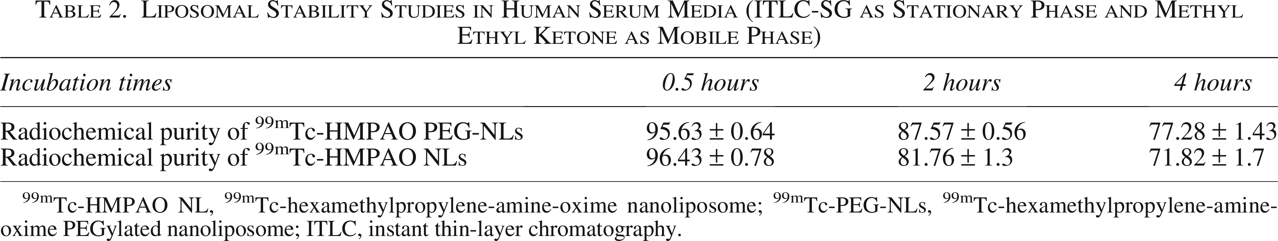
Formulation and Characterization of PEG-NLs and NLs
DSPE-mPEG-2000, 1,2-distearoyl-sn-glycero-3 phosphoethanol amine-N-[methoxy (polyethylene glycol)]-2000; HSPC, hydrogenated soy phosphatidylcholine; NLs, nanoliposome; PDI, polydispersity index; PEG-NLs, PEGylated nanoliposomes.
To remove unencapsulated patent blue dye and GSH, the liposomes were dialyzed for 48 hours at 2–8°C against PBS, with the dialysis buffer being replaced every 4 hours.
The particle size of the liposomes was measured in triplicate using dynamic light scattering on a Nano-ZS instrument (Malvern, UK). The zeta potential was also measured on the same device, with the average value calculated from 20 individual measurements. 26
Radiolabeling of liposomes
The liposomes were radiolabeled using a method adapted from Phillips et al. 27 A solution of liposomes at a concentration of 70 mM was mixed with 370 MBq of freshly prepared 99mTc-HMPAO. The mixture was incubated at room temperature for 30 minutes. After incubation, unbound 99mTc was separated from the liposomes by passing the mixture through a PD-10 chromatography column (Sephadex G-25). The column was eluted with 5% dextrose, and the radiolabeling efficiency was determined by measuring the radioactivity of the liposomes before and after purification using a dose calibrator (Capintec, CRC-15R). 21,28
In vitro stability of the formulations
The stability of the 99mTc-HMPAO-PEG-NLs and 99mTc-HMPAO-NLs formulations was assessed by incubating them in human serum at 37°C for up to 24 hours. At different time points (1, 2, and 24 hours), the radiochemical purity of the liposomes was analyzed using instant thin-layer chromatography (ITLC-SG) with methyl ethyl ketone as the mobile phase and silica gel as the stationary phase. 21
Biodistribution study by lymph node drainage
Animal experiments were conducted in accordance with the NIH Animal Use and Care Guidelines and approved by the Institutional Animal Care and Use Committee of Mashhad University of Medical Sciences. For the lymph node drainage study, 99mTc-HMPAO-Blue-dye-liposomal formulations (37 MBq in 100 µL) were injected into the dorsum of BALB/c mice (three groups, three mice per group). Mice were sacrificed at 0.5, 1, and 4 hours postinjection. Inguinal lymph nodes and foot pads were excised, weighed, and analyzed for radioactivity using a gamma counter (Delshid, DL100). The percentage of the injected dose per gram of organ tissue (%ID/g) was calculated by dividing the activity in the organ by its weight. 17
Animal scintigraphic imaging
BALB/C mice were anesthetized by an intraperitoneal injection of ketamine. A subcutaneous injection of 0.5 mL of 99mTc-HMPAO-Blue-dye-NLs (34 MBq in 100 µL) was administered to the foot pad. Planar scintigraphic imaging was performed using a Siemens dual-head gamma camera equipped with a low-energy high-resolution collimator, set to 140 keV with a ±20% window. Images were acquired with a 256 × 256 matrix and a zoom factor of 2, totaling 300,000 counts at 0.5 and 1 hour postinjection.
Statistical analysis
Data are expressed as mean ± standard deviation. Means were compared using Student’s t-test. The p values of less than 0.05 were considered statistically significant.
Result
Liposome characterization
The final liposomal formulations encapsulated blue dye at concentrations of 14.25 mg/mL for PEG-NLs and 17.5 mg/mL for NLs. The average particle size, zeta potential, and polydispersity index (PDI) for PEG-NLs were 130.7 ± 0.348 nm (n = 3), −22.4 ± 0.54 mV, and 0.118 ± 0.12, respectively. For NLs, the corresponding values were 120.46 ± 0.506 nm (n = 3), 7.5 ± 0.65 mV, and 0.055 ± 0.009. The two formulations had similar particle sizes (Table 1). However, the zeta potential of PEG-NLs was significantly more negative than that of NLs, which can be attributed to the presence of DSPE-mPEG-2000 in the PEGylated formulation. Both formulations exhibited a monomodal size distribution, and the low PDI values indicated a narrow particle size distribution for both types of liposomes.
Radiochemical purity and stability
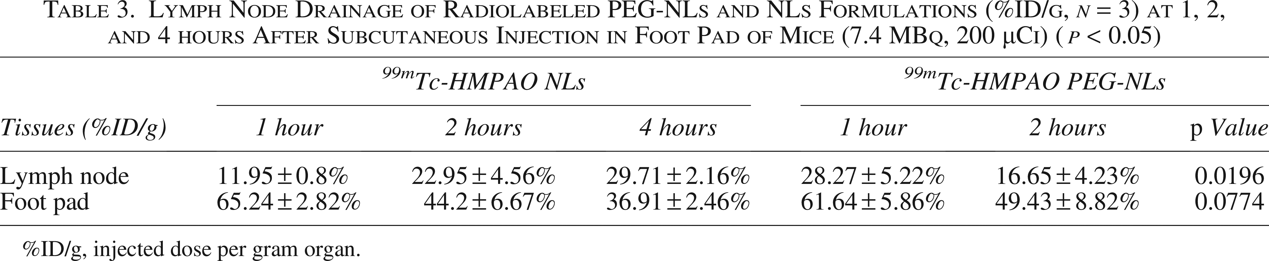
The radiochemical purity of the 99mTc-HMPAO PEG-NLs and NLs formulations was 82.87 ± 1.47% and 93.82 ± 2.4%, respectively. This high radiochemical purity was achieved by passing the liposomes through a PD-10 column (Sephadex G25) to separate the 99mTc-HMPAO-labeled liposomes from free 99mTc-HMPAO. To assess the entrapment efficiency of 99mTc-HMPAO in the liposomes, an in vitro stability test was conducted by exposing the radiolabeled liposomes to human serum. The labeling stability of 99mTc-HMPAO PEG-NLs, as measured by ITLC, was 95.63 ± 0.64%, 87.57 ± 0.56%, and 77.28 ± 1.43% at 0.5, 2, and 4 hours postlabeling, respectively. For 99mTc-HMPAO NLs, the labeling stability was 96.43 ± 0.78%, 81.76 ± 1.3%, and 71.82 ± 1.7% at the same time points (Table 2).
Liposomal Stability Studies in Human Serum Media (ITLC-SG as Stationary Phase and Methyl Ethyl Ketone as Mobile Phase)
99mTc-HMPAO NL, 99mTc-hexamethylpropylene-amine-oxime nanoliposome; 99mTc-PEG-NLs, 99mTc-hexamethylpropylene-amine-oxime PEGylated nanoliposome; ITLC, instant thin-layer chromatography.
Tissue distribution in mice
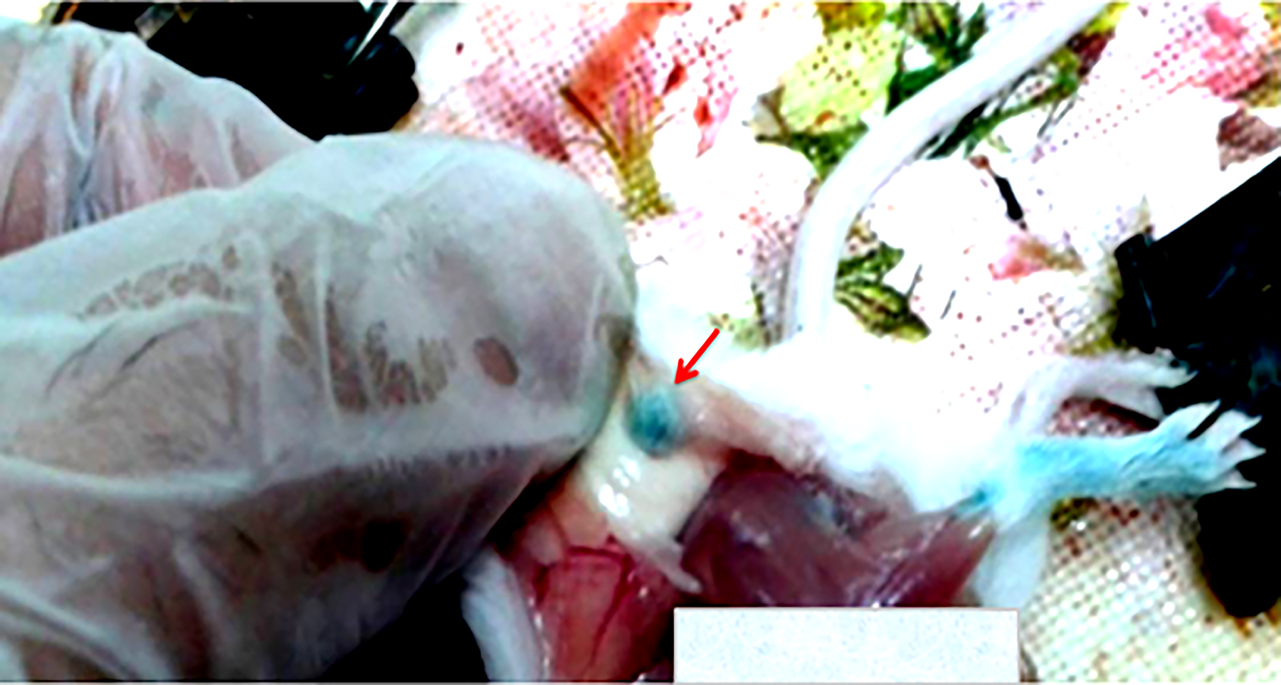
The biodistribution of radiolabeled liposomes was evaluated in mice at various time points (n = 3 per time point). As shown in Table 3, 99mTc-HMPAO-PEG-NLs demonstrated significantly higher lymph node uptake shortly after foot pad injection, with values of 28.27 ± 5.22%ID/g and 16.65 ± 4.23%ID/g at 1 and 2 hours, respectively. However, at 4 hours postinjection, there was a marked decrease in lymph node uptake and a reduction in blue dye staining, indicating rapid migration of PEGylated NLs through the lymphatic system. In contrast, 99mTc-HMPAO-NLs exhibited increased lymph node accumulation over time, with values of 11.95 ± 0.8%ID/g, 22.95 ± 4.56%ID/g, and 29.71 ± 2.16%ID/g at 1, 2, and 4 hours, respectively (Fig. 1). In addition, foot pad uptake for both PEG-NLs and NLs decreased over time, suggesting that the liposomes were draining through the lymphatic system.

Lymph node accumulation of radiolabeled PEG-NLs and NLs formulations (%ID/g, n = 3) at 1, 2, and 4 hours after subcutaneous injection in foot pad of mice (7.4 MBq, 200 µCi) (p < 0.05).
Lymph Node Drainage of Radiolabeled PEG-NLs and NLs Formulations (%ID/g, n = 3) at 1, 2, and 4 hours After Subcutaneous Injection in Foot Pad of Mice (7.4 MBq, 200 µCi) (p < 0.05)
%ID/g, injected dose per gram organ.
Scintigraphic studies in mice
Planar imaging of lymphatic drainage was performed following subcutaneous injection of 99mTc-HMPAO Blue-dye liposomal formulations into the foot pads of each mouse. The kinetic profile revealed rapid migration of the labeled NLs from the injection site toward the inguinal lymph nodes, with peak concentrations observed at 1 hour for both nodes. The inguinal lymph nodes were located approximately 15 mm from the foot pad injection site (Fig. 2). In later images, lymphatic drainage to higher lymph nodes was noted, along with the presence of radioactivity in the heart and liver, indicating the excretion of NLs into the bloodstream (Fig. 3).
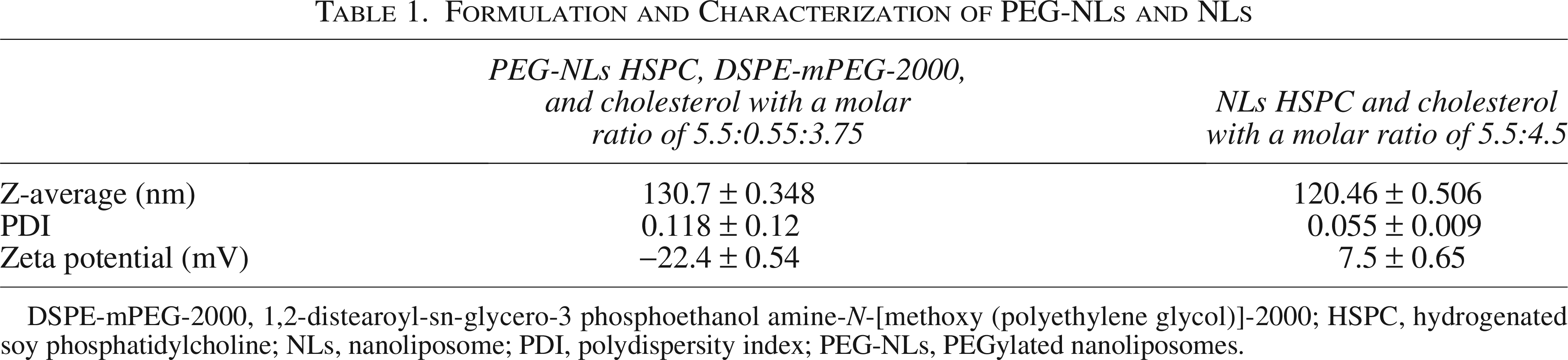
Blue dye visual inspection toward inguinal lymph node after 99mTc-HMPAO-nanoliposome injection in foot pad of a BALB/c mouse.
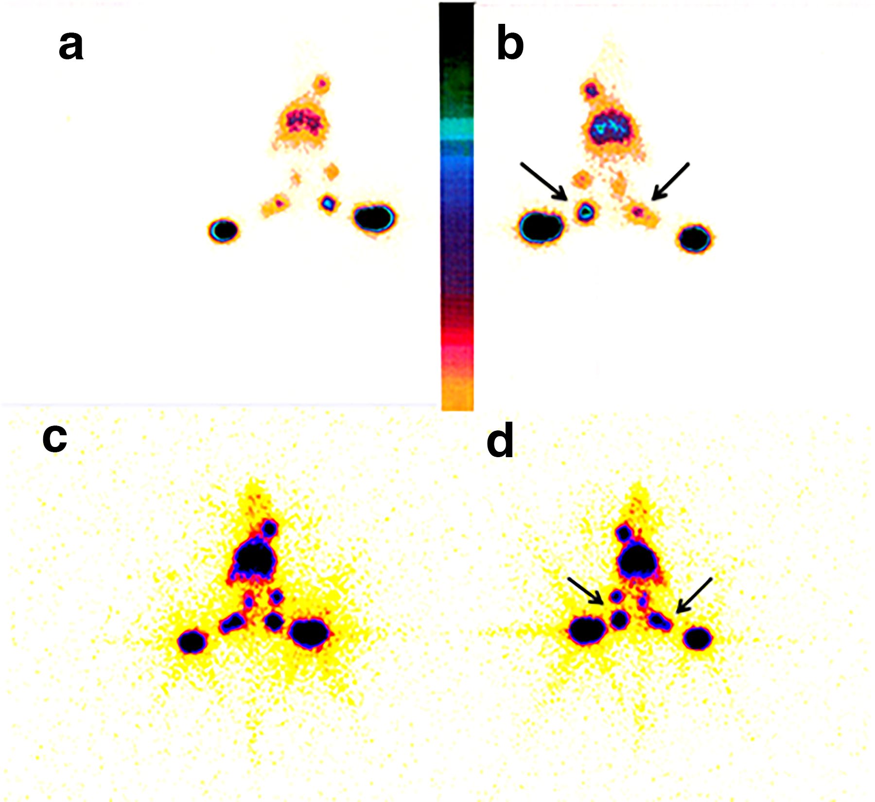
Anterior and posterior images of mouse at 0.5 hours
Discussion
The development of precise imaging agent to distinguish involved lymph nodes plays an important role in patients’ diagnosis and treatment. SLN mapping is a standard, minimally invasive diagnostic method in the surgical treatment of many solid tumors. 1,29,30 The success rate of SLN detection depends largely on the type of radiopharmaceuticals used for its detection. 31 Particle size and surface characteristics can influence the rate of colloid drainage from the injection site to the dermal lymphatic capillaries and, furthermore, phagocytosis by lymph node macrophages. 32,33
The retention time of trapped liposomes in SLNs is limited and influenced by the structure of the NLs. Phagocytosis of NPs typically begins with opsonization, which can take from 30 minutes to several hours, depending on the cell type and particle surface characteristics. The uptake via phagocytosis is determined by the physicochemical properties of nanoparticles, including size, shape, and surface characteristics. 24,34,35
Colloidal radiopharmaceuticals with a particle size of 2–200 nm can easily enter the lymphatic system by endocytosis through the epithelial cell membrane. 32,33 NLs with a particle size of 100–200 nm generally pass through lymphatic system like colloids. Studies have reported that particles in the size range of 14–150 nm are biologically active in macrophage immune response. 24,33,36 In this study, no significant difference between two groups with respect to their particle size was noted (Table 3). 24,34,35 Smaller nanoparticles have low uptake due to their inability to bind to multiple receptor sites or to establish strong associations with receptors before being internalized by the cell membrane. Although larger nanoparticles are capable of multivalent receptor binding, they are too large for efficient membrane engulfment. 24
In our study the uptake of PEGylated NLs peaked in the first hour (28.27 ± 5.22%), which would decline from the lymph nodes in the second hour (16.65 ± 4.23%) (Table 3). PEGylated NLs demonstrated faster migration to the lymph nodes and bloodstream, resulting in no detectable radioactivity in sentinel nodes after 4 hours. In comparison, non-PEGylated NLs migrated much slower than PEG-NLs through lymphatic vessels, which leads to higher accumulation in sentinel nodes over time. Their uptake was 11.95 ± 0.8% in the first hour, which increased exceedingly to 29.71 ± 2.16% in the fourth hour. This difference is largely due to the surface properties of liposomes, influencing phagocytosis, opsonization, and interactions with cell membrane receptors that prolong retention time for non-PEGylated liposomes in 4 hours.
From a clinical and translational perspective, understanding these differences between PEGylated and non-PEGylated liposomes is important for optimizing SLN imaging and surgery. The rate and duration of lymph node retention directly determine the optimal time window for imaging and intraoperative detection. PEGylated liposomes, with their faster lymphatic transit and shorter nodal retention, may be advantageous when imaging or surgery is performed shortly after injection. Conversely, non-PEGylated formulations that remain longer within the SLN can improve detection sensitivity when there is a delay between injection and surgery. Moreover, lymphatic transport can vary among patients due to tumor-induced lymphangiogenesis, obstruction of lymphatic channels, or inflammatory responses, all of which may alter tracer migration and uptake patterns. Recognizing these factors may help clinicians tailor tracer selection and imaging timing to individual patient conditions, improving the accuracy and reproducibility of SLN mapping in clinical practice.
PEGylation of nanoparticles can hinder opsonization by reducing protein adsorption on their surfaces, thereby extending their circulation half-life from minutes to several hours by evading from the reticuloendothelial system (RES). 24,25,37
A key differentiating factor between our formulations was their surface charge. The non-PEG-NLs possessed a cationic charge (+7.5 mV), whereas the PEG-NLs were anionic (−22.4 mV). Cationic particles often exhibit stronger electrostatic interactions with negatively charged cell membranes of phagocytic cells in the lymph nodes, potentially enhancing uptake and leading to the prolonged retention we observed with non-PEG-NLs. 38,39 The anionic charge and steric hindrance from PEGylation reduce nonspecific interactions, facilitating faster transit through the lymphatic vessels but resulting in lower long-term retention within the nodes. Positively charged nanoparticles may enhance membrane wrapping due to their adhesive properties. In comparison, liposomes with negative surface charges are quickly cleared from the bloodstream and phagocytosed by the RES. Although most studies showed phagocytosis differences between cationic and anionic liposomes are mostly dependent on absolute values of zeta potentials. 24,38,39
Therefore, when surgery is scheduled for gamma probe lymphoscintigraphy close to the injection time, the use of PEG-NLs is recommended. On the other hand, if there is a significant time gap between the injection and surgery, non-PEG-NLs are preferred.
Although the general structure and physiological function of the lymphatic system are conserved between mice and humans, several species-specific differences should be considered when interpreting lymphoscintigraphy results. Mice exhibit a higher frequency of lymphatic contractions and lower sensitivity to hydrostatic pressure compared with humans, partly due to their small size and posture during imaging. Furthermore, anatomical drainage patterns differ; for instance, both inguinal and popliteal lymph nodes drain the hind limb in mice, whereas in humans the drainage is more regionally compartmentalized. At the molecular level, lymphatic endothelial cells share conserved gene expression profiles across species, but notable differences exist in genes related to immune modulation and lymphatic transport. These physiological and anatomical distinctions may influence tracer kinetics and nodal retention, and therefore should be considered when extrapolating preclinical data to human applications. 40,41
Future studies should investigate various nanoparticle formulations with different surface charges, sizes, and zeta potentials for lymphoscintigraphy. In addition, analyzing liposome biodistribution in ex vivo models and exploring alternative labeling methods to enhance liposome stability would be valuable. This study also has some limitations. It was conducted in a murine model, which may not fully replicate the complexity of human lymphatic physiology, particularly in pathological conditions such as cancer-related lymphangiogenesis or inflammation. Moreover, the relatively short observation period (up to 4 hours) may not reflect delayed clearance or systemic redistribution that could occur at later time points. Variations in plasma protein composition and immune response in humans may further influence nanoparticle behavior. Taken together, these findings provide valuable insight into how surface chemistry and formulation parameters influence the lymphatic trafficking of liposomes. Future studies—including long-term imaging and validation in large-animal or clinical settings—are warranted to confirm the translational applicability of these findings.
Conclusion
This study highlights the promising role of 99mTc-HMPAO NLs as an innovative imaging agent for SLN diagnosis during surgical procedures. Our results demonstrate that conventional non-PEGylated NLs exhibit superior and sustained retention in lymph nodes over time, while PEGylated NLs provide a strong initial signal. This prolonged retention of non-PEGylated NLs makes them particularly suitable for surgical procedures where there is a significant delay between injection and gamma probe detection. Notably, non-PEGylated NLs and PEGylated NLs successfully identified the first draining lymph node within 30 minutes postinjection, both visually and with gamma probe. This rapid identification, coupled with the prolonged retention time and high detection rate due to slow clearance from lymphatic vessels, underscores the potential of labeled NLs to enhance SLN mapping and improve surgical outcomes. These findings may guide the rational design of next-generation dual-modality SLN tracers for improved intraoperative guidance and patient outcomes.
Authors’ Contributions
N.H. and H.A.: Investigation, formal analysis, and tabulation; P.S. and A.Aghaee.: Literature search and writing—original draft; S.B. and A.Abbasi.: Resources and investigation; A.B. and S.R.Z.: Supervision; M.R.J.: Validation and review and editing; R.S.: Supervision, validation, and visualization; and K.S.: Conceptualization, supervision, data curation, review and editing, methodology, and project administration. All authors have read and agreed to the published version of the article.
Footnotes
Author Disclosure Statement
The authors declare that they have no conflicts of interest.
Funding Information
The research has been part of a pharmacy doctorate thesis and was supported and approved by Research Department of Mashhad University of Medical Sciences (grant number: 900543).