



Abstract
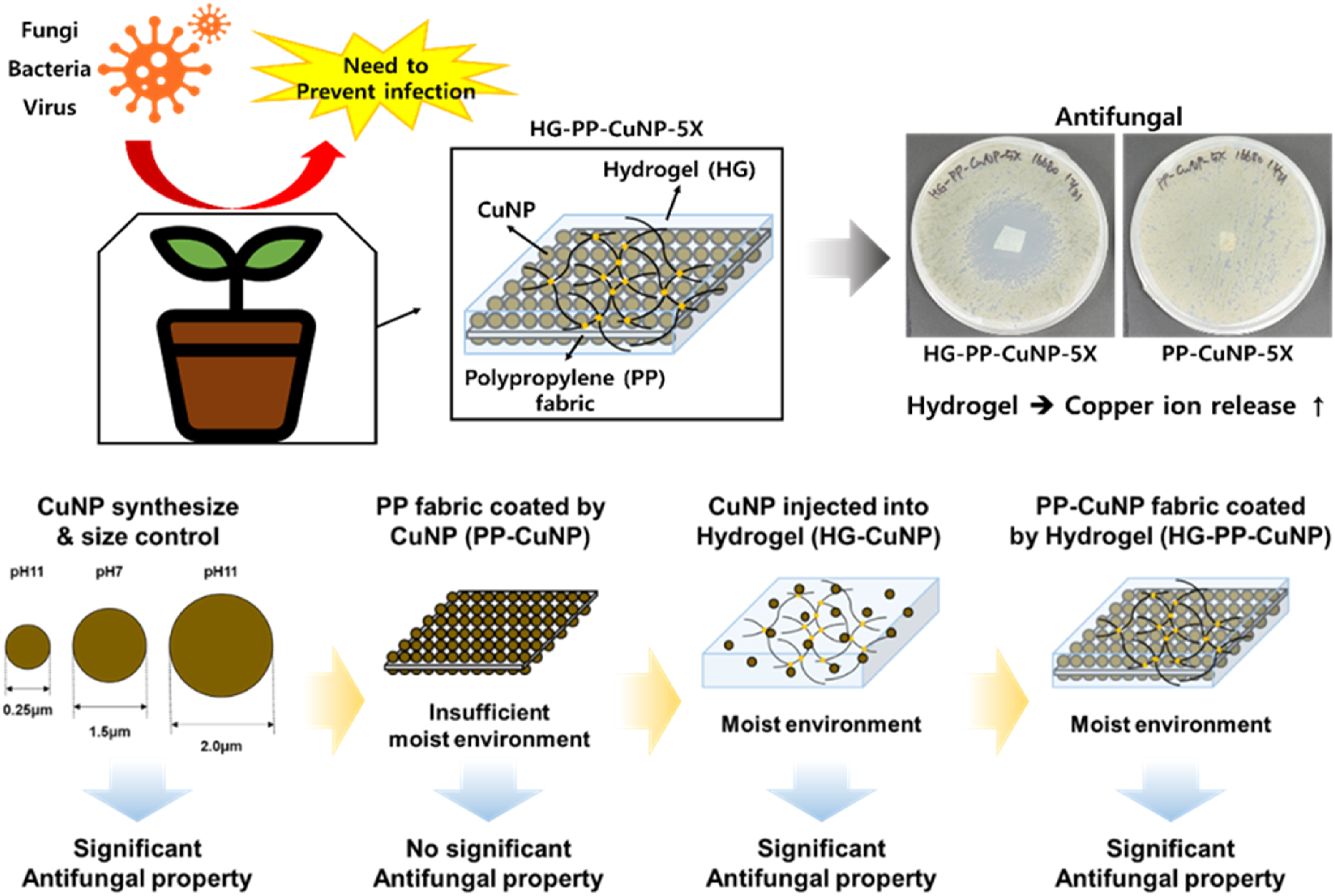
Fungal contamination on textile surfaces can contribute to disease transmission in agricultural and biomedical environments. Although copper nanoparticles (CuNPs) exhibit broad antimicrobial activity, CuNP-coated hydrophobic fabrics may show limited antifungal performance because ion-mediated activity is restricted under dry surface conditions. In this study, we report a hydrogel-assisted strategy to enhance the antifungal function of CuNP-coated polypropylene (PP) fabrics by creating a moisture-retaining interface around the nanoparticles. CuNPs with controlled particle sizes were synthesized by an ascorbic-acid-based reduction method and deposited onto PP nonwoven fabrics through a surface activation process. A thin hydrogel layer was then formed on the CuNP-coated fabric to provide a hydrated environment and continuous diffusion pathway. Compared with PP–CuNP fabrics without hydrogel, hydrogel-coated PP–CuNP fabrics exhibited markedly improved antifungal activity against Botryotinia fuckeliana and Cladosporium cladosporioides, while maintaining antibacterial activity against Escherichia coli. These results demonstrate that the local moisture environment is a critical factor governing the antifungal performance of CuNP-coated textiles. This work provides a structure–environment–function strategy for designing antifungal textile surfaces for agricultural and biomedical applications.
Highlights
► Copper nanoparticles were uniformly coated on polypropylene fabric. ► Hydrogel composite layers were added to enhance moisture-mediated antimicrobial activity. ► Both fabrics with or without hydrogel composite layer coating were antibacterial. ► Hydrogel composite layer coated fabric showed outstanding antifungal properties.
Introduction
Fungal pathogens represent a persistent and growing threat to agriculture, medical textiles, and public health due to their eukaryotic cell structures, chitin-rich cell walls, and increasing resistance to conventional fungicides.1–3 In agricultural systems, fungal infections such as gray mold and blossom blight cause substantial crop losses, raising concerns regarding food security and sustainable cultivation.4–6 Despite extensive efforts to develop antifungal strategies, effective and durable antifungal textile surfaces remain challenging to realize. 7
Metal nanoparticles have been widely investigated as alternative antimicrobial agents owing to their broad-spectrum activity and oligodynamic effects.8,9 Among them, copper nanoparticles (CuNPs) have attracted particular attention because of their low cost, high availability, and well-documented antibacterial and antifungal properties.10–12 Numerous studies have demonstrated that CuNP-based systems can inhibit microbial growth through ion-mediated mechanisms, including Cu2+ release and subsequent oxidative stress imposed on microbial cells.13–15 Accordingly, CuNP-coated textile substrates have been proposed for medical, agricultural, and protective applications.16,17
However, despite their strong antibacterial performance, CuNP-coated fabrics often exhibit inconsistent or limited antifungal activity, particularly under dry or hydrophobic conditions.18,19 This discrepancy suggests that antifungal performance is not governed solely by nanoparticle composition, size, or surface loading density. Instead, the surrounding physicochemical environment that regulates ion mobility and biological availability may play a decisive role in determining antifungal efficacy.20,21
Recent studies have increasingly highlighted the importance of moisture availability and ion transport pathways in metal nanoparticle–based antimicrobial systems.22–24 Hydrogels, which can retain large amounts of water and provide continuous diffusion pathways, have emerged as promising matrices for regulating ion-mediated biological activity.25–27 Nevertheless, the role of moisture-retaining environments in activating or suppressing antifungal performance of CuNP-coated textile systems has not been systematically examined. In particular, it remains unclear how identical CuNP systems behave under dry, hydrophobic, and hydrogel-assisted conditions, and how these environmental differences translate into antifungal outcomes. While previous studies have explored CuNP-coated textiles for antimicrobial applications, the role of environmental moisture regulation as an independent design parameter has remained largely underexplored.
In this study, we hypothesize that the antifungal performance of CuNP-coated textiles is governed primarily by the local moisture-mediated ion transfer environment rather than by nanoparticle presence alone. To test this hypothesis, CuNPs with controlled particle sizes were applied to polypropylene (PP) fabrics, followed by the introduction of hydrogel composite layers to create a moisture-rich interface. By comparing antifungal behaviors of identical CuNP systems under dry, dispersed, and hydrogel-assisted environments, this work demonstrates that environmental engineering of the nanoparticle interface is a critical design parameter for antifungal textile functionality. This study establishes a structure–environment–function relationship that provides new mechanistic insights into antifungal textile design beyond conventional material substitution or increased nanoparticle loading.
Materials and methods
Materials
Polypropylene (PP) nonwoven fabrics were purchased from Hangeulbujigpo (Seoul, Republic of Korea). The PP fabric was a melt-blown nonwoven fabric with a basis weight of 40 g/m2 and a thickness of 0.38 mm. These specifications were used to ensure reproducibility of the coating and antimicrobial evaluation experiments. Copper nitrate trihydrate (Cu(NO3)2·3H2O), trisodium citrate dihydrate, ascorbic acid, sodium hydroxide, sodium carbonate, ammonium persulfate, N,N′-methylenebisacrylamide (MBAA), N,N,N′,N′-tetramethylethylenediamine (TEMED), sodium alginate, stannous chloride (SnCl2), palladium chloride (PdCl2), and other analytical-grade reagents were obtained from Sigma-Aldrich or Daejung Chemicals and used as received. Deionized (DI) water was used throughout all experiments. Luria-Bertani (LB) and Potato dextrose broth were purchased from BD Difco.
Synthesis of copper nanoparticles (CuNPs)
Copper nanoparticles were synthesized using an aqueous reduction method based on ascorbic acid as a reducing agent. 27 Briefly, copper nitrate trihydrate 0.1 mol/L and trisodium citrate 0.2 mol/L were dissolved in DI water to prepare a copper precursor solution (CS). Then, a 1 M ascorbic acid solution (AA) were prepared. The pH of copper precursor solution (CS) and ascorbic acid solution (AA) were adjusted to pH 5, 7, 11 using sodium hydroxide to regulate particle size. Solutions were stirred together at room temperature to initiate reduction.
The reaction was allowed to proceed for 3 h, after which the resulting CuNPs were collected by centrifugation at 4000 rpm for 20 min. The nanoparticles were washed three times with DI water to remove residual reactants and dried at 50 °C. Particle size and morphology were characterized by dynamic light scattering (DLS) and scanning electron microscopy (SEM), respectively.
Surface activation and CuNP coating of polypropylene fabrics
Because pristine PP fabrics possess chemically inert and hydrophobic surfaces, a surface activation process was performed prior to CuNP deposition. Before chemical activation, the PP fabrics were thermally flattened at 110 °C for 5 min to flatten the fabrics and immersed in an alkaline solution containing NaOH (8 g/L) and Na2CO3 (4 g/L) at 80°C for 20 min to increase surface wettability. Subsequently, the fabrics were treated sequentially with a sensitizing solution containing SnCl2 (2 g/L)/HCl (0.65 mol/L) and an activation solution containing PdCl2 (0.01 mol/L)/HCl (0.32 mol/L) to generate catalytic sites on the fiber surface.
Activated PP fabrics were immersed in the copper precursor solution (CS) for 1 h, followed by the addition of 10 min treatment of ascorbic acid solution (AA) to induce in situ reduction and deposition of CuNPs onto the fabric surface. This coating procedure was repeated up to five times to control surface coverage, and the resulting samples were designated as PP–CuNP–nX, where n indicates the number of coating cycles. After coating, the fabrics were thoroughly rinsed with DI water and dried at room temperature.
Preparation of hydrogel and Hydrogel–CuNP composite
The hydrogel matrix was prepared using acrylamide (121.932 g/L) as the primary monomer, MBAA (0.18 g/L) as a crosslinker, sodium alginate (4.068 g/L) as a secondary polymer, and ammonium persulfate (1.224 g/L) as an initiator. All components were dissolved in DI water under stirring to obtain a homogeneous precursor solution.
For hydrogel–CuNP composite samples, pre-synthesized CuNPs were dispersed into the hydrogel precursor solution at predefined concentrations, followed by sonication to ensure uniform dispersion. Gelation was initiated by ammonium persulfate/TEMED redox activation and allowed to proceed at room temperature until complete polymerization.
Hydrogel coating on CuNP-Coated PP fabrics
To fabricate hydrogel-coated CuNP fabrics, PP–CuNP samples were first immersed in an aqueous TEMED solution (11.12 µL/25 mL) to promote polymerization at the fabric surface. The samples were then transferred to the hydrogel precursor solution and allowed to react for 1 h, forming a thin hydrogel layer encapsulating the CuNP-coated fibers.
After coating, the fabrics were rinsed thoroughly with DI water to remove unreacted monomers and dried at ambient conditions. The resulting samples are denoted as HG–PP–CuNP.
Preparation of CuNP-Coated and hydrogel-modified PP samples
Experiment details for antimicrobial assay.

Antifungal activity evaluation
Antifungal activity was evaluated using Botryotinia fuckeliana (KCTC 6973) and Cladosporium cladosporioides (KCTC 16680). Escherichia coli (E. coli) (KCTC 1682) was used as a representative Gram-negative model for supplementary antibacterial assessment. Potato dextrose agar (PDA) media were prepared according to standard protocols, and fungal spores were inoculated at the center using cork bore (6.5 mm) or spore suspensions (5 × 10⁵cells/mL) uniformly spread depending on the experimental configuration.
CuNPs, PP–CuNP fabrics, hydrogel–CuNP composites, and hydrogel-coated PP–CuNP fabrics were placed on PDA plates and incubated at 25 °C. Antifungal performance was assessed by measuring inhibition zones and fungal growth areas. All antifungal experiments were performed in triplicate, and results are reported as mean ± standard deviation.
Antibacterial activity evaluation
Antibacterial activity was assessed using Escherichia coli (KCTC 1682). Bacterial cultures were grown in Luria–Bertani (LB) broth, adjusted to a standardized optical density, and spread uniformly onto LB agar plates using cotton swab. Test samples were placed on the agar surface and incubated overnight at 37 °C.
Inhibition zones were measured and calculated using Equation (2) to evaluate antibacterial efficacy. All antibacterial experiments were conducted in triplicate, and results are presented as mean ± standard deviation.
Characterizations
The size and morphology of CuNPs were observed using scanning electron microscopy (SEM). Particle size was evaluated with a particle size analyzer (90plus, Brookhaven) using the dynamic light scattering (DLS) technique. The mean particle size was determined from three measurements.
The antifungal and antibacterial properties were measured in terms of inhibition efficiency % and inhibition zone as shown in Equations (1) and (2), respectively. When CuNP-0.25 was added to potato dextrose agar (PDA) media and both fungi were inoculated in PDA media, the inhibition efficiency % was calculated as follows:

Results and discussion
Physical characteristics of CuNPs
The physical characteristics of CuNPs in three experimental cases, CuMP-2.0, CuMP-1.5, and CuNP-0.25, were evaluated, as shown in Table 1. Nearly spherical particles were observed in the SEM images (Figure 1(a)). The mean particle sizes varied according to the pH of the reactants (Figure 1(b)–(e)). As the pH increased, particle size decreased. The mean particle size was measured three times by DLS. The mean particle sizes were 2092.83 nm at pH 5, 1618.97 nm at pH 7, and 228.52 nm at pH 11 (Figure 1(e)). The sample name was determined based on DLS measurements. Scanning electron microscopy (SEM) images and dynamic light scattering (DLS) analysis of copper nanoparticles (CuNPs) (a) SEM images of CuMP-2.0, CuMP-1.5, CuNP-0.25. (b) CuMP-2.0, synthesized at pH 5, (c) CuMP-1.5, synthesized at pH 7, (d) CuNP-0.25, synthesized at pH 11. (e) The average size of CuNPs according to pH.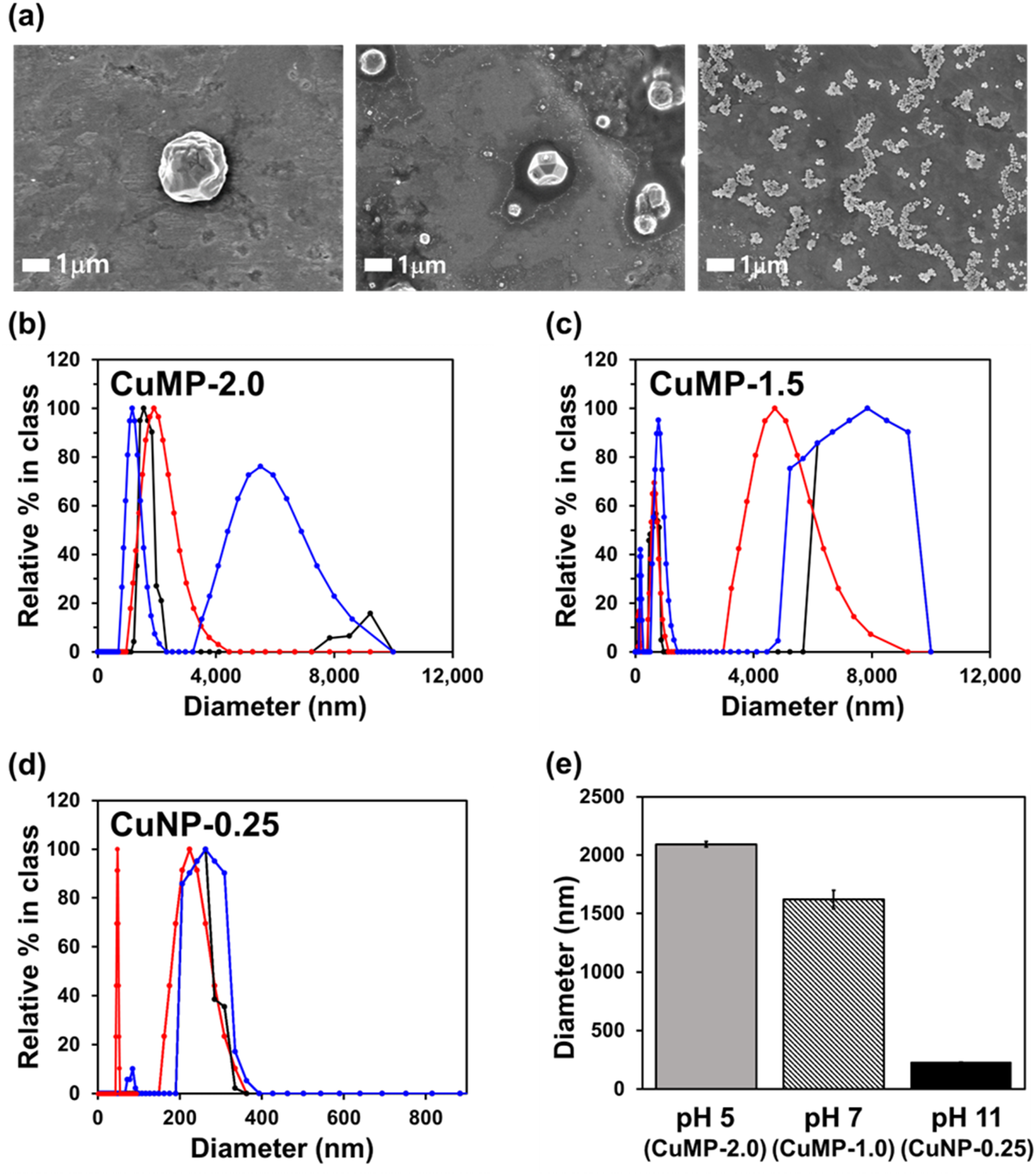
Physical characteristics of PP-CuNP and HG-PP-CuNP
The physical characteristics of PP-CuNPs produced in five different experimental cases, PP-CuNP-1X to PP-CuNP-5X, were evaluated, as listed in Table 1. SEM images were obtained by varying the number of coatings (Figure 2). As a result, when the number of coatings increased from 1X to 5X, more CuNPs were densely attached to the surface of the PP fabric, and CuNPs were more agglomerated (Figure 2). In addition, the color of PP-CuNP became darker with increasing number of coatings (Figure 2). The physical characteristics of the HG-PP-CuNPs were also identified (Figure 2). The TEMED and HG solutions were applied to PP-CuNP-5X to form hydrogel films on the PP-CuNP-5X fabric. As a result, the hydrogel film was effectively coated onto the fabric surface without significantly damaging the CuNP coating layers (Figure 2). SEM images of polypropylene (PP) fabric coated with copper nanoparticles (CuNPs) and hydrogel (HG).
HG-CuNP antibacterial properties
The antibacterial properties of HG-CuNP-PC4 (Table 1) were evaluated against the gram-negative bacterium E. coli (KCTC 1682). The control represents bacterial growth without HG-CuNP-PC4 (Figure 3(a) and (b)). The inhibition zone was calculated by using Equation (2). The results showed an average inhibition zone of 1.02 cm for HG-CuNP-PC4 (Figure 3(b)), indicating observable antibacterial activity against E. coli under the tested conditions. Antibacterial analysis. (a) and (b) HG-CuNP-PC4 against E. coli (KCTC 1682). (c) and (d) PP-CuNP-5X and HG-PP-CuNP-5X against E. coli (KCTC 1682).
PP-CuNP and HG-PP-CuNP antibacterial properties
The antibacterial properties of PP-CuNP-5X and HG-PP-CuNP-5X (Table 1) were determined using the gram-negative bacterium E. coli (KCTC 1682). The bacterial solution and control represent bacterial growth without PP fabric and with untreated PP fabric, respectively (Figure 3(c)). The results showed an average inhibition zone of 0.4 cm for HG-PP-CuNP-5X (Figure 3(d)), indicating observable antibacterial activity against E. coli. In contrast, PP-CuNP-5X did not show a comparable level of antibacterial activity.
CuNP antifungal property
After CuNP-0.25 was mixed with PDA media at concentrations of 15 mg/L, 150 mg/L, and 1500 mg/L (Table 1), the antifungal properties against B. fuckeliana (KCTC 6973) and C. cladosporioides (KCTC 16680) were evaluated (Figure 4). The growth patterns of B. fuckeliana and C. cladosporioides were different. B. fuckeliana grew radially from the center starting from the center of the fungal inoculation, but C. cladosporioides grew irregularly as spores spread (Figure 4(c) and (f)). Because the growth patterns of the two fungi were different, the diameter of the fungal growth was measured for B. fuckeliana and the growth area was measured using ImageJ for C. cladosporioides. The inhibition efficiency % was calculated using Equation (1). Antifungal analysis. (a)–(c) CuNP-0.25 against B. fuckeliana (KCTC 6973). (d)–(f) CuNP-0.25 against C. cladosporioides (KCTC 16680).
Figure 4 shows the antifungal activity against B. fuckeliana and C. cladosporioides at different concentrations of CuNP (CuNP-0.25-PC1, CuNP-0.25-PC2, and CuNP-0.25-PC3 in Table 1). CuNP-0.25-PC3 effectively inhibited both B. fuckeliana and C. cladosporioides, with 65% inhibition efficacy against B. fuckeliana (Figure 4(b)) and nearly 100% inhibition efficacy against C. cladosporioides (Figure 4(e)). C. cladosporioides did not grow after inoculation with CuNP-0.25-PC3, whereas B. fuckeliana did grow; however, the growth morphology differed from that of the control (Figure 4(c) and (f)). The results indicate that CuNPs had a stronger antifungal effect against C. cladosporioides than against B. fuckeliana.
Additional antifungal experiments were conducted to confirm the antifungal properties of CuNP-0.25 not dispersed in the PDA media. CuNP-0.25 was deposited directly on the center of the PDA medium where the fungal suspensions were spread. As a result, the abnormal growth of the two fungi around CuNP-0.25, which turned yellow for B. fuckeliana and white for C. cladosporioides (Figure 5(a) and (b)), indicated that CuNPs have antifungal properties but are ineffective, unlike CuNPs dispersed in PDA media. This finding suggests that the dispersing media can enhance the antifungal effects of CuNPs. Antifungal analysis. (a) and (b) CuNP-0.25 against B. fuckeliana (KCTC 6973) and C. cladosporioides (KCTC 16680). (c) and (d) HG-CuNP-PC3 against B. fuckeliana (KCTC 6973) and C. cladosporioides (KCTC 16680).
HG-CuNP antifungal properties
The antifungal properties of HG-CuNPs were evaluated using two concentrations of CuNPs inside the HG: HG-CuNP-PC3 and HG-CuNP-PC4 (Figure 5(c) and (d), Figure 6, and Table 1). As a result, HG-CuNP-PC3 did not show significant antifungal properties against either B. fuckeliana or C. cladosporioides (Figure 5(c) and (d)), although C. cladosporioides (Figure 5(d)) showed slight inhibition around HG-CuNP-PC3 on the second day (Figure 5(d)). Because C. cladosporioides is more sensitive to CuNPs than B. fuckeliana, the antifungal effects of HG-CuNP-PC3 (Figure 5(d)) and CuNP-0.25-PC3 (Figure 4) were similar. HG-CuNP-PC4 showed significant antifungal effects against both fungi (Figure 6(a) and (b)). For B. fuckeliana, the color of the mycelium changed from white to yellow (Figure 6(a)), indicating antifungal properties. However, for C. cladosporioides, an inhibition zone was observed (Figure 6(b)). Therefore, we measured the diameter of the inhibition zone of HG-CuNP-PC4. The average inhibition zone was 2.31 cm on the second day and 0.92 cm on the seventh day (Figure 6(b) and (c)). Although the diameter of the inhibition zone decreased as the fungi grew, we found that HG-CuNP-PC4 showed effective antifungal properties against both B. fuckeliana and C. cladosporioides (Figure 6(a) and (b)). Antifungal analysis of HG-CuNP-PC4. (a) and (b) B. fuckeliana and C. cladosporioides. (c) The inhibition zone sizes.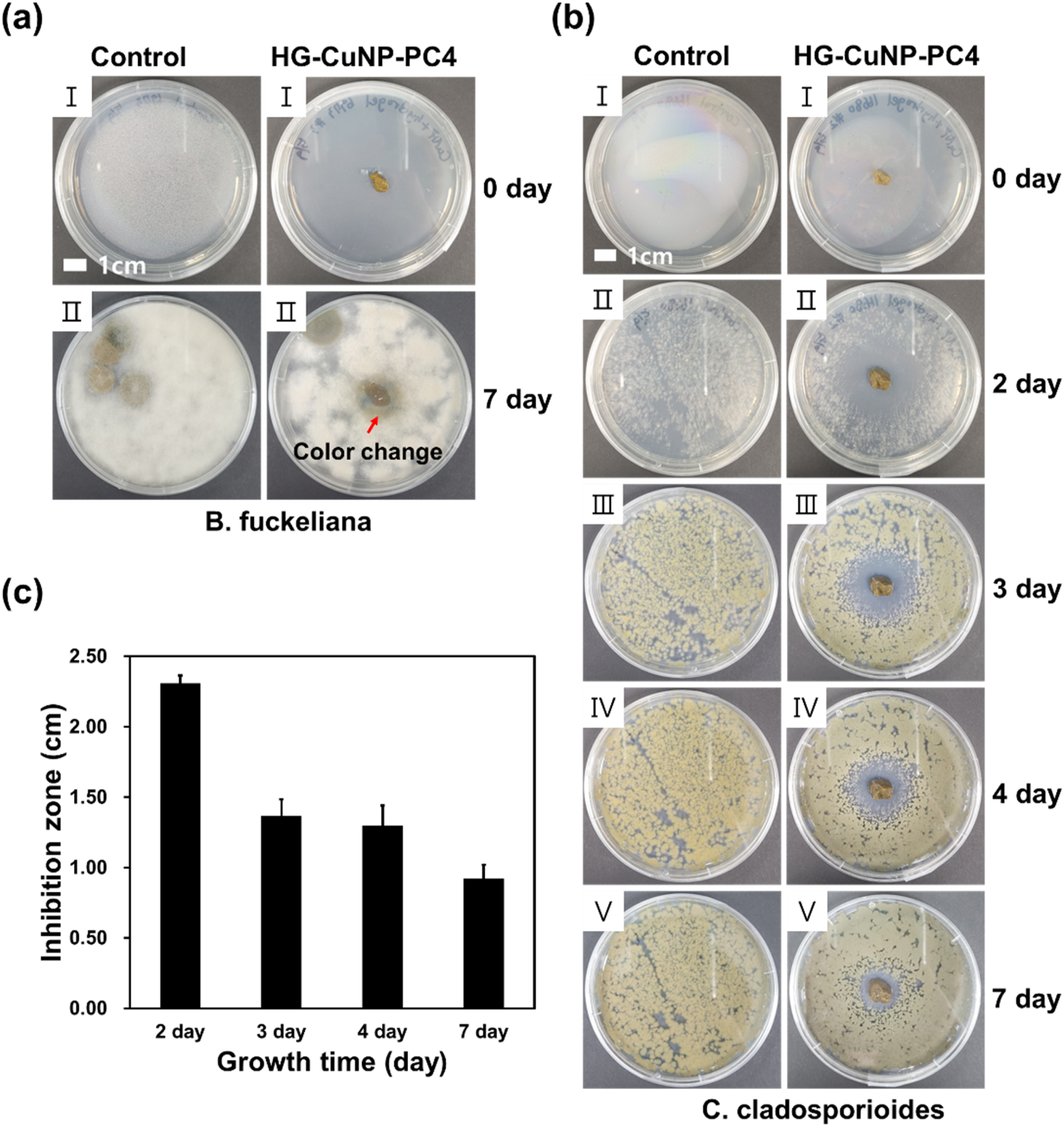
PP-CuNP and HG-PP-CuNP antifungal properties
To evaluate the antifungal properties of PP-CuNP-5X and HG-PP-CuNP-5X (Table 1), the fabrics were placed on PDA media where B. fuckeliana and C. cladosporioides spore suspensions were spread evenly. The fungal spores and control in Figure 7(a) and (b) represent the fungal growth results without PP fabric and with untreated PP fabric, respectively. Notably, outstanding antifungal performance was obtained with HG-PP-CuNP-5X, which produced an average inhibition zone of 2.15 cm against C. cladosporioides on the third day (Figure 7(b) and (c)). Furthermore, PP-CuNP-5X showed fungal growth in the contact area with B. fuckeliana; however, in HG-PP-CuNP-5X, the fungal mycelium color changed from white to yellow (Figure 7). These results demonstrate that the antifungal properties against both fungi were significantly improved by the hydrogel film layer (Figure 7). Antifungal analysis of PP-CuNP-5X and HG-PP-CuNP-5X against C. cladosporioides (KCTC 16680) (a) and B. fuckeliana (KCTC 6973) (b). (c) The inhibition zone sizes.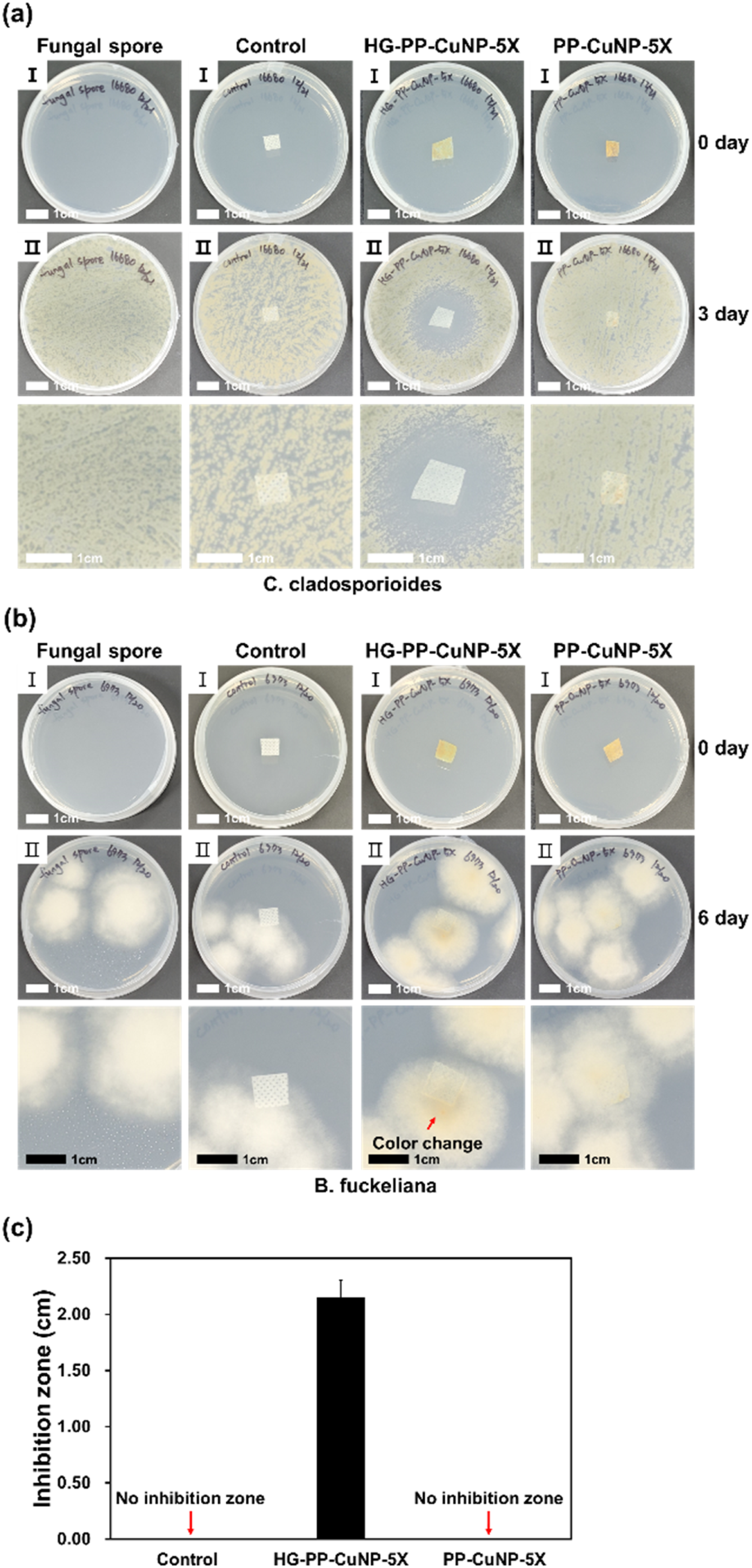
Antimicrobial mechanisms of CuNPs
The antifungal activity of copper nanoparticles (CuNPs) has generally been attributed to ion-mediated mechanisms, particularly the release of Cu2+ ions and the induction of oxidative stress in fungal cells.13,14 The present findings extend this understanding by demonstrating that antifungal efficacy is not determined solely by intrinsic nanoparticle properties such as size, composition, or surface density, but is strongly influenced by the surrounding physicochemical environment that regulates ion mobility and bioavailability.20,22
When CuNPs were dispersed in aqueous PDA media, pronounced antifungal activity was observed against Botryotinia fuckeliana and Cladosporium cladosporioides. In contrast, identical CuNPs deposited on agar surfaces or immobilized on hydrophobic polypropylene (PP) fabrics exhibited markedly reduced antifungal effects. Because nanoparticle composition, size, and loading were maintained constant, these differences are best explained by variations in moisture availability and ion transport rather than intrinsic nanoparticle characteristics.18,21
Role of hydrogel matrix in enhancing moisture-mediated ion transport
The introduction of hydrogel matrices substantially altered this behavior. By providing a moisture-rich microenvironment and continuous diffusion pathways, hydrogels facilitate ion mobility and prolong the local availability of biologically active copper species.25,26 Both hydrogel-dispersed CuNPs and hydrogel-coated CuNP fabrics exhibited significantly enhanced antifungal performance compared with their dry counterparts, despite unchanged nanoparticle chemistry. These results indicate that environmental regulation functions as a primary activating factor in this system.
Although direct quantification of Cu2+ release was not performed in the present study, the enhanced antifungal activity observed under moisture-rich hydrogel-assisted conditions is consistent with previously reported ion-mediated antimicrobial mechanisms of copper-based nanoparticles.13,15,22 Collectively, the findings demonstrate that antifungal functionality can be activated or suppressed through environmental engineering, without modifying nanoparticle composition or increasing loading.24,27 Therefore, the present results should be interpreted as biological evidence that the hydrogel environment facilitates moisture-mediated antifungal activity, rather than as direct quantitative proof of Cu2+ release kinetics. Because the antibacterial assay was performed only against E. coli, the antibacterial result should not be interpreted as evidence of broad-spectrum antibacterial activity. Future studies will include Gram-positive bacteria, such as Staphylococcus aureus, to evaluate the broader antibacterial applicability of the hydrogel-coated CuNP textile system.
Comprehensive physicochemical characterization, including detailed ion-release kinetics and crystallographic analysis such as X-ray diffraction (XRD), would further strengthen the mechanistic interpretation. However, the primary objective of the present work was to elucidate the environmental activation of antifungal function rather than to optimize nanoparticle synthesis. Such analyses will be considered in future studies aimed at correlating structural parameters with moisture-mediated activation behavior.
Environmental engineering as a design strategy for antifungal textiles
From an application perspective, the present work should be regarded as a mechanistic proof-of-concept for antifungal textile design. While standardized washing durability, long-term stability, and regulatory safety assessments remain essential for practical deployment, the current findings demonstrate that antifungal performance of CuNP-coated textiles can be significantly enhanced through environmental regulation without increasing nanoparticle loading.16,24 These results provide a conceptual framework for the development of antifungal textile systems in which moisture-mediated ion availability is treated as a primary design parameter in medical and agricultural applications. By distinguishing mechanistic clarification from subsequent durability and safety optimization, this approach enables a more systematic and rational development of functional antimicrobial textiles guided by a clearly defined governing mechanism.
Conclusions
In this study, we demonstrated that PP fabric coated with CuNPs and hydrogel enhances the antifungal properties of CuNPs by establishing a moist environment that enhances the availability of ion-mediated antimicrobial activity. To understand the antifungal process, two representative fungi that cause disease in strawberry plants were used. Interestingly, we found that the PP fabric coated with CuNP lacked adequate antifungal capabilities due to its hydrophobicity, which reduced ion transfer to the fungi. To enhance the ion transfer efficiency, we fabricated a composite layer of hydrogel and CuNP on the PP fabric. Our findings indicate that the hydrogel film layer on the PP fabric can facilitate the transfer of ions from the CuNPs to fungi, resulting in strong antifungal effects. The following is a summary of the findings of this study: I. Cu particles were synthesized using environmentally friendly ascorbic acid as the reducing agent, without using hazardous reducing agents such as sodium borohydride. The size of the Cu particles was controlled between 200 nm and 2 µm by adjusting the pH. II. CuNPs (200 nm) showed significant antifungal activity at high concentrations against B. fuckeliana and C. cladosporioides, which are representative strawberry fungi, and the inhibition efficiency % were 65% and nearly 100%, respectively. We confirmed that the CuNPs used in this study had sufficient antifungal properties at the nanoscale. III. CuNPs were uniformly coated onto the PP fabric (PP-CuNPs) using a unique surface activation process. However, PP-CuNPs showed insufficient antifungal properties, even at a high CuNP surface density. We hypothesized that the ions released from CuNPs do not effectively transfer to fungi because of the hydrophobicity of the PP-CuNP surfaces. IV. To create an efficient ion transfer environment around the CuNPs, we employed a hydrogel, which can contain more than 80% water. We produced hydrogel containing uniformly dispersed CuNPs (HG-CuNP), and HG-CuNP showed outstanding antifungal properties. The results indicate that a humid environment is essential for increasing the ion transfer efficiency, resulting in high antifungal performance. V. Finally, we developed a two-step coating process to produce hydrogel-CuNP composite layers on the PP fabric (HG-PP-CuNP). PP-CuNP were first immersed in the catalyst solution for polymerization and then in the hydrogel monomer and cross-linker solution. As a result, the PP fabric coated with CuNPs covered with a thin layer of hydrogel showed significant antibacterial and antifungal properties.
This study aimed to develop a coating method that maximizes the antifungal properties of PP fabrics coated with CuNPs. The results showed that the hydrogel is an effective matrix for establishing a moist environment that promotes ion transfer, which is critical for increasing antifungal properties. The developed antifungal fabric can be used in various medical and agricultural applications that require solid antimicrobial properties. In agricultural applications, the hydrogel-coated CuNP fabric may be considered for antifungal nonwoven covers, greenhouse liners, protective sheets, and post-harvest handling surfaces where moisture-rich conditions favor fungal growth. The present work should be regarded as a proof-of-concept for antifungal textile design, and further durability, washing stability, phytotoxicity, and field-level safety evaluations will be required before practical deployment. Furthermore, this study will help to suppress the spread of diseases by inhibiting the survival of fungi, which can protect the health of animals and plants.
Footnotes

Consent for publication
All the authors gave their consent to publish this work.
Author contributions
Y.J.K.: Investigation, Methodology, Data collection, formal analysis, visualization, writing–original draft. Y.S.J.: Conceptualization, Methodology, Validation, Funding acquisition, project administration, Supervision, Writing - Review & Editing.
Funding
The authors disclosed receipt of the following financial support for the research, authorship, and/or publication of this article: This work was supported by the Ministry of Education of the Republic of Korea and the National Research Foundation of Korea (NRF-RS-2024-00348608).
Declaration of conflicting interests
The authors declared no potential conflicts of interest with respect to the research, authorship, and/or publication of this article.
Data Availability Statement
The authors confirm that the data supporting the findings of this study are available within the article. All of the data and materials are owned by the authors and no permissions are required.
Human and animal rights statement
This research did not involve any Human Participants and/or Animals.