
Abstract
Objectives
This study aims to valorize Fagonia arabica (Dhamasa booti) herbal waste by synthesizing bioactive iron oxide (FeO) and zinc oxide (ZnO) nanoparticles for anticancer applications.
Methods
Aqueous extracts of F. arabica were utilized to green-synthesize FeO and ZnO NPs. The nanoparticles were characterized for morphology, size, crystallinity and zeta potential. Their antioxidant activity, brine shrimp lethality, and hemolytic assays were performed to determine bioactivity and cyto-compatibility.
Results
FeO and ZnO NPs were spherical, monodispersed and polycrystalline with particle sizes of 28.8 nm and 30.4 nm, respectively. ZnO NPs showed stronger antioxidant activity (80.81% scavenging at 150 μg/mL) and higher cytotoxicity in the brine shrimp assay (LC50 = 0.98 μg/mL), compared to FeO NPs and the crude extract. Minimal hemolytic activity, confirmed their favorable biocompatibility. Zeta potential values of -36 mV (FeO) and +33.6 mV (ZnO) indicate moderate colloidal stability. In vitro anticancer analysis revealed superior performance of ZnO NPs with IC50 values of 43.79 µg/mL (HepG2) and 66.71 µg/mL (HT-29).
Conclusion
Fagonia Arabica-derived ZnO NPs exhibit potent antioxidant, cytotoxic and anticancer activities, along with favorable biocompatibility. Their significant therapeutic performance highlights their role as effective plant-mediated antineoplastic agents and supports the value-added utilization of F. arabica herbal waste.
1. Introduction
Cancer carved its presence into the lives of 20 million individuals worldwide and put an end to nearly 10 million lives in the year 2022, a tragedy expected to escalate to 35 million new cases and over 18 million deaths annually by 2050. It is also evident from Globocan, 2022 data that roughly 1 out of 5 people develop cancer during their lifetime.1-5
Developing countries are experiencing >70% cancer deaths as per world health organization (WHO) statistics such as Pakistan, India, Bangladesh, Nepal, Indonesia, Myanmar, Philippines and Cambodia. Never-endingly Kenya, Nigeria, Ghana, Zambia from Africa and Bolivia, Honduras and Nicaragua of Latin America, where the availability of life-saving therapies is rare, infrastructure for early diagnosis is underdeveloped or absent and have relatively higher exposure to carcinogens.6-10 Despite advances in chemotherapy, radiotherapy, surgery, immunotherapy, and targeted agents, the majority of conventional interventions remain expensive, highly cytotoxic and inadequate particularly for resistant cancers. Therapy-induced toxicity, tumor relapse, multidrug resistance, and lack of specificity in treatment remain unresolved.11-14
For centuries, Fagonia arabica (F. arabica) has found reverence in Unani, Ayurvedic, and folk medicine systems, recommended by herbalists and hakims for fever, liver disorders, gynecological conditions, and enigmatic “swellings” long suspected to be tumors.1,15,16 This study unites the therapeutic legacy of medicinal plants and the engineered specificity of nanotechnology. By merging ethnopharmacology with green chemistry, we reached to a new frontier of medicine that is not only effective but also sustainable.17,18
This study focusses on green synthesis, an innovative technique where bioactive plant extracts play a dynamic part in reduction, stabilization and capping in the fabrication of metal oxide nanoparticles (MO NPs). Conventional chemicals and physical approaches for NPs synthesis frequently involve toxic reagents, high energy consumption and generation of hazardous wastes, which limit their biochemical applicability. Whereas, green synthesized NPs are both environmentally gentle and biologically compatible. Notably, NPs synthesized by naturally occurring biochemical pathways often retain surface bound phytochemicals, including alkaloids, saponins, flavonoids, tannins and phenolic compounds, that impart intrinsic bioactivity, including anti-inflammatory, antimicrobial, antioxidant and anticancer effects.19,20
In current research, Fagonia arabica was employed for the biosynthesis of FeO and ZnO NPs. Iron oxide NPs are well recognized for their magnetic effects which reinforces their applications in MRI and targeted drug delivery system. ZnO NPs, on the other hand, are known for their ability to disrupt mitochondrial function, activate apoptotic signaling cascade and preferentially provoke cytotoxicity in cancer cells while displaying lower toxicity towards normal tissues.
When synthesized using aqueous extract of F. arabica (DOSE) both metal oxide NPs yielded within a size range suitable for therapeutic treatment. LC-MS profiling revealed a rich composition of phytochemicals, many of these constituents are reported to suppress tumor angiogenesis promotes programmed cell death, attenuate inflammatory responses and modulate the expression of oncogenes and tumor suppressor genes. By integrating this phytochemicals reservoir with nanotechnology, the present work aims to translate that traditional medicinal value of Dhamasa booti into advanced nanomedicines with combined diagnostic relevance and therapeutic efficacy.
So, this study was designed to utilize herbal biomass (Dhamasa booti) in accordance with green chemistry principle focusing on development of F. arabica mediated FeO and ZnO NPs for biomedical applications. Different characterization techniques were employed to evaluate prepared NPs. Anticancer activity of the green-synthesized NPs against HepG2 and HT-29 cell lines, measuring IC50 values, and apoptotic indices. Further, brine shrimp lethality assays, antioxidant capacity, antibacterial profile and hemolytic behavior with human RBCs are examined to assess general biocompatibility. We also employed a benzene-induced chronic myeloid leukemia (CML) rat model to evaluate therapeutic response. Parameters include complete blood count (CBC), histopathological analysis of spleen, and body weight dynamics.
2. Materials and Method
2.1. Extraction
F. arabica (Zygophyllaceae family) was collected from Akbari Market, Lahore, Pakistan. The plant leaves and stalks were thoroughly rinsed with distilled H2O to eliminate impurities, then shade dried for approximately 14–21 days. To prepare the F. arabica aqueous extract abbreviated as DOSE (Dhamasa Overall Stalk Extract), 25 g of powdered sample was added to 250 mL distilled water, agitated continuously, and heated at 70 °C for about 90 min. It was cooled down and filtered. The purified aqueous extract was stored at low temperature (4 °C) for subsequent use.
2.2. Metallic Nanoparticles Synthesis
FeO NPs were synthesized by mixing 100 mL of FeCl3 (1 M) with 200 mL of sieved F arabica aqueous extract. The mixture was stirred at 70 °C for 30 min, during which a visible color change signified nanoparticle formation. After cooling, adjustment of pH and the solution was centrifuged. The resulting material was washed with distilled H2O oven-dried on 75 °C for 2 hrs. The dry material was finely ground and kept at 600 °C for 3 hrs to yield pure, deep red FeO NPs.
ZnO NPs were prepared using a green method involving DOSE extract. Exactly 200 mL of the extract was taken in a 250 mL conical flask, then heated and stirred at 60 °C for 1 hr. Separately, 21.94 g of Zn(CH3COO)2.2H2O was dissolved in 50 mL distilled H2O and agitated at 35 °C for 20-25 min. The two mixtures were combined, at (pH∼12), forming a milky white suspension. After cooling, the fusion was centrifuged at 5000 rpm. The precipitate was washed, dried at 90 °C for 24 hrs, and ground into fine powder. The dried material was subsequently calcinated at 350 °C for 4 hrs followed by gentle grinding to obtain phase-pure ZnO NPs. The final product was stored in airtight glass containers under ambient condition until further characterization and analysis.
2.3. LC-MS Analysis of Extract
To evaluate key phyto-chemicals in the aqueous extract of F. arabica, standard qualitative assays were conducted. We found saponins, cardiac glycosides, flavonoids, tannins and phenolic compounds. 21 For LC-MS analysis, 0.5 g of powdered dhamasa booti extract was sonicated in 10 mL distilled water for 30 min, homogenized, and filtered through a 0.2 µm membrane. The filtrate was analyzed using a Shimadzu LCMS-8040 Triple Quadrupole system equipped with a Shim-pack XR-ODS III column at temperature of 40 °C (2.2 µm, 150 mm × 3.0 mm). The mobile phase comprised of formic acid (0.1%) in acetonitrile and water, with 0.6 mL/min gradient flow. The auto-sampler was maintained at 15 °C, and 10 µL of sample was injected. Mass spectrometry was carried out in both positive and negative ion modes under MRM conditions, with an acquisition range of 50–1200 Da. Natural product’s spectral library was used to identify the compounds. 21
2.4. Characterization of Synthesized Material
UV-Visible spectroscopy was used to examine the optical behavior and confirm NPs formation. Spectral analysis of MO NPs was performed using a PerkinElmer Lambda-2 spectrophotometer covering wavelength range of 200 to 800 nm, the characteristics absorption maximum (λmax) confirm successful synthesis. Fourier transform infrared (FTIR) spectroscopy (400 to 4000 cm-1) was employed to identify surface functional groups and chemical bonding. The spectra indicated interaction between phytochemicals and NPs surface supporting effective capping and stabilization.
XRD analysis was conducted to determine crystalline structure and estimate average crystallite size. Measurements were obtained using Cu Kα radiation (λ = 1.542 Å) at 30 kV and 15 mA, with a scanning range of 10°–80°, step size of 0.02 °, and scan rate of 1° min-1. Crystallite size was using Debye Scherer equation:

Particle size and morphology were investigated using electron microscopy (SEM & TEM), and elemental composition as well as purity were verified using energy-dispersive X-rays spectroscopy (EDX). Based on Brownian motion and the stroke-Einstein equation, hydrodynamic particle size and polydispersity index (PDI) in colloidal suspension were measured using dynamic light scattering (DLS). Surface charge and colloidal stability were assessed using zeta potential measurements, which provide information about how NPs behave in biological settings. 22 A Q50 analyzer (TA Instruments, USA) was used to perform thermogravimetric analysis (TGA) in conjunction with thermal decomposition analysis. Samples were heated in a nitrogen atmosphere from room temperature to 1000 °C. The resulting thermograms confirmed the thermal integrity of the biosynthesized NP by showing distinct mass loss stages corresponding to moisture removal, degradation of organic components derived from plants, and stabilization of metal oxide phases.
2.5. Biological Applications of Metallic Oxide NPs
The antimicrobial potential for MO NPs was determined by disc diffusion methodology against selected Gram-positive and Gram-negative strains. Fresh bacterial cultures were uniformly spread on nutrient agar plates and sterile 6 mm filter paper discs loaded with 300 µg of MO NPs were placed on agar surface. Gentamicin (30 µg) was used as positive control. After 24 hrs of incubation at 37 °C, the zones of inhibition were measured to evaluate antibacterial efficacy.
22
The antioxidant activity of MO NPs was evaluated using DPPH assay. A DPPH solution (10 mg) was prepared in methanol (10 mL) and diluted to concentrations of 50, 100, and 150 µg/mL. Absorbance was measured at 517 nm. Likewise, varying strengths of NPs & ascorbic acid (50, 100, and 150 µg/mL) were tested for their radical-scavenging capacity. The percentage of DPPH inhibition was calculated using the standard formula.

2.6. In Vitro Cytotoxicity
DOSE fabricated metal oxide NPs were evaluated for cytotoxicity using brine shrimp nauplii. Ovum were hatched in 3.8 g/L synthetic seawater under light (30 °C) for 24 hrs. Groups of 10 nauplii were exposed to MO NPs at concentration of 10, 100 and 1000 µg/mL. Vincristine sulfate as a positive-control, while seawater with negative-control (dimethyl sulfoxide) was used. After incubation at 30 °C for 24 hrs, mortality rates were recorded, and LC50 values calculated via Probit analysis. LC50 values below 1000 µg/mL indicated cytotoxicity. 23
HepG2 (hepatocellular carcinoma) and HT-29 (colorectal carcinoma) cells were cultured in DMEM/F12 medium supplemented with 10% FBS, 1% L-glutamine, 1% non-essential amino acids, and 1% penicillin/streptomycin. Cells were preserved at 37 °C in a moistened incubator with carbon dioxide (5%).
MTT assay was utilized to estimate the cytotoxicity of integrated MO NPs anti HepG2 and HT-29 cells. Cells were plated in 96-well plates and treated with 10–100 µg/mL nanoparticles for 72 hrs at 37 °C with 5% CO2. Unprocessed cells alongside MTT-mixture & solubilized buffer both worked as positive control, while MTT-solution beside dimethyl sulfoxide functioned as negative-control. Cell viability was determined by measuring absorbance at 570 nm, and IC50 values were calculated.
Plant extracts may contain metabolites that disrupt red blood cell (RBC) membranes, causing hemolysis. Thus, assessing RBC compatibility is vital for the safety evaluation of plant extracts and plant-mediated NPs.24,25 The hemolysis capability of integrated MO NPs was tested on human RBCs at concentrations of 10–100 µg/mL. Blood sample from a healthy volunteer was incubated with NPs at 37 °C for 1 hr. Hemoglobin release was gauged at 590 nm. Sodium dodecyl sulfate (1% SDS) served as a positive control for comprehensive breakdown and PBS as a negative control. Hemolysis percentage was calculated to assess nanoparticle safety.
26

2.7. Flow Cytometry
Apoptosis was evaluated by flow cytometry using Annexin V-FITC and propidium iodide (PI) staining. HepG2 and HT-29 cells (1 × 10⁵) were seeded in 24-well plates and incubated at 37 °C with 5% CO2 for 48 hrs. The medium was then replaced with serum-free media containing DOSE-mediated MO NPs. After one day, cells were trypsinized, rinsed with PBS and centrifuged. Pellets were re-suspended in binding-buffer, stained with Annexin-V/FITC versus propidium iodide, fixed with 1% paraformaldehyde, and incubated in the dark at 25 °C for 15 min. Briefly, all in vitro assays were performed in triplicate (n = 3), and data are presented as mean ± SD.
2.8. In Vivo Study
Wister rats (20 to 25 g) were obtained from Tollinton market Lahore and maintained in sterilized cage under controlled environmental conditions (25 ± 2 °C) with a standard light dark cycle and unrestricted access to a regular laboratory diet. Animals were acclimatized for 15 days prior to experimentation in accordance with established ethical and experimental guidelines. 27 Chronic myeloid leukemia (CML) was induced by intravenous administration of benzene (0.2 mL of a 1:10 diluted solution) on alternate days for 3 consecutive weeks as previously described. 28
Animals were randomly assigned into 5 groups each containing six rats (n = 6), as per standard toxological and anticancer evaluation protocols. One group served as normal control while the remaining four groups received benzene to induce CML. Body weights were recorded at baseline and on day 21 to assess treatment related changes in hematological parameters and body weight alterations. Hematological parameters including hemoglobin concentration, packed cell volume (PCV), red blood cells (RBCs) count, white blood cells (WBCs) count and platelet levels were analyzed using an automated hematology analyzer. Blood samples were collected by a cardiac puncture into EDTA-coated tubes for complete blood profiling.
2.9. Statistical Analysis
The entire experimental data obtained from antimicrobial, antioxidant, cytotoxicity and hematological assessments was statistically analyzed using GraphPad prism version 6. Whereas, two-way analysis of variance (ANOVA) followed by Dunnett’s and Tukey’s post hoc tests was applied to determine significance. Results were presented as mean ± standard deviation (SD) with statistical significance defined at p < 0.05.
3. Results and Discussion
Iron oxide nanoparticles were produced through reduction of iron (III) chloride FeCl3 using aqueous extract of F. arabica. A gradual color change from dark brown to reddish-black at 70 °C indicated the formation of FeO NPs, attributed to surface plasmon resonance. 29 Phytochemicals in DOSE extract includes alkaloids, sterols, terpenes, glycosides, carbohydrates, sulfur-containing organic compounds, and flavonoids such as quercetin, kaempferol, beta-sitosterol, salvadoricine, catechin, and salvadoral. Similarly, ZnO NPs formed when Zn (CH3COOH)2.2H2O was mixed with DOSE, as evidenced by a color change from red to white at 60 °C.
Fifteen bio-active compounds were found in crude aqueous dhamasa booti extract by LCMS. Furthermore, LCMS data of DOSE indicated that these metallic NPs could be utilized in nutraceutical preparations for cancer treatment. The isolated compounds are illustrated in Figure 1 and Table 1. LC-MS base peak chromatograph of aqueous crude DOSE Phytoconstituents Isolated From Dhamasa Booti Aqueous Extract (DOSE) by LC-MS

3.1. Characterization of MO NPs
To characterize the synthesized nanoparticles, UV-Visible spectroscopy was employed and wavelengths ranging from 200-800 nm was scanned to confirm the formation of MO NPs. A peak absorbance at 355 nm indicated successful synthesis of both FeO and ZnO NPs, consistent with the surface plasmon resonance. Figure 2 shows the UV-Visible spectrum of the FeO/ZnO nanoparticles. UV-Vis absorption peak of (A) FeO NPs and (B) ZnO-NPs synthesized from DOSE
Fourier transform infra-red was used to explore the functional-molecular vibrations and potential bio-molecules accountable for production & successful stabilization of MO NPs. The peaks in spectra indicate -OH stretch, alkynes, primary amines, Aliphatic Amines, phenols, primary alcohols and alkenes, respectively. While the band appearance at 598 cm−1 illustrates the existence of Fe-O bond vibrations from ∝-hematite (Fe2O3) phase. Whereas, in the FT-IR spectra of DOSE mediated ZnO NPs peak at 482 cm−1 illustrates the existence of Zn-O vibrations indicating zincite (ZnO) phase. The slight change in peaks indicates the contribution of DOSE functional-groups in the MO NPs growth. The results are illustrated in Figure 3. Functional group spectrum of DOSE mediated (A) FeO NPs and (B) ZnO NPs
The XRD spectra was achieved to assess the crystallinity of DOSE assisted metal oxide NPs. The X-rays diffraction analysis, as shown in Figure 4, suggests crystal-like description of FeO/ZnO NPs. In XRD spectrum, the DOSE-assisted peaks for FeO NPs are in close promise with ∝-hematite, with JCPD card No: 96-101-1268. On the basis of 2θ and FWHM details, the calculated average size of FeO NPs by Scherer’s equation was ∼28.46 nm. The XRD analysis of FeO NPs is well aligned with reported literature.
30
On the same pattern, peaks data of DOSE mediated ZnO NPs suggest complete agreement with pure phase zincite, corresponding with JCPD card No: 96-101-1663. The sharp peaks show the high crystalline nature of ZnO NPs with an average size of ∼30.17 nm. The XRD analysis of ZnO NPs is also in line with previous studies.31,32 XRD spectra of FeO and ZnO NPs synthesized from DOSE
Figure 5 presents SEM images of DOSE-mediated MO NPs confirming their spherical shape and average size of 28.8 nm for FeO NPs and 30.4 nm for ZnO NPs, very much aligned with the data with DLS obtained diameter. SEM photographs and size dispersal graphs with statistical analysis of DOSE-mediated MO NPs
EDX analysis of FeO NPs showed strong peaks for iron and oxygen, confirming their purity with no other elements detected (Figure 6). Similarly, EDX spectra of ZnO NPs displayed prominent zinc and oxygen signals, verifying their pure composition (Figure 6). Correspondingly, graphs substantiate the presence of iron and zinc in prepared NPs. EDX photograph of (A) FeO NPs and (B) ZnO NPs synthesized from DOSE
TEM analysis of FeO NPs declared spherical shape and confirmed the average particle size of ∼28.5 nm. Similarly, TEM characterizes ZnO NPs with an average size of ∼30.3 nm with a comparable morphology. TEM micrographs for both MO NPs with statistical analysis is shown in Figure 7. TEM photographs of DOSE mediated (A) FeO & (B) ZnO NPs
DLS analysis revealed the average size of FeO NPs as 34 nm, slightly larger than calculated by XRD. The zeta potential was -36 mV with a PDI of 0.630, indicating excellent stability against aggregation. These results are well in alignment with the reported literature. Similarly, DLS analysis of ZnO NPs showed an average particle size of 34 nm, slightly larger than XRD measurements. The zeta potential was +33.6 mV with a PDI of 0.921, indicating low aggregation and excellent stability. Detailed results are presented in Figure 8A and B. Whereas, Comparative DLS data is presented in Table 2. Graphical presentation of zeta potential and size distribution of DOSE (A) FeO NPs and (B) ZnO NPs respectively Comparative DLS Data of DOSE MO NPs

Minimal weight loss occurs, mainly due to evaporation of physically adsorbed water or moisture. A minor weight loss can be observed in this region. This is commonly linked with the presence of moisture in the sample. This could suggest thermal decomposition or the evaporation of bound water (chemically bonded) and residual volatile organic compounds. This phase indicates the start of some degradation of the sample’s primary components.
There is a noticeable weight loss at this stage, likely pointing to the degradation of the core organic components or polymer chains present in your sample. Decomposition of primary material is common in this range, with carbonaceous residues remaining. As the temperature increases further, the sample undergoes more weight loss, potentially due to the breakdown of more stable components or conversion of residual carbonaceous matter into gases (like CO2 or CO). A continuous drop indicates ongoing decomposition or oxidation reactions.
TGA revealed multiple weight loss stages corresponding to distinct thermal events (Figure 9). An initial minor weight loss at lower temperatures was due to the loss of moisture and low boiling constituents. This was followed by a sharper decline indicating the degradation of weekly bound water molecules and low energy organic bonds. A pronounced weight loss in the mid temperature range reflected thermal decomposition of larger plant derived organic constituents beyond 450 °C, gradual mass reduction suggested the continued breakdown and oxidation of residual carbonaceous matter leaving thermally stable metal oxide nanoparticles (MO NPs). Generally, the profile confirms effective removal of organic moieties and stabilization of the inorganic phase. Graphic illustration of TGA report aimed at DOSE-mediated FeO & ZnO NPs
3.2. Applications of Synthesized MO NPs
3.2.1. Antimicrobial Activities
Researchers are actively exploring new strategies to combat resistance and limit the spread of diseases.
22
Advances in nano-biotechnology offer promising antimicrobial solutions. The antimicrobial potential of DOSE-mediated MO NPs was evaluated to address the rising challenge of antibiotic resistance, using Kirby-Bauer disc diffusion method 300 μg/disc). FeO and ZnO NPs has shown remarkable antibacterial activity against bacterial strains. Both types of fabricated NPs were highly effective against B. subtilis indicating higher susceptibility of gram-positive bacteria (Figure 10A). These outcomes are very close to previous studies reported on plant-derived NPs.26,33 The antimicrobial activity of MO NPs is primarily linked to ROS production and membrane damage, with phytochemicals on the NPs surface, enhancing these effects. Graphic appearance of (A) microbial-inhibition and (B) DPPH-assay of DOSE MO NPs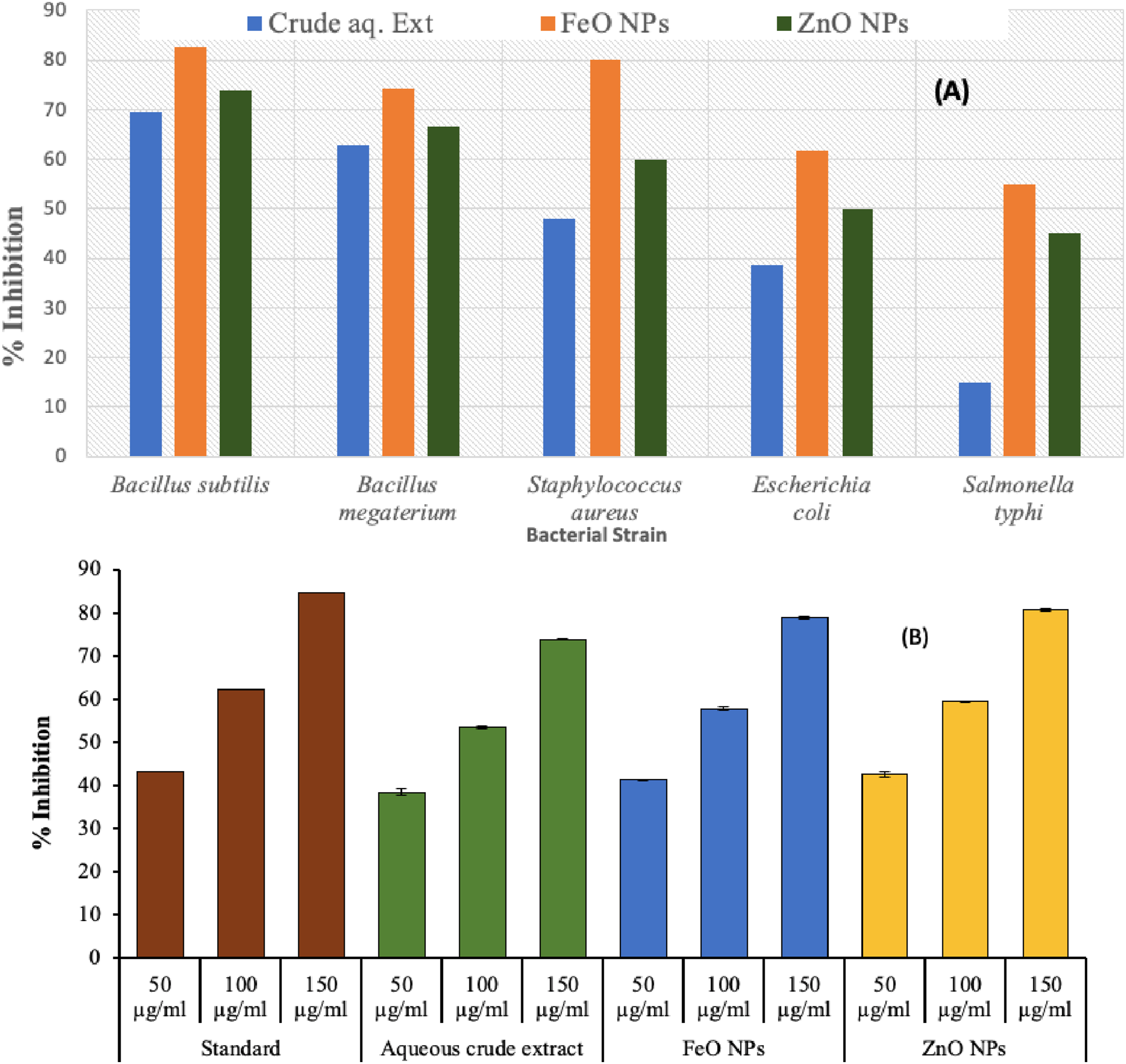
Overall, ZnO NPs and FeO NPs demonstrated the strongest antibacterial activity, while the crude extract was comparatively less effective. Two-way ANOVA demonstrated high significant effect of treatment type and bacterial strain on inhibition percentage (p < 0.0001), with a noteworthy interactive effect (p = 0.013), indicating strain specific sensitivity to NPs. ROS production, membrane disruption, and the cooperative action of surface-bound phytochemicals are probably the mechanisms underlying the antimicrobial action.
3.2.2. Antioxidant Assay
The antioxidant activity of synthesized nanoparticles was determined using DPPH essay at concentration range of 50-150 μg/mL. FeO NPs exhibited maximum scavenging activity of 79.02% at 150 μg/mL, exceeding that of DOSE alone (73.91%). Similarly, ZnO NPs achieved highest scavenging activity (80.81%) at the same concentration, confirming enhanced antioxidant performance (Figure 10B). The improved activity is attributed to flavonoids and phenolic compounds. These findings align with previous plant-mediated FeO NPs studies, 34 with variations attributed to synthesis methods, environmental factors, and nanoparticle characteristics. Two-way ANNOVA revealed that both treatment types and concentration significantly influenced antioxidant activity (p < 0.0001), with a strong dose-dependent response. A significant interaction effect (p = 0.0003) further indicated that the extent of scavenging varied with NPs type and concentration. Among the tested samples, FeO NPs and ZnO NPs exhibited the highest antibacterial effects, surpassing both the aqueous extract and standard drug at equivalent concentrations, particularly at 150 µg/mL, where inhibition levels exceeded 78%.
3.2.3. Brine Shrimp Lethality Assay (BSLA)
Dose-dependent Increase in Mortality for DOSE and MO NPs by BSL Assay

3.2.4. RBCs Biocompatibility Assay
Comparative % Hemolysis of DOSE Facilitated MO NPs

3.2.5. Anticancer Activity
Current analysis evaluated the anticancer outcomes of FeO NPs on HT-29 & HepG2 cell lines, presenting cytotoxicity based on dose dependence with IC50 data of 45.61 µg/mL and 96.02 µg/mL, respectively. ZnO NPs demonstrated superior cytotoxicity by IC50 estimates 43.79 µg/mL (HepG2) and 66.71 µg/mL (HT-29). These results are well aligned with the literature already reported.
37
Overall, ZnO NPs showed greater antineoplastic activity than FeO NPs. The superior efficacy of ZnO NPs aligns with the hypothesis that Zn2+-mediated ROS generation promotes apoptosis in hepatocellular and colorectal carcinoma cells. Data is presented in Figure 11. The cytotoxic potential of the nanoparticles was statistically validated using a one-way ANOVA, which demonstrated a highly significant influence of concentration on HT-29 cell viability. The analysis showed that the mean differences between the six tested concentrations were much bigger than the differences within each group. This was shown by a high F-value (F = 240.30) and a p-value of (p < 0.0001). The reduction in cell viability was concentration-dependent and statistically significant, confirming that the cytotoxic response was directly linked to NP exposure rather than experimental variability. The significant decrease in metabolic activity with higher doses shows a strong dose-response relationship, where cell survival decreased significantly with higher concentrations. These results highlighting the potent anticancer efficacy of the NPs against HT-29 cells even at relatively low concentration. Graphic presentation of the anticancer ability of DOSE-mediated MO NPs vs HepG2 and HT-29 cell-lines
3.2.6. Apoptosis Analysis
The apoptotic and necrotic defense of DOSE-mediated synthesized MO NPs was assessed using flow cytometry.
38
Analysis of Annexin-V and propidium iodide (PI) mark exhibited that DOSE FeO NPs induced 25.74% cell death, while ZnO NPs caused 26.15% in HepG2 cells. In HT-29 cells, FeO NPs triggered 17.91% and ZnO NPs 25.03% cell death after 48 hrs. These results confirm the superior apoptotic activity of DOSE ZnO NPs over FeO NPs. The visual results are shown in Figure 12, where readings were taken in triplicate at the IC50 value. Sustainable cells are in the inferior left-quadrant, early apoptotic cells are in the inferior right-quadrant, late apoptotic cells are in the higher right-quadrant and non-viable necrotic cells are in the higher left-quadrant. Analysis of Annexin-V & PI staining showed that DOSE FeO NPs induced 25.74% cell death, while ZnO NPs caused 26.15% in HepG2 cells. Flow cytometry sketch of HepG2 & HT-29 cell lines, (A) Untreated cells, (B) DOSE FeO NPs treatment (C) DOSE ZnO NPs treatment
The apoptotic induction demonstrates the therapeutic potential of MO NPs, which induce programmed cell death (PCD) by activating apoptotic pathways and producing reactive oxygen species (ROS). This enhanced cytotoxic effect is likely driven by the combined influence of dhamasa booti-derived phytochemicals, NPs concentration and exposure time as previously reported. 39 Importantly, the induction of apoptosis, rather than necrosis, offers a controlled mode of cell death minimizing inflammatory responses and collateral tissue damage which is advantageous in cancer therapy. Furthermore, DOSE-mediated green synthesis improves biocompatibility by incorporating natural antioxidant, anti-inflammatory and pro-apoptotic compounds onto the nanoparticle surface. 40 Mutually, these results underline the potential of phytochemical-functionalized NPs as targeted anticancer agents that selectively induce opposes while preserving healthy cells.
3.3. Hematological Analysis of Experimental Rats
Hematological parameters (complete blood count) were evaluated in male Wistar rats to assess the therapeutic impact of DOSE-mediated MO NPs in a benzene induced chronic myeloid leukemia (CML) model (Figure 13A and B). Graph exhibiting records of benzene-induced CML rat model for Miswak for MO NPs (A) Hematological parameters and (B) Platelets count
3.3.1. Normal Control Group (NC)
Normal control rats exhibited hematological values within physiological range confirming normal erythropoiesis profile, hemoglobin (14.43 g/dL), RBCs (8.26 x 10⁶/µL), hematocrit (44.81%), MCV (55 fL), MCH (18 pg) and MCHC (32 g/dL). Whereas, platelets count (786.5 x 10³/µL), and WBCs values (7.86 x 10³/µL) confirmed physiological immune and clotting function. These baseline values served as reference for disease and treatment comparison 41 Similarly, differential WBC counts viz neutrophils (23.66%), lymphocytes (68.65%), monocytes (5.8%), eosinophils (1.44%), and basophils (0.45%), were also within normal limits.
3.3.2. Disease Control Group (DC)
In contrast, benzene treated disease control rats showed marked hematological disruption, characterized by severe anemia, reduced hemoglobin (6.88 g/dL), lessened RBC count (3.01 × 10⁶/µL) and hematocrit value (17.81%) and pronounced leukocytosis. Hallmark feature of CML progression. 42 These alterations demonstrated bone marrow dysfunction linked to leukemia and confirmed the successful disease induction. With a differential count of 91.47% lymphocytes and 39.92% neutrophils, the white blood cell count was markedly elevated (27.35 x 10³/μL), which is consistent with leukocytosis in CML. 43 The platelets (348.78 × 10³/μL) are within a reasonable range. The disease model and typical CML pathology were supported by all of these findings.
3.3.3. Standard Drug Control Group (PC)
Treatment with standard drug (Gleevec, 20 mg/kg) resulted in substantial recovery of hematological parameters with 13.25g/dL hemoglobin, 6.72 x 10⁶/μL RBCs levels approached normal values, indicating effective restoration of erythropoiesis and immune balance, thereby serving as a benchmark for NPs efficacy. Neutrophils (24.99%), lymphocytes (66.27%), and WBC count (9.17 × 10³/μL) all demonstrated immune normalization. 44 Platelets count of 635.55 x 10³/µL were somewhat less than normal but higher than in disease group. All these values serve as reference point values for the assessment of therapeutic efficacy of DOSE-mediated MO NPs in experimental rat groups.
3.3.4. DOSE-Mediated FeO NPs Control Group
Rats treated with DOSE-meditated FeO NPs showed partial hematological improvement. While WBCs counts were moderated hemoglobin (9.94 g/dL) and RBCs (3.98 × 10⁶/µL) levels remained below normal suggesting mild anemia possibly due to suppressed erythropoiesis or oxidative stress linked to NPs exposure. 45 Slight elevation in neutrophils (34.92%) and moderate lymphocytes (55.62%) indicating a low grade inflammatory or immune response. 46 Though platelet count (452.56 × 10³/µL) were moderately reduced compared to NC & DC, but within physiological limits, indicating minimal thrombopoietic toxicity. 47 CBCs reports showed mildly elevated Monocytes (6.04%) echoing immune surveillance. Whereas, 2.84% eosinophils, 0.58% basophils were in normal range indicating no allergic or parasitic reaction. 48
3.3.5. DOSE-Mediated ZnO NPs Control Group
In comparison, DOSE-mediated ZnO NPs demonstrated superior hematological recovery, hemoglobin (10.47 g/dL) and RBCs (4.86 × 10⁶/µL) count approached near normal levels. WBCs count (10.13 × 10³/µL) was significantly reduced relative to disease control. Neutrophils (30.38%) and lymphocytes (59.96%) in the differential leukocyte count (DLC) profile indicated a more balanced immune response, indicating improved biocompatibility and therapeutic efficacy. On the other hand, 0.58% basophils and 2.84% eosinophils were within normal limits, suggesting no parasitic or allergic reaction. 48 However, increased phagocytic activity associated with nanoparticle clearance may be the cause of slightly elevated monocytes (6.64%). As part of a supportive inflammatory or immune response, platelets (494.89 × 10³/μL) demonstrated better restoration than FeO NPs. 49
3.3.6. Comparative Assessment
Treatments with DOSE-mediated FeO & ZnO NPs improved hematological parameters in comparison to disease control, according to a comparative analysis. However, when it came to minimizing immunological dysregulation and restoring erythroid and platelet indices, ZnO NPs consistently demonstrated greater normalization of hematological parameters than FeO NPs. These outcomes are aligned with previous reports on the immunomodulatory and hematological effects of MO NPs, which may be caused by immune engagement and oxidative stress.50,51 To confirm the long-term safety and effectiveness of these biogenic NPs in CML treatment, ongoing hematological monitoring is crucial.
Statistical analysis confirmed highly significant difference across all hematological parameter (p < 0.0001), confirming that disease induction and succeeding treatments produced physiological responses, Both MO NPs showing the improved restorative effect. Collectively, these findings indicate that DOSE-mediated ZnO NPs offers strongest hematological and therapeutic potential in CML. The ANOVA outcomes revealed a highly substantial effect of handling on platelet count (F + 182.6; p < 0.0001). CML induction triggered a sharped drop in platelet sum, whereas exposure to FeO and ZnO NPs significantly increased platelet level. As mentioned above ZnO NPs showed most pronounced restorative effect, approaching normal values closely than FeO NPs, suggesting superior hematological protection.
3.3.7. Gross Body Weight Changes after MO NPs Treatments
Rats treated with DOSE-mediated FeO NPs showed a moderate increase in body weight rising from 154.43 g to 163.45 g. In contrast the ZnO NPs treated group exhibited greater improvement with body weight increasing to 166.56 g from 158.72 g. This enhanced recovery indicates that DOSE-mediated ZnO NPs were more effective in mitigating disease-associated cachexia demonstrating efficacy comparable to standard drug treatment. These may also enhance appetite and stimulate anabolic pathways, supporting muscle maintenance while lowering energy expenditure. 52

3.3.8. Histopathology of CML-Induced Rat Spleen
Rats were anesthetized with ether and perfused with saline via the cardiac route. For histological examination, spleen tissues were removed and placed in 10% neutral buffered formalin. Samples underwent standard processing for dehydration, clearing, and paraffin embedding using an automatic tissue processor. 4 μm-thick sections were prepared via microtome, marked with hematoxylin and eosin (H&E) and histopathological analysis were performed using optical studies.
3.3.9. Histopathological Changes in DOSE-Treated Groups
Histopathology of rat spleen sections was done to appraise the efficacy of DOSE-assisted MO NPs anti-benzene-induced chronic myeloid leukemia (CML) model in vivo. Panel (A) shows the Normal Control (NC) group with healthy spleen architecture, intact lymphoid follicles, and normal sinusoidal spaces, reflecting preserved immune function. In contrast, the Disease Control (DC) group (B) exhibits disrupted lymphoid follicles and loss of sinusoidal spaces, indicating severe spleen damage due to CML and infiltration by abnormal blast cells. Panel (C), representing the Positive Control (PC) treated with a standard drug, shows partial restoration of spleen structure. Panel (D) showed improved red pulp architecture and improved lymphoid tissues revealing moderate improvement of lymphoid tissues. Lastly, panel (E) depicts a section of the spleen from rats given DOSE-mediated ZnO NPs; it displays distinct red and white pulp regions as well as a noticeable restoration of lymphoid follicles. This structural recovery indicates a strong reversal of CML-induced splenic damage (Figure 14A-E) and highlights the superior therapeutic efficacy of ZNO NPs. These observations are consistent with earlier reports demonstrating histopathological recovery in vital organ, including spleen, liver and kidney, following NPs based interventions.
53
In CML, leukemic cell infiltration commonly leads to splenomegaly, architectural disruption, extramedullary hematopoiesis and nuclear abnormalities with irregular morphology.
54
This structural recovery indicates a strong reversal of CML-induced splenic damage (Figure 14A-E) and highlights the superior therapeutic efficacy of ZNO NPs. Wister rat’s histopathology, microscopic photographs of spleen sections of different experimental groups for DOSE (A) NC group, (B) DC group, (C) PC group, (D) Experimental control (treated with DOSE FeO NPs) and (ES) Experimental control (treated with DOSE ZnO NPs), (Magnification 200×)
These pathological characteristics were successfully replicated in benzene-induced CML in recent research, supporting the disease model. Treatment outcomes showed clear disease persistence in control group, substantial recovery with standard drug and significant improvement following MO NPs administration. Notably, DOSE-mediated ZnO NPs produced the most pronounced histological restoration, achieving effects comparable to standard therapy. These results sustenance the potential of plant-mediated ZnO nanotherapeutics as a cost effective and less toxic strategy for CML management. Future studies should extend to advanced animal models and clinical investigations to further validate efficacy, address drug resistance and explore their utility in early diagnosis and disease monitoring. Treatment outcomes showed clear disease persistence in control group, substantial recovery with standard drug and significant improvement following MO NPs administration.
4. Conclusion
FeO & ZnO NPs were produced using an aqueous extract of F. arabica (DOSE), and phytochemicals were detected using LC-MS. FeO and ZnO NPs were found with an average size of 28.8 nm and 30.4 nm, respectively. These biosynthesized nanoparticles exhibited potent antimicrobial activity against Bacillus subtilis, with ZnO NPs showing superior antioxidant capacity through enhanced DPPH scavenging. ZnO NPs showed good biocompatibility in toxicity tests by effectively killing brine shrimp and causing little hemolysis of red blood cells. Strong colloidal stability was indicated by zeta potential values of -36 mV for FeO NPs and +33.6 mV for ZnO NPs. In terms of anticancer activity, ZnO nanoparticles performed better than FeO NPs, with IC IC50 value of 43.79 µg/mL for HepG2 and 66.71 µg/mL for HT-29. Higher apoptosis induction by ZnO NPs was confirmed by flow cytometry, with early and late apoptotic rates of 26.15% in HepG2 and 25.03% in HT-29 cells. In vivo, rats treated with ZnO NPs showed near-normal hemoglobin (10.47 g/dL) and RBC counts (4.86 × 10⁶/µL), along with stabilized WBC counts (10.13 × 10³/µL), indicating improved erythropoiesis and moderated immune response. ZnO NPs effectively mitigated cachexia and restored spleen histology, reversing CML-induced damage, with efficacy comparable to standard treatment. These findings feature the potential of integrating traditional herb Dhamasa Booti, with modern nanotechnology to develop effective anticancer nanomedicine. Though additional research is required to speak to toxicity concerns, ensure biodistribution studies, stability and scale-up challenges in green synthesis at an affordable price.
Footnotes
Acknowledgments
The authors express their gratitude to Princess Nourah bint Abdulrahman University Researchers Supporting Project number (PNURSP2026R158), Princess Nourah bint Abdulrahman University, Riyadh, Saudi Arabia. The authors are thankful to the Deanship of Graduate Studies and Scientific Research at University of Bisha for supporting this work through the Fast-Track Research Support Program.
Ethical Considerations
For the cytotoxicity (hemolytic) assay, blood sample was collected from young volunteer, the objective of blood collection was discussed. Blood sample was collected in the University medical center (The University of Lahore), under the supervision of medical officer.
Author Contributions
Funding
The authors disclosed receipt of the following financial support for the research, authorship, and/or publication of this article: The authors express their gratitude to Princess Nourah bint Abdulrahman University Researchers Supporting Project number (PNURSP2026R158), Princess Nourah bint Abdulrahman University, Riyadh, Saudi Arabia. The authors are thankful to the Deanship of Graduate Studies and Scientific Research at University of Bisha for supporting this work through the Fast-Track Research Support Program.
Declaration of Conflicting Interests
The authors declared no potential conflicts of interest with respect to the research, authorship, and/or publication of this article.
Data Availability Statement
The datasets used and/or analyzed during the current study are available from the corresponding author on reasonable request.