



Abstract
Purpose
We aimed to examine the effects of sildenafil and n-acetylcystein on ischemia/reperfusion injury in femoral artery endothelium and gastrocnemius muscle.
Basic methods
32 rats of Sprague-Dawley breed were randomly divided into four groups (n = 8). Median laparotomy was performed, then a 120-minute ischemia was created by microvascular clamping of infrarenal aorta, followed by the release of clamping. In sildenafil group, 1 mg/kg of sildenafil infusion and in the n-acetylcystein group, 100 mg/kg of n-acetylcystein infusion was administered after release of clamps. Blood samples and tissue samples of femoral artery and gastrocnemius muscle were extracted for a histopathological evaluation.
Principal findings
Serum levels of malondialdehyde in ischemia/reperfusion group (6.16 ± 0.79) were higher compared to the control group (4.69 ± 0.33), whereas a significant decrease was detected in sildenafil (5.17 ± 0.50) and n-acetylcystein (4.96 ± 0.49) groups. Femoral artery tissue sections of the control group, mean tumor necrosis factor alpha and hypoxy-induced factor-1 alpha immunoreactivity were found to be negative. In the ischemia/reperfusion group, mean tumor necrosis factor α immunoreactivity was intense and mean hypoxy-induced factor-1 alpha immunoreactivity was 51–75%. In the ischemia/reperfusion + Sildenafil and ischemia/reperfusion + NAS groups, mean tumor necrosis factor α immunoreactivity was slight and mean hypoxy-induced factor-1 alpha immunoreactivity was 26–50%.
Conclusions
In conclusion, sildenafil and n-acetylcystein may reduce femoral artery endothelium and gastrocnemius muscle injury following lower extremity ischemia/reperfusion.
Introduction
Ischemia is defined as impairment of delivery of oxygen and other metabolites that are required by the tissues due to a decrease or cessation of blood flow to the tissue or organ. 1 Ischemia/reperfusion (I/R) injury in the lower extremities occur in acute femoral artery occlusions, traumatic and iatrogenic arterial injuries, and in transient abdominal aortic clamping, especially during aortic surgery. 2 Local effects of I/R injury of lower extremities are observed in skeletal muscle and vascular endothelium, while systemic effects can be seen in any organ and especially in lungs, heart, brain and kidneys. 3 Skeletal muscle plays a major role in lower extremity I/R damage, since it is the most sensitive tissue to ischemic injury. Inflammatory response to reperfusion is correlated directly with the amount of reversible damage and inversely with the amount of muscle necrosis. 3 We investigated the effects of sildenafil whose effects has not yet been evaluated adequately in the I/R injury on vascular endothelium and skeletal muscle and a mucolytic agent n-acetylcysteine that has been demonstrated to be a powerful antioxidative in experimental studies on the acute ischemia of the lower extremity. The aim of this study is to investigate the effects of sildenafil and n-acetylcysteine on I/R injury in femoral artery endothelium and gastrocnemius muscle after infrarenal abdominal aorta occlusion-reperfusion.
Material and methods
Thirty two Sprague-Dawley rats, approximately 3.5–4 months old, weighing 190–250 g were used in the study. Rats were kept in transparent cages in rooms with automatically lightening to provide 12-hour day and 12-hour night environment, with a room temperature of 20–22℃ and moisture of 45–50%. Nutrition of the rats was provided using pellet feed and tap water. Adaptation of rats to the environment was provided for a week before the experiment. All rats to be used in the experiment were weighed before the intervention and their body weights were recorded. Local ethical board approval was obtained for the study.
All rats were administered intramuscular anesthesia to the left forefoot after 8 hours of fasting using ketamine HCl 40 mg/kg (Alfamin 10%® 100 mg/ml flacon, Alfasan International B.V., Woerden, Holland) + xylazine hydrochloride 5 mg/kg (Basilazin 2%® 23.32 mg/ml, 25 ml flacon, aniMedica GmbH, Senden-Bösensel, Germany). Interventions were continued maintaining spontaneous respiration of the rats. Rats were laid on the table in supine position under a warming lamp. After aseptic preparation of the skin of the rats, midline laparotomy was performed starting from just under the xiphoid up to 0.5 cm superior to the pubis. Intestines were deviated to the left with the help of a moist gauze. Infrarenal abdominal aorta was explored using blunt dissection. All rats received lowdose heparin (100 U/kg) for anticoagulation; 0.9% NaCl in a dose of 10 ml/kg was administered during the experiment from the tail vein to provide fluid resuscitation. All infusions were administered using a perfusator. Atraumatic microvascular clamps (Novaclip® 12 mm Angle, Plymouth, USA) were applied on to the infrarenal abdominal aorta. Approximately 5 ml of warm serum physiologic solution was administered to the peritoneal space after clamping. Abdominal wall was approximated using 3 silk stitches to prevent fluid loss. Atraumatic microvascular clamp on the infrarenal abdominal aorta was released after 2 hours of ischemia and 2 hours of reperfusion was allowed. After the end of the reperfusion period, the midline laparotomy incision was extended upwards to open the mediastinal cavity, the heart was reached and blood samples were taken from the right ventricular space using a 5-ml syringe. Similarly, the midline incision was extended to the right inguinal area, and a 1-cm part of the right femoral artery was excised with the help of atraumatic surgical instruments. A right gastrocnemius skeletal tissue sample was obtained afterwards. Rats were sacrificed after the procedure. Femoral artery and gastrocnemius tissue samples were stored in 10% formaldehyde solution until immunohistochemical (IHC) and hematoxylene eosin (HE) evaluations were performed. Blood drawn from the rats were centrifuged at 4000 r/min for 10 minutes after keeping at room temperature for 30 minutes, and rat plasma samples were then stored in −80℃.
Rats were randomly divided into four groups, each including an equal number of rats (n = 8).
Group (Control Group) (n = 8): Aorta was circumferentially dissected by blunt dissection. No clamp was applied on to the aorta. Group (I/R Group) (n = 8): Aorta was circumferentially dissected by blunt dissection, and atraumatic microvascular clamp was applied on to the aorta. The microvascular clamp was released after 2 hours of ischemia. Group (I/R + Sildenafil Group) (n = 8): Aorta was circumferentially dissected by blunt dissection, and atraumatic microvascular clamp was applied on to the aorta. The microvascular clamp was released after 2 hours of ischemia. Sildenafil was prepared for intravenous injection by dissolving it in 25 ml of serum saline to a final concentration of 1 mg/ml. Sildenafil infusion was started through the tail vein in a dose of 1 mg/kg after the removal of the clamp and at the start of reperfusion; the infusion was continued during the 2-hour reperfusion period. Group (I/R + N-Acetylcysteine Group) (n = 8): Aorta was circumferentially dissected by blunt dissection, and atraumatic microvascular clamp was applied on the aorta. The microvascular clamp was released after 2 hours of ischemia. N-acetylcysteine infusion was started through the tail vein in a dose of 100 mg/kg after the removal of the clamp and at the start of reperfusion; the infusion was continued during the 2-hour reperfusion period.
Serum MDA levels were measured according to the spectrophotometric measurement of the pink-red compound composed of MDA which is one of the final products of peroxidation of polyunsaturated fatty acids and thiobarbituric acid (TBA) in hot environment at 532-nm wave length. Serum MDA concentration was calculated using the molar absorptivity MDA at 532 nm. Results were expressed in mmol/l.
For IHC examinations of femoral artery and gastrocnemius muscle tissue sections, tumor necrosis factor (TNFα; [4E1]:sc-130349. Santa Cruz Biotechnology, Inc. Santa Cruz, California, USA) and hypoxy-induced factor-1 alpha (HIF-1α; [H1alpha 67]:sc-53546. Santa Cruz Biotechnology, Inc. Santa Cruz, California, USA) antibodies were used. Density and intensity of dye uptake of the TNFα antibody was evaluated as described by Khandoga et al. 4 TNFα was evaluated as no staining (−), slight staining (+) and intense staining (++). IHC findings according to the groups were calculated as 0 points for no staining (−), 1 point for slight staining (+) and 2 points for intense staining (++). HIF-1α was evaluated as (+) for 1–25% staining, (++) for 26–50% staining, (+++) for 51–75% staining and (++++) for 76–100% staining and they were given degrees as 0,1,2,3 and 4, respectively. For both antibodies, staining in the vascular endothelium of the femoral arterial sections and muscle fibers of the gastrocnemius muscle sections were evaluated. Cytoplasmic staining for TNFα and nuclear and cytoplasmic staining for HIF-1α was accepted as positive. All pathological examinations were performed by pathologists blinded to the groups using microscopes (Olympus BX51).
Results were expressed as mean ± standard deviation. Kruskal Wallis test was used in the comparisons between the groups. When differences were found between the groups, Mann Whitney U test with Bonferroni correction was used; p < 0.05 was accepted as statistically significant. SPSS 19.0 (License no = 10240642 SPSS Inc, Chicago, IL, USA) was used performing the statistical analyses.
Results
Serum malondialdehyde levels in all groups (nmol/ml).

I/R: ischemia reperfusion; I/R + S: ischemia reperfusion + Sildenafil; I/R + NAS: ischemia reperfusion + N-acetylcysteine.
Tumor necrosis factor alpha and hypoxy-induced factor-1 alpha scores for femoral artery in all groups (mean ± standard deviation).

I/R: ischemia reperfusion; I/R + S: ischemia reperfusion + Sildenafil; I/R + NAS: ischemia reperfusion + N-acetylcysteine; TNFα: tumor necrosis factor alpha; HIF-1α: hypoxia-induced factor-1 alpha
TNFα and HIF-1α immunoreactivities were found to be significantly increased in the I/R, I/R + Sildenafil and I/R + NAS groups compared to the control group (p < 0.05). Immunoreactivity in I/R + Sildenafil and I/R + NAS groups were significantly decreased compared to the I/R group (p < 0.001).
Tumor necrosis factor alpha and hypoxy induced factor-1 alpha scores for gastrocnemius muscle in all groups (mean ± standard deviation).

I/R: ischemia reperfusion; I/R + S: ischemia reperfusion + Sildenafil; I/R + NAS: ischemia reperfusion + N-acetylcystein; TNFα: tumor necrosis factor alpha; HIF-1α: hypoxia-induced factor-1 alpha.
Significant differences were demonstrated between the I/R group and I/R + Sildenafil group and between I/R group and I/R + NAS group (p < 0.001). TNFα and HIF-1α immunoreactivity was found to be significantly increased in the I/R, I/R + Sildenafil and I/R + NAS groups compared to the control group (p < 0.05). Immunoreactivity was significantly reduced in I/R + Sildenafil and I/R + NAS groups compared to the I/R group (p < 0.001). No significant differences were found between I/R + Sildenafil and I/R + NAS groups.
No staining was detected in the control group (Figure 1a). Diffuse and intense cytoplasmic staining with TNFα was found in the I/R group in general. Narrow arrows show endothelial fields of varying intensity demonstrating positive reaction with TNFα immunohistochemically. Thick arrows show clustering intraluminal erythrocytes and their relationship with the endothelial surface (Figure 1b); whereas injury in the femoral artery endothelium due to ischemia-reperfusion in I/R + Sildenafil and I/R + NAS groups was observed as a decreased rate of staining in both groups.
Features of immunohistochemical staining of tumor necrosis factor alpha of femoral artery endothelial sections; (a) Control group, (b) I/R group, (c) I/R + Sildenafil group, (d) I/R + NAS group (400×, tumor necrosis factor alpha antibody).
Narrow arrows show endothelial fields positively reacting with TNFα immunohistochemically and thick arrows show intraluminal clustering erythrocytes and their relationship with the endothelial surface (Figure 1c and d) (400 × , TNFα antibody).
There was no staining in the control group (Figure 2a). Staining with HIF-1α in a pattern of diffuse cytoplasmic nuclear positivity was seen in the I/R group. Narrow arrow shows endothelial fields in varying intensity, with positive IHC reaction to HIF-1α. Thick arrows show intraluminal erythrocytes in clusters and their relation with the endothelial surface and adhesion to the endothelial surface (Figure 2b). On the other hand, slight and intermediate degree cytoplasmic nuclear staining with HIF-1α was seen in I/R + Sildenafil and I/R + NAS groups. Narrow arrows show endothelial fields with a positive IHC reaction to HIF-1α and thick arrows show intraluminal clustering erythrocytes (Figure 2c and d) (400×, HIF-1α antibody).
Features of immunohistochemical staining of hypoxia induced factor-1 alpha of femoral artery endothelial sections; (a) Control group, (b) I/R group, (c) I/R + Sildenafil group, (d) I/R + NAS group (400×, hypoxia induced factor-1 alpha antibody).
No staining was seen in the control group (Figure 3a). An I/R damage was developed in the I/R group as a general myofibrillary irregularity in the skeletal muscle which was demonstrated as a generally diffuse, intense cytoplasmic staining with TNFα. Arrows show myofibrillary regions in varying intensity, immunohistochemically positively reacting with TNFα (Figure 3b). On the other hand, the rate of staining was decreased in both I/R + Sildenafil and I/R + NAS groups and myofibrillary irregularity was present in both groups. Arrows show myofibrillary regions in varying intensity, immunohistochemically positively reacting with TNFα (Figure 3c and d) (200×, TNFα antibody).
Features of immunohistochemical staining of tumor necrosis factor alpha of gastrocnemius muscle sections; (a) Control group, (b) I/R group, (c) I/R + Sildenafil group, (d) I/R + NAS group (200×, tumor necrosis factor alpha antibody).
No staining was seen in the control group (Figure 4a). Staining with HIF-1α in a pattern of diffuse cytoplasmic nuclear positivity was seen in the I/R group. Arrow shows myofibrillary fields in varying intensity, with positive IHC reaction to HIF-1α (Figure 4b). On the other hand, slight and intermediate degree cytoplasmic nuclear staining with HIF-1α was seen in I/R + Sildenafil and I/R + NAS groups. Arrows show myofibrillary fields with a positive IHC reaction to HIF-1α (Figure 4c and d) (200×, HIF-1α antibody).
Features of immunohistochemical staining of hypoxia induced factor-1 alpha of gastrocnemius muscle sections; (a) Control group, (b) I/R group, (c) I/R + Sildenafil group, (d) I/R + NAS group (200×, hypoxia induced factor-1 alpha antibody).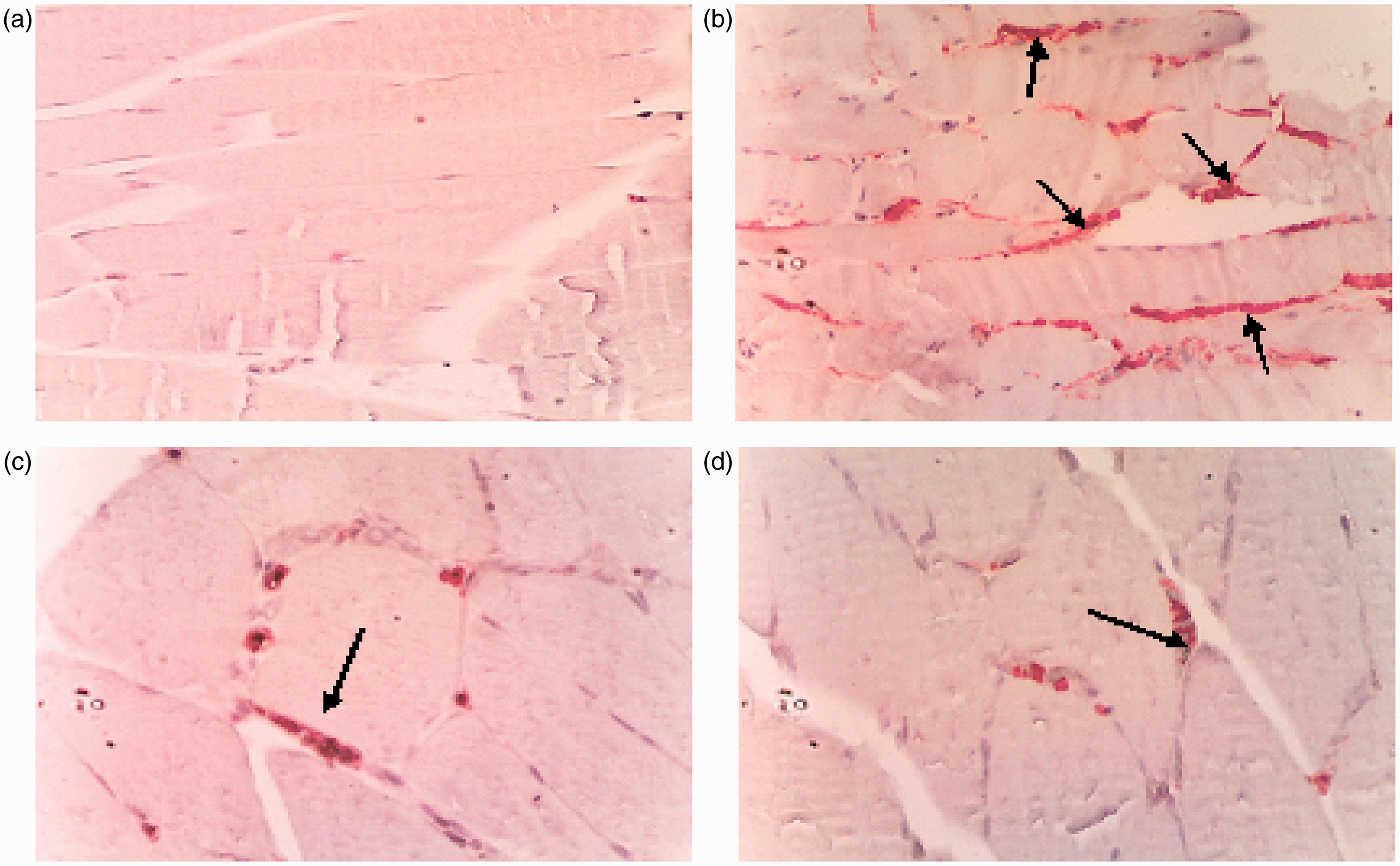
Discussion
Acute ischemia of an extremity is a condition encountered during peripheral vascular surgery, aortic surgery, reimplantation of the extremities, crushing injury, peripheral vascular trauma or acute arterial obstructions. 5 Even in the case of complete reperfusion of the extremity, the systemic inflammatory response may result in morbidities such as extremity loss, acute renal and respiratory failure, multiple organ dysfunction and even mortality. Delay in surgical intervention increases those risks. 5 Since skeletal muscle is one of the most vulnerable tissue to ischemic injury, it plays a central role in the lower extremity I/R injury. Prognosis in reperfusion injury depends on the amount of muscle damage. 3
Antioxidative agents, antithromboxanes, antileukotrienes and antiplatelet activating factors have been demonstrated to prevent the systemic effects of reperfusion in some experimental studies. 3 These antioxidant substances were shown to exert their preventive effect on the distant organ damage that is produced after ischemia and reperfusion by either increasing microvascular permeability and preventing neutrophil accumulation or activating the antioxidative system.5,6
Infrarenal aortic clamping and clamp release to provide circulation during abdominal aorta surgery is the cause of aortic I/R. 7 After this event, a spectrum of consequences such as systemic inflammatory response syndrome starting with inflammatory cytokines such as interleukin-1, interleukin-6 and TNFα and multiple organ dysfunction may ensue. 8 Rat abdominal aorta was clamped, lower extremity ischemia was produced and the clinical picture of I/R occurring in the aneurysm surgery was mimicked in this study.
Duration of the development of the tissue damage after lower extremity I/R is correlated with the duration of I/R. Gunduz et al., 8 in their I/R study on isolated endothelial cell culture, demonstrated that endothelial cytosolic calcium concentration increased in a two-step way during a 30-minute ischemic period and increased even more during the course of a 40-minute reperfusion compared to the ischemic period. Increase in calcium results in endothelial cell contraction and increases the distance between the endothelial cells and thus edema. Kiris et al. 7 found significant increases in the levels of malonydialdehyde, catalase and superoxide dismutase and myeloperoxidase activity in rats in the aortic I/R group in which 60 minutes of reperfusion was applied after a 30-minute ischemia compared to the control group. Narin et al. 9 found significant increases in the levels of malonydialdehyde, catalase and superoxide dismutase and myeloperoxidase activity in rats in the aortic I/R group in which 120 minutes of reperfusion was applied after a 120-minute ischemia compared to the control group. We applied 120 minutes of ischemia followed by a 120 minutes of reperfusion in the study groups of this study.
Sildenafil is a PDE-5 enzyme inhibitor which increases cGMP levels and thus results in relaxation in smooth muscle, with an increasingly widening clinical use in the treatment of erectile dysfunction and pulmonary hypertension due to its vasodilator effect. In addition to its vasodilating effects, sildenafil has been known to have inhibitory effects on thrombocyte aggregation and anti-inflammatory and antioxidative effects as well. Sildenafil is known to increase endothelium-dependent vasodilation and exercise tolerance in patients with coronary artery disease.10,11
In recent human and animal studies, positive effects of sildenafil on congestive heart failure, pulmonary hypertension and I/R injury of the heart have been demonstrated. It has been shown to decrease the endothelial dysfunction, apoptosis, necrosis, oxidative injury and thrombocyte activation in addition to a decrease in the amount of infarct area in heart I/R injury.10–12 Sildenafil is reported to have a direct inhibitory effect on necrosis and apoptosis mediated by NO and to decrease the number of apoptotic cells which are increased due to I/R in-vitro rat cardiomyocytes. Increase in the endothelial nitric oxide synthase (eNOS) and inducible nitric oxide synthase (iNOS) expression and hence NO bioavailability have been shown to play roles on these effects of sildenafil. 13 This supports the therapeutic effect of sildenafil against cell death which occurs following the I/R.
Vasodilator effects of sildenafil might trigger a signal pathway which in turn will result in NOS phosphorylation followed by NO production that demonstrate a preconditioning effect like adenosine and/or bradykinine. 14 Subsequently, NO was suggested to produce cGMP by activating guanylate cyclase and the cGMP produced was suggested to have a cardioprotective effect by opening the mitochondrial potassium-ATP channels by protein kinase G (PKG) activity.14,15 It may be the same mechanism by which sildenafil demonstrates a repairing effect on the impaired muscle and femoral artery endothelial function due to low extremity I/R. On the other hand, cardiovascular preventive effect of sildenafil has been demonstrated to be primarily by increasing tissue cGMP levels. 16 Potassium-ATP channels have been shown to exist not only in the heart but also in the endothelial cells. 16
Malondialdehyde is a stable end product occurring as a result of peroxidation of polyunsaturated fatty acids. In tissue I/R injury, xanthine oxidase enzyme, mitochondrial oxidation, cyclooxygenase-mediated unsaturated fatty acid oxidation, catecholamine oxidation, cytochrome p 450-mediated oxidation, leukocyte NADPH oxidase activation, iron release and redox cycle all contribute to local and systemic free oxygen radical production. 1 However, dominant system for each tissue is different. Increased free oxygen radicals cause peroxidation of cell membrane phospholipids and result in the impairment of cellular membrane integrity, cellular swelling and arachidonic acid/lipid peroxide release. 1 During this process, a chain reaction of free oxygen and fatty acid radical production and a progressive cellular wall damage occurs. 1 Hence, measurement of MDA, a stable molecule which is produced during this vicious cycle, might give an idea about the reactive oxygen derivatives and degree of the membrane damage. 17 Gurcun et al. 18 pointed out that although the preventive effect of ischemic preconditioning on the I/R injury of skeletal muscle is partially inhibited by NAS use, it is not completely blocked. Since free radicals are demonstrated to occur rapidly in 15–20 seconds after reperfusion, if a radical catcher is to be used, it is known to be effective when given 15 minutes before reperfusion, with no preventive effects when given after reperfusion.
Serum MDA levels were found to be significantly increased in the I/R group in this study compared to the control group. Serum MDA levels were significantly decreased in the I/R + Sildenafil and I/R + NAS groups compared to the I/R group. Serum MDA levels were identified to be significantly decreased in the control group compared to the I/R + Sildenafil and I/R + NAS groups. Similarly, increased plasma MDA levels were shown in some experimental studies.
Hypoxia and HIF-1 expression are tissue-specific. HIF-1α expression in heart tissue with 12-hour daily intervals of hypoxia for 12 days was not found to be increased, instead, nuclear translocation was shown. Thus, leukocyte HIF-1α expressions were found to be very high in patients with coronary collateralization in a study in patients with coronary artery disease and this pathway was suggested to be effective in ischemic angiogenesis. 19 In our study, the significantly increased HIF-1α immunoreactivity in the I/R group points out HIF-1α synthesis which is an important marker of increased damage after I/R and hypoxia. In this study, histopathologically, HIF-1α immunoreactivity in both femoral artery endothelium and skeletal muscle tissue sections were detected to be significantly increased in the I/R group compared to the control group. Immunoreactivity was found to be significantly decreased in I/R + Sildenafil and I/R + NAS groups compared to the I/R group.
TNFα causes tissue damage by neutrophil aggregation or activation and by producing proteolytic enzyme release from mesenchymal cells. Secretion of cytokines such as TNFα is stimulated by endotoxins, immune complexes, toxins, physical trauma and some inflammatory mediators. The most important sources of TNFα are macrophages and mast cells. 20 Healing effects in the I/R damage are produced by the inhibition of synthesis or receptor interactions of some mediator inflammatory substances such as TNFα. 21
In an experimental extremity I/R model produced by the tourniquet model by Mizusawa et al., 22 posterior root ganglion cytokine, neurotropin, neurotropin receptor and NOS mRNA expressions were evaluated using polymerase chain reaction method and TNF-α mRNA expression in the I/R group was found to be significantly increased compared to the control group.
The effects of iloprost combined with cardioplegic solution were evaluated on myocardial performance in an experimental cardiopulmonary by-pass model published by Katircioglu et al. 23 and plasma TNF-α level was found to be significantly decreased in the iloprost group compared to the control group.
In this study, the markedly increased TNF-α immunoreactivity suggests increase in the local TNF-α synthesis after I/R. We found histopathologically in both femoral artery endothelium and skeletal muscle tissue sections that TNF-α immunoreactivity was increased significantly in the I/R group compared to the control group. Immunoreactivity was detected to be significantly decreased in I/R + Sildenafil and I/R + NAS groups compared to the I/R group.
In this study, a marked decrease was not found histopathologically between the groups in femoral artery endothelium and gastrocnemius muscle tissue sections by HE staining. Statistical analysis of the biochemical and histopathological data were found to be significant. The results we obtained were similar with the previously published studies.
The powerful antioxidative effect of n-acetylcysteine has been demonstrated in various I/R models. However, the effects of sildenafil in experimental I/R model on femoral artery endothelium and skeletal muscle damage was encountered in the literature. We suggest that this study will enlighten future clinical and experimental studies on sildenafil, which we suppose has an efficacy equal to n-acetylcysteine in reducing the reperfusion injury.
In conclusion, the I/R injury occurring in femoral artery endothelium and gastrocnemius muscle is significantly reduced by sildenafil through its vasodilating effects and inhibitory effects on lipid peroxidation, oxidative stress, cytokine production and neutrophil accumulation, and by n-acetylcysteine through its antioxidative effects preventing tissue damage.
Footnotes
Funding
The study was funded by Trakya University Scientific Research Department.
Conflict of interest
None declared.