



Abstract
Objective
Neutrophil-to-lymphocyte ratio (NLR) value has emerged as a cardiovascular prognostic marker. Although several recent studies suggested NLR was associated with arterial stiffness, it was still controversial. The aim of this study was to investigate the correlation between NLR and arterial stiffness by measuring of brachial-ankle pulse wave velocity (baPWV) in an apparently healthy population.
Methods
This retrospective study enrolled 5612 participants during the health examinations from 1 October 2007 to 30 September 2011. Arterial stiffness was measured by baPWV. NLR was calculated as the ratio of the absolute neutrophil count to the absolute lymphocyte count in peripheral blood. According to the quartiles of NLR, the patients were categorized into four groups in males and females, respectively. Associations between NLR and baPWV were evaluated using partial correlation and multivariate logistic regression analysis.
Results
Both female and male subjects with increased arterial stiffness (baPWV ≥ 1400 cm/s) were likely to be older (females: P < 0.001, males: P < 0.001) and have higher systolic blood pressure (females: P < 0.001, males: P < 0.001), diastolic blood pressure (females: P < 0.001, males: P < 0.001), fasting plasma glucose (females: P < 0.001, males: P < 0.001), serum total cholesterol (females: P < 0.001, males: P = 0.028), triglyceride (females: P < 0.001, males: P = 0.031), urea nitrogen (females: P < 0.001, males: P < 0.001) than those without increased arterial stiffness. In addition, compared to those without increased arterial stiffness, body mass index (P < 0.001), waist circumference (P < 0.001), low-density lipoproteins cholesterol (P < 0.001), creatinine (P < 0.001), uric acid (P < 0.001) and lymphocytes (P = 0.001) were higher in females with increased arterial stiffness. However, males with increased arterial stiffness had higher NLR value (2.0 ± 0.7 vs. 2.1 ± 0.9, P < 0.001) and neutrophils (4.3 ± 1.4 vs. 4.5 ± 1.5, P < 0.001) than those without increased arterial stiffness, while the difference was not found in females. ANCOVA showed that males with quartile 3 and quartile 4 of NLR had greater levels of baPWV. NLR was correlated to baPWV in males by partial correlation analysis (r = 0.110, P < 0.001), but not in females. In multiple logistic regression analysis, the quartile 4 of NLR was positively associated with increased arterial stiffness in males (OR = 1.43, 95% confidence intervals [CI]=1.12–1.82, P = 0.004), but there was no obvious correlation in females.
Conclusions
Our findings suggest that there is a gender difference in the relationship between arterial stiffness and NLR. After adjusting for other confounders, the risk of increased arterial stiffness in apparently healthy adult males (rather than females) is independently associated with the highest quartile of NLR.
Keywords
Introduction
Arterial stiffness, an important determinant of cardiovascular risk, has been shown to be independently associated with stroke, retinal, autonomic disease, cognitive decline and mortality in various cohort studies.1–7 It may be directly involved in the process of atherosclerosis.8 Pulse wave velocity (PWV) is considered as an important parameter for assessing arterial stiffness.9,10 The Framingham Heart Study found that one standard deviation increment of PWV was associated with a 48% increase in cardiovascular disease risk. 3 Among a variety of measures, the brachial-ankle pulse wave velocity (baPWV) has become increasingly important for measurement of arterial stiffness, because it is easily used, reproducible, effective and less dependent on operators.11,12 Furthermore, researches have demonstrated that baPWV is comparable with carotid-femoral PWV, 11 which is the gold standard for measuring arterial stiffness. Both of them exhibit similar extent of associations with cardiovascular disease risk.
Inflammation and immunity have been demonstrated to affect the pathogenesis and progression of vascular stiffness and atherosclerosis.13–15 A variety of studies showed that inflammatory markers, such as interleukin-6, interleukin-1β, tumor necrosis factor-α and C-reactive protein were associated with arterial stiffness.16–21 Neutrophil-to-lymphocyte ratio (NLR), a simple marker of systemic inflammatory response, has recently been proposed as an independent prognosticator for some cardiovascular diseases.22–26 NLR can be easily obtained from routine complete blood counts in peripheral blood. Investigation of association between the NLR and atherosclerosis at early stage is of great importance for the prevention of atherosclerosis. Although the association of NLR with arterial stiffness has been investigated in some studies, most of them were inconsistent in different populations.27–31 Recent studies have suggested that NLR value is positively associated with arterial stiffness in postmenopausal females with osteoporosis, patients with type 1 or type 2 diabetes, and patients with peritoneal dialysis,27,30–32 but not in postmenopausal females and subjects without type 2 diabetes.30,31 To date, few studies are conducted to investigate the relationship between NLR and arterial stiffness in an apparently healthy population undergoing a health examination. Therefore, it is unknown whether there is an association between NLR with arterial stiffness in this population. Furthermore, arterial stiffness has been demonstrated to vary by gender, the arterial stiffness described in men is greater compared to women in China.33,34 However, the gender-related differences between arterial stiffness and NLR still remain unclear. Therefore, this study was designed to determine whether elevated NLR was associated with an increased arterial stiffness in a large, apparently healthy adult population of both genders.
Patients and methods
Study population
This study was approved by the Ethics Committee of the Third Xiangya Hospital of the Central South University, China. It was conducted in accordance with the Declaration of Helsinki and was consistent with applicable guidelines for good clinical practice. Each personal identification information was anonymized, so written informed consent was not required.
We designed a retrospective study to establish the relationship between NLR and arterial stiffness. Data were drawn from individuals who underwent a health examination in the Third Xiangya Hospital of the Central South University from 1 October 2007 to 30 September 2011. The medical history and symptoms of each subject were confirmed by the consulting physicians. Exclusion criteria were as follows: participants aged under 18 or over 70, and those with a history of smoking or drinking, atherosclerotic cardiovascular disease, stroke, cardiomyopathy, hypertension, diabetes mellitus, chronic kidney disease (estimated glomerular filtration rate (eGFR) < 60 ml/min/1.73m2 or proteinuria) and cancer. eGFR was calculated according to the Modification of Diet in Renal Disease (MDRD) equation
35

In addition, patients taking medications known to influence blood pressure, plasma glucose and lipid profile were also excluded. Furthermore, we also excluded subjects with missing data for essential variables. According to the above exclusion criteria, the study was finally included in 5612 participants (3303 males and 2309 females, age range from 18 to 70 years).
Assessment of brachial-ankle pulse wave velocity
The baPWV was measured via the ColinVP-1000 (Colin, Komaki, Japan), a non-invasive arterial atherosclerosis measuring system. Measurements were taken between 8 o’clock a.m. and 10 o’clock a.m. on the examination day. Participants were asked to prevent drinking of coffee, tea for at least 3 h and quit exercising for 30 min before measurement of arterial properties. Subjects were placed in supine position without a pillow for at least 5 min in a room with controlled temperature of 22°C to 25°C and then measurements were performed. During the examination, subjects should keep quiet. The cuffs were wrapped on both brachia and ankles, simultaneously measuring blood pressure (BP) and pulse waves in the bilateral brachial and tibial arteries and were connected to an oscillometric sensor that determines volume pulse form and to an oscillometric pressure sensor that measures BP. The baPWV was calculated using the formula: baPWV= (La−Lb)/Tba (La: the path length from the heart to the ankle; Lb: the path length from the heart to the brachium; Tba: the time delay between the arrival of the pulse wave at the brachium and ankle). The average baPWV values of the left and the right sides were used for statistical analysis. According to previously researches, increased arterial stiffness was defined as baPWV ≥ 1400 cm/s. 36
Other measures of interest
Other measures of interest included age, sex, current smoking status, history of hypertension, coronary artery disease and diabetes mellitus, medical history and family medical history were collected. Body weight and height were measured and then body mass index (BMI) calculated as weight in kg/height in meters squared was obtained. Waist circumference (WC) was measured according to the WHO recommended method. The systolic blood pressure (SBP) and diastolic blood pressure (DBP) were measured on the right upper arm in the sitting position after subjects rested 10 to 15 min in clinic using a validated digital automatic BP monitor between 7 o’clock a.m. and 9 o’clock a.m. Blood pressure was measured three times then averaged. In addition, levels of fasting plasma glucose (FPG), high-density lipoproteins cholesterol (HDL-C), low-density lipoproteins cholesterol (LDL-C), triglyceride (TG), total cholesterol (TC), creatinine (Cr), urea nitrogen (BUN), uric acid and complete blood count were determined by standard laboratory assays. NLR was calculated as the ratio of the absolute neutrophil count to the absolute lymphocyte count in peripheral blood. The quartiles of NLR were calculated in males and females, respectively. The results of quartiles of the NLR were: quartile 1: NLR ≤ 1.52, quartile 2: 1.52 < NLR ≤ 1.86, quartile 3: 1.86 < NLR ≤ 2.31, quartile 4: NLR > 2.31 for males and quartile 1:NLR ≤ 1.46, quartile 2: 1.46 < NLR ≤ 1.82, quartile 3: 1.82 < NLR ≤ 2.32, quartile 4: NLR > 2.32 for females.
Statistical analysis
All data were expressed as means ± SD (
Results
The comparison of the clinical and laboratory characteristics of subjects with and without increased arterial stiffness is described in Table 1. There were significant differences in SBP, DBP, FPG, BMI, WC, TC, TG, HDL-C, LDL-C, eGFR, Cr, BUN, uric acid, neutrophils, lymphocytes and baPWV between males and females (all P values are less than 0.001) except for age and NLR. Compared with females, males had higher values for baPWV, neutrophils, lymphocytes, SBP, DBP, FPG, BMI, WC, TC, TG, LDL-C, Cr, BUN and uric acid and lower values for HDL-C and eGFR (all P values are less than 0.001). In addition, individuals with increased arterial stiffness were more likely to be older and have higher BP (SBP, DBP), FPG, TC, TG and BUN in both genders (all P values are less than 0.05 and are listed in Table 1). Furthermore, female subjects with increased arterial stiffness had a higher BMI, WC, LDL-C, Cr, uric acid and lymphocytes and lower HDL-C (all P values are less than 0.001). Males with elevated arterial stiffness were observed to be featured with significantly higher neutrophil and NLR compared to those without elevated arterial stiffness (P < 0.001, P < 0.001, respectively). However, there were no significant differences of neutrophils and NLR in women (P = 0.92, P = 0.14, respectively) or BMI, WC, HDL-C, LDL-C, Cr, uric acid and lymphocytes in men (P = 0.66, P = 0.19, P = 0.24, P = 0.80, P = 0.63, P = 0.13, P = 0.12, respectively) between the baPWV < 1400 cm/s group and baPWV ≥ 1400 cm/s group.
Comparisons of clinical characteristics between subjects with and without increased arterial stiffness by gender (

Note: Data are presented as mean ± SD or number.
SBP: systolic blood pressure; DBP: diastolic blood pressure; FPG: fasting plasma glucose; BMI: body mass index; WC: waist circumference; TC: total cholesterol; TG: triglyceride; HDL: high-density lipoprotein cholesterol; LDL: low-density lipoprotein cholesterol; eGFR: estimated glomerular filtration rate; Cr: creatinine; NLR: neutrophil-to-lymphocyte ratio; baPWV: brachial-ankle pulse wave velocity.
#P < 0.05(female vs. male); *P < 0.05((baPWV < 1400 cm/s) vs. (baPWV ≥ 1400 cm/s)).
In addition, we conducted a further analysis to reveal the association between NLR and baPWV when subjects were divided into four groups based on NLR quartiles by gender (Figure 1). Compared with quartile 4 of NLR, males with quartile 1, quartile 2 and quartile 3 had lower baPWV values. Additionally, males with quartile 3 had higher baPWV than those with quartile 1. However, baPWV values were not significantly different in four quartile groups for females.

Comparison of baPWV levels among subjects with different quartiles of NLR values by gender. Data were shown as mean ± SD. *P < 0.05 (quartile 1 vs. quartile 3 or quartile 4); #P < 0.05(quartile 2 vs. quartile 4); &P < 0.05 (quartile 3 vs. quartile 4). NLR: neutrophil-to-lymphocyte ratio;baPWV: brachial-ankle pulse wave velocity.
The partial correlation coefficients of NLR and baPWV are illustrated in Table 2. The strongest correlation was observed between NLR and baPWV in the total subjects (r = 0.058; P < 0.001) and this correlation was also presented in males showed in Figure 2 (r = 0.110; P < 0.001). After adjusting for age, BMI, WC, SBP, DBP, FPG, TC, TG, HDL, LDL, Cr, BUN, uric acid and eGFR, NLR was positively correlated with baPWV in total subjects (r = 0.051; P < 0.001) and in males (r = 0.078; P < 0.001), respectively, but had no significant correlation with baPWV in females(r = 0.025; P = 0.23).The relationship between NLR and baPWV in total subjects may be ascribed for the correlations between NLR and baPWV in males.
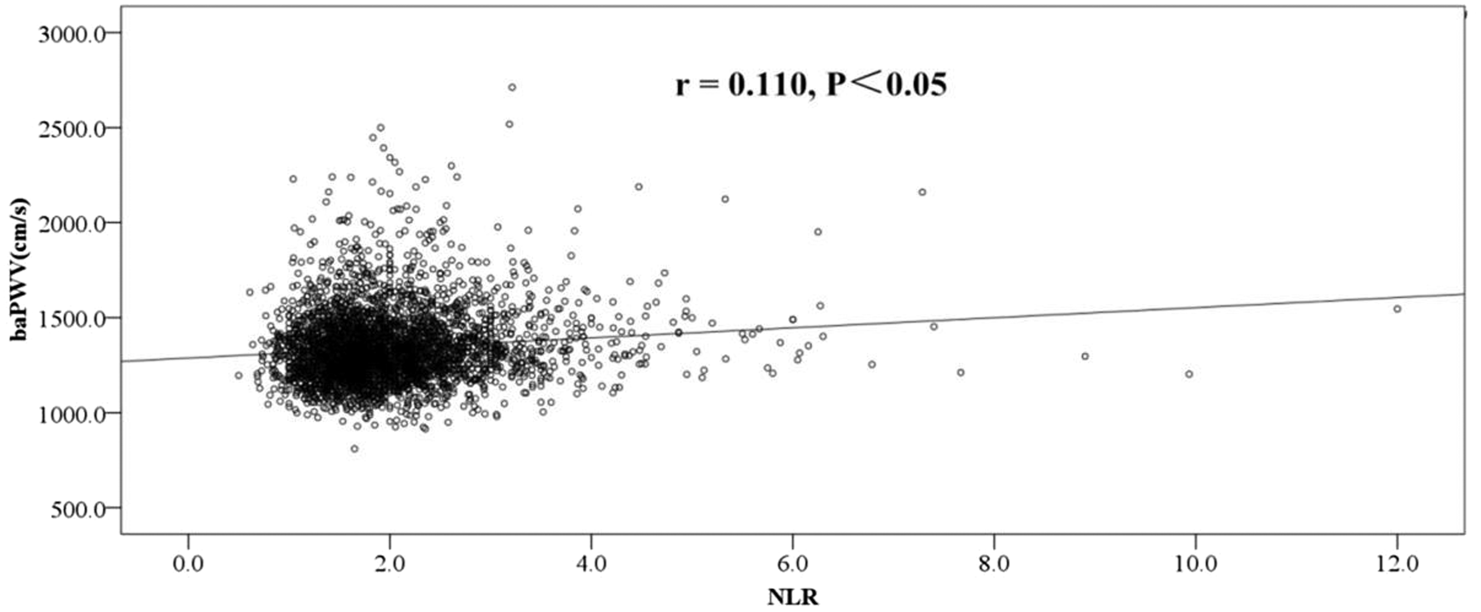
The relationship between NLR and baPWV in males.
Partial correlation coefficient (r) for NLR in relation to baPWV.

Note: Model 1, unadjusted. Model 2, adjusted for age, BMI and WC. Model 3, adjusted for age, BMI, WC, SBP, DBP, Cr, BUN, eGFR, aric acid, TC, TG, HDL-C, LDL-C and FPG. Abbreviations: see Table 1.
As shown in Table 3, multivariate logistic regression models (Model 1 to Model 6) were created and quartile 1 of NLR was used as the reference to further examine the association between increased arterial stiffness and different quartiles of NLR. Considering confounder factors including age, BMI, WC, SBP, DBP, Cr, BUN, eGFR, uric acid, TC, TG, HDL-C, LDL-C and FPG might affect the results, we performed variable-adjusted logistic regression models (model 2, model 3, model 4, model 5, model 6) to retest the results. Whether in the crude model (model 1) or variable-adjusted models showed that the fourth quartile of NLR in males, but not the second or third ones, was associated with a higher risk of increased arterial stiffness (P values for model 1 to 6 were <0.001, 0.01, 0.01, 0.006, 0.005, 0.004, respectively). Those in the fourth quartile of NLR in males had 1.43-fold increased odds of increased arterial stiffness as compared with those in the reference group after adjustment for all confounders. However, the relationship between all quartiles of NLR and increased arterial stiffness did not reach statistical significance in females.
Odds ratios and 95% confidence intervals for increased arterial stiffness according to NLR quartiles by gender.

Note: Model 1, unadjusted. Model 2, adjusted for age. Model 3, adjusted for age, BMI and WC. Model 4, adjusted for the same independent parameters as model 3 and plus SBP, DBP. Model 5, adjusted for model 4 variables plus Cr, BUN, uric acid, and eGFR. Model 6, adjusted for model 5 variables plus TC, TG, HDL-C, LDL-C and FPG. Abbreviations: see Table 1.
Discussion
In our study, we found that the NLR, a new inflammatory marker, was significantly higher in males with increased arterial stiffness participants than those without increased arterial stiffness participants, but in females the differences were not statistically significant. In addition, the current study also found that NLR was positively correlated with baPWV in males, but not in females. Moreover, multiple logistic regression analysis showed that the highest quartile of NLR is associated with a higher risk of increased arterial stiffness in apparently healthy males after adjustment for several confounding factors. However, each quartile of NLR exhibited no obvious association to the increased arterial stiffness in females.
A literature-based meta-analysis has shown that every 3.85 m/s increase of the baPWV was associated with a 1.21-fold increase in the risk of cardiovascular disease in a population with a relatively low to intermediate risk of development of cardiovascular disease (general population and hypertensive/diabetic patients) and another study also has shown that every 10 m/s increase of the baPWV was associated with a 3.1-fold increase in the risk of cardiovascular disease, which corresponds to a 12% increase of cardiovascular disease occurrence per every 1 m/s.37,38 There is increasing evidence that inflammation is associated with increased baPWV. The NLR has been proposed as a novel inflammation marker. To date, several studies have researched the relationship between baPWV and NLR. Our findings are similar to those previously researches, in which elevated NLR has been shown to be strongly and positively associated with a higher risk of arterial stiffness. Huguet et al.39 reported that NLR was independently associated with PWV in adults undergoing cardiovascular screening tests. Arterial stiffness in patients with coronary artery disease was also associated with NLR. 29 There was a positive correlation between NLR and baPWV in type 2 diabetes patients, and no obvious correlation in control subjects without type 2 diabetes.30 Ayhan et al. showed that there was a significant negative correlation between NLR and aortic dilatation, aortic strain in patients with type 1 diabetes, indicating a positive association between NLR and arterial stiffness. 27 Recently, a cross-sectional study also revealed that elevated NLR was associated with baPWV in all hypertension patients, whereas no statistically significant correlation was observed in participants without hypertension. 40 In addition, Lu et al.41 suggested a positive correlation between NLR and arterial stiffness in patients undergoing peritoneal dialysis. Although many previous studies showed that increased NLR is a risk factor for increased arterial stiffness,28–31 the exact mechanism for this association remained unclear. Consistent with the results, some reports have documented the association between arterial stiffness and inflammatory markers, such as interleukin-6, interleukin-1β, tumor necrosis factor-α, and C-reactive protein.16–20 Many studies have demonstrated a state of chronic inflammation may promote the development and progression of atherosclerosis.39,42–45 Neutrophils are found to be involved in the pathophysiology of atherosclerosis via multiple pathways.46,47 Firstly, activated neutrophils secreted a variety of granule proteins that induce expression of adhesion molecules and permeability changed and disturbed the bioavailability of nitric oxide, causing further endothelial dysfunction. Additionally, the granule proteins were deposited onto the endothelium and were released at the site of inflammation, leading to adhesion and recruitment of inflammatory monocytes. Neutrophils are also implicated in macrophages activation, foam cell formation and plaque destabilization. The above-mentioned facts could increase the formation of atherosclerotic lesion, and subsequently elevate arterial stiffness. Hence, neutrophils may play an important role in the development of arterial stiffness. The other component of the NLR is the lymphocytes. Hedrick48 indicated that lymphocytes have a great influence on atherosclerosis. However, different lymphocyte subsets may play a different role in atherosclerosis. For example, CD4+ T lymphocytes have been shown to gradually aggravate atherosclerosis. 49 In contrast, CD4+Foxp3+Tregulatory cells and B1 lymphocyte cells have been shown to be highly protective in atherosclerosis.50,51 In short, the roles of lymphocyte cells in the pathogenesis of atherosclerosis remain largely unknown and should be further investigated in future studies.
To the best of our knowledge, the current research is the first study to find that the highest quartile of NLR (Q4) is associated with a higher risk of increased arterial stiffness in healthy men without any history of medical disease and medications. But this relationship was not found in women. Although Yu’s studies showed that NLR was positively associated with baPWV in women, they enrolled 512 postmenopausal women and the correlation between NLR and arterial stiffness was only found in postmenopausal women with osteoporosis. 31 In addition, Yang’s52 also revealed a positive relationship between NLR and arterial stiffness in women, but in this study they only included 90 young female patients with systemic lupus erythematosus. Consistent with our findings, males had a higher incidence of cardiovascular disease and atherosclerosis than women of similar ages. 53 Also, in a study with 599 healthy Korean subjects, the NLR of males, but not females, was significantly associated with coronary artery calcification score.54 This gender difference may be caused by different sex hormones between males and females. Studies had shown that estradiol, a human sex hormone and steroid, strongly inhibited cytokine-induced expression of E-selectin, vascular cell adhesion molecule-1, and intercellular adhesion molecule-1 in vascular endothelial cells, which was associated with reduced adhesiveness of lymphocytes and monocytes to stimulated endothelial cells.55 Moreover, it has been reported that estradiol induces up-regulation of annexin A1 on human neutrophils, and inhibits neutrophil adhesion to endothelial cells under the shear stress, prompting that it may also contribute to the anti-inflammatory effect of estradiol.56 Recently, Aomatsu et al. 57 demonstrated that male neutrophils are more responsive to LPS and IFN-γ stimulation than female neutrophils. The gender difference in neutrophil responsiveness may partly explain our findings. To our knowledge, few studies have investigated the effects of estrogen on arterial stiffness. One study demonstrated a negative correlation between estrogen receptor α content and collagen concentration, indicating that activated estrogen receptor α by estrogen prevented against vascular collagen accumulation making the vessel more distensible.58 Therefore, estradiol exerted the beneficial effects possibly via modulation of the vascular endothelial cells, monocytes/macrophages, and neutrophils functions and accumulation of vascular collagen.
In fact, this study has some potential limitations in this study. First, the retrospective study design failed to explain the causal relationship between NLR and arterial stiffness exhibited by baPWV. Second, we only performed measurements of the brachial-ankle rather than the carotid-femoral PWV, which is currently the gold standard of measuring arterial stiffness, and most hypertension and arterial guidelines recommend carotid-femoral PWV measurements. Finally, the study only included the subjects from the Third Xiangya Hospital of Central South University, and thus the findings may not be generalizable to other regions.
In summary, the results of current work indicate that the highest quartile of NLR is strongly and positively associated with greater risk of increased arterial stiffness in these apparently healthy males after adjusting for other confounders. However, no statistically significant correlation between NLR and increased arterial stiffness was observed in apparently healthy females.
Footnotes
Acknowledgements
The authors wish to acknowledge Ren Guo for his contributions to this manuscript.
Declaration of conflicting interests
The author(s) declared no potential conflicts of interest with respect to the research, authorship, and/or publication of this article.
Funding
The author(s) disclosed receipt of the following financial support for the research, authorship, and/or publication of this article: This study was supported by the New Xiangya Talent Project of the Third Xiangya Hospital of Central South University (No.20150218), Program for New Century Excellent Talents in University (NCET-13–0605), Provincial Natural Science Foundation of China (2018JJ6051) and National Clinical Pharmacy Key Specialty Construction Project.