



Abstract
Objective
To investigate the safety and efficacy of endovascular treatment for totally occlusive lesions of the subclavian artery (SCA).
Methods
A retrospective study was performed on 57 patients treated with angioplasty and stenting, including 42 males and 15 females, with an average age of 61.8 years (range: 49 to 81 years). Efficacy, safety, and complications were evaluated.
Results
Procedural success was achieved for 47/57 patients and symptoms were relieved. Rat-tail occlusion is the most common type, and all cases were successfully recanalized. Plain type occlusion is less common with a recanalization rate of 55.6%. Hilly and plain occlusions are the main types of stent implantation failure. Through univariate analysis and trend matching analysis, the type of SCA occlusion and surgical approach had statistical significance on the success rate of surgery. The mean follow-up time was 34.6 ± 16.2 months. The cumulative stent patency rates at 1, 3, and 5 years were 95.5%, 86.4%, and 77.3% in the calcified plaque group and 92.0%, 76.0%, and 68.0% in the non-calcified plaque group, respectively. The 3-year and 5-year patency rates in the calcified plaque group were higher than those in the non-calcified plaque group (p < .05).
Conclusion
Different occlusion types and surgical approaches can affect the surgical success rate. The combined femoral and brachial approach can improve the rate of recanalization of SCA occlusions. The patency rates at 3 and 5 years in the calcified plaque group were higher than those in the non-calcified plaque group.
Introduction
Subclavian artery (SCA) occlusion is one of the most common diseases in vascular surgery with hidden clinical manifestations, which can cause subclavian steal syndrome (SSS). 1 Very few patients are caused by innominate artery stenosis or occlusion. Most patients present with bilateral asymmetrical blood pressure (BP), upper limb weakness after exercise, weak pulse, and even dizziness, blurred vision, articulation disorder, and a series of vertebral basilar artery insufficiency in severe cases. The most common causes are atherosclerosis, arteritis, vascular injury, and thoracic outlet syndrome. Endovascular treatment for SCA occlusion is the most important method,2,3 making it difficult to improve the success rate of surgery and the long-term patency rate in clinical treatment. The data of 57 patients with SCA occlusion receiving endovascular treatment from April 2010 to June 2019 were retrospectively analyzed to explore its clinical efficacy.
Patients and methods
This retrospective study included patients from The First Affliated Hospital of Wannan Medical College. The requirement of ethical approval for this was waived by the Institutional Review Board of The First Affliated Hospital of Wannan Medical College. The need for written informed consent was waived by the Institutional Review Board of The First Affliated Hospital of Wannan Medical College due to the retrospective nature of the study. All methods were performed in accordance with the relevant guidelines and regulations.
Inclusion criteria: (1) Patients diagnosed with total SCA occlusion by CTA; (2) patients with SSS, VBI, and upper extremity ischemia; (3) patients with inter-arm BP difference >20 mm Hg. Exclusion criteria: (1) patients with cerebrovascular accidents within 6 months; (2) patients with severe heart, lung, or kidney failure who cannot tolerate surgery; (3) patients with contraindications for anticoagulant and anti-platelet contraindications; (4) patients with contrast agent allergy.
Endovascular treatment
Following successful puncture via the common femoral artery, coordinate the loach guide wire and angiographic catheter to thoracic aorta. Aortic arch and intracranial arteries angiography were performed with intraarterial digital subtraction to identify the site of occlusion, the shape of the stump, and the situation of coronary collateral circulation and steal. After the exchange of a stiff guidewire, an 8F long sheath was put to the stump of SCA. Using digital road mapping, a 0.018-inch Abbott connect guidewire or 0.035-inch Terumo loach guidewire was used, and then MPA-1 catheter or VER catheter was put into SCA carefully. If not successful, we will return the long sheath to the descending aorta and implant the loach guidewire to the ascending aorta to observe the tangent position of the top of the bow and the SCA. In order to catheterize the SCA, the left brachial artery is directly punctured. Then 4F catheter and 0.018-inch Abbott connect guidewire or 0.035-inch Terumo loach guidewire were used to pass through the lesion of the SCA. It should also be ensured that the guidewire was in the true lumen when the guidewire and catheter passed through the tangential position. If it passes blindly, it could cause arterial dissection. After the successful establishment of access, the 7F long sheath deliver stent system should be replaced if necessary. Generally, self-expanding nitinol stents (approximately 10% greater than the measured value) and balloon-expandable stents (approximately 10%–20% greater than the measured value) were used. Under the imaging of DSA, the stent was transported to the stenosis, accurately positioned, and released.
The success of the operation was evaluated in three aspects by postoperative angiography: (1) stent position covered all stenosis sites; (2) residual stenosis rate <20%, (3) vertebral artery flow restored. Stents were used in 47 patients, including 6 Luminex (Bard), 28 Acculink (Abbott), and 13 Ominilink (Abbott).
Definition
The types of SCA occlusion 4 : (1) rat-tail type occlusion: CTA revealed mild stenosis at the initial segment of the SCA and subtotal occlusion of the distal end; (2) hilly type occlusion: CTA revealed totally occlusive ostia of the SCA with a visible stump; (3) peak type occlusion: CTA revealed totally occlusive SCA and the initial segment is slightly convex on the arch; (4) plain type occlusion: CTA revealed totally occlusive ostia of the SCA without stump.
The patients were divided into calcified plaque and non-calcified plaque groups. Calcified plaque group: calcified plaque can be clearly seen at the occlusion site, and the intima thickness was ≥ 2 mm. Non-calcified plaque group: no obvious calcified plaque at the occlusion site.
Statistical analysis
The quantitative data were presented as mean ± standard deviation in this study. In testing of statistical hypothesis, the significance level was set at 0.05. The Shapiro–Wilk test was used for assessment of normality. A t-test was used for pairwise comparison. Qualitative characteristics were compared using Pearson’s chi-squared test or Fisher’s exact test. The primary patency (defined as no restenosis of the target lesion at follow-up) was measured by the Kaplan–Meier method. Endovascular treatment failure was analyzed using single factor and propensity matching. The Statistical Package for the Social Sciences (SPSS) version 19.0 was used to perform the calculations.
Result
General clinical data
Baseline data of 32 patients with SCA occlusion.

Mean ± SD, Mean ± Standard Deviation; VBI, vertebrobasilar insufficiency; BP, blood pressure.
Treatment results of SCA occlusion
Occlusion type, aortic arch classification, surgical approach, and success rate of SCA.


Left SCA occlusion, plain type. A. aortic CTA suggests occlusion of the SCA; B. aortography shows no stump in the left SCA; C. the SCA stump is searched by 8F long sheath and guide wire; D. the guide wire passed smoothly and the stent is inserted without residual stenosis. SCA, subclavian artery; CTA, computed tomography angiography.

Left SCA occlusion, peak type. A. aortic CTA suggests occlusion of the SCA; B. aortography shows stump in the left SCA; C. the femoral artery approach cannot be performed, and brachial artery approach was successfully performed; D. the guide wire passed smoothly and the stent is inserted without residual stenosis. SCA, subclavian artery; CTA, computed tomography angiography.
SCA dissection occurred in 3 patients during the surgery, including 2 cases that gave up treatment because the guidewire entered into the subintimal space via brachial artery access, but the true lumen could not be entered. No contrast extravasation was observed after angiography, and no arterial dissection formed. In another patient, the guidewire was inserted into the subintimal space and reached the true lumen after repeated attempts. The arterial dissection disappeared after stent implantation. One patient had a transient decrease in heart rate and BP during balloon dilation but improved after symptomatic treatment, which may be caused by vagus nerve reflex.
Postoperative TIA occurred in 2 cases, dizziness, and headache in 2 cases. Cerebral infarction was excluded by imaging examination, which was suspected to be caused by postoperative hyper-perfusion. They improved after dehydration and brain protection. Two patients presented with a right femoral artery pseudoaneurysm, which was improved after compression dressing. Secondary thrombosis occurred in 2 patients and stent restenosis in 1 patient, which was improved by thrombolysis and bulbous expansion.
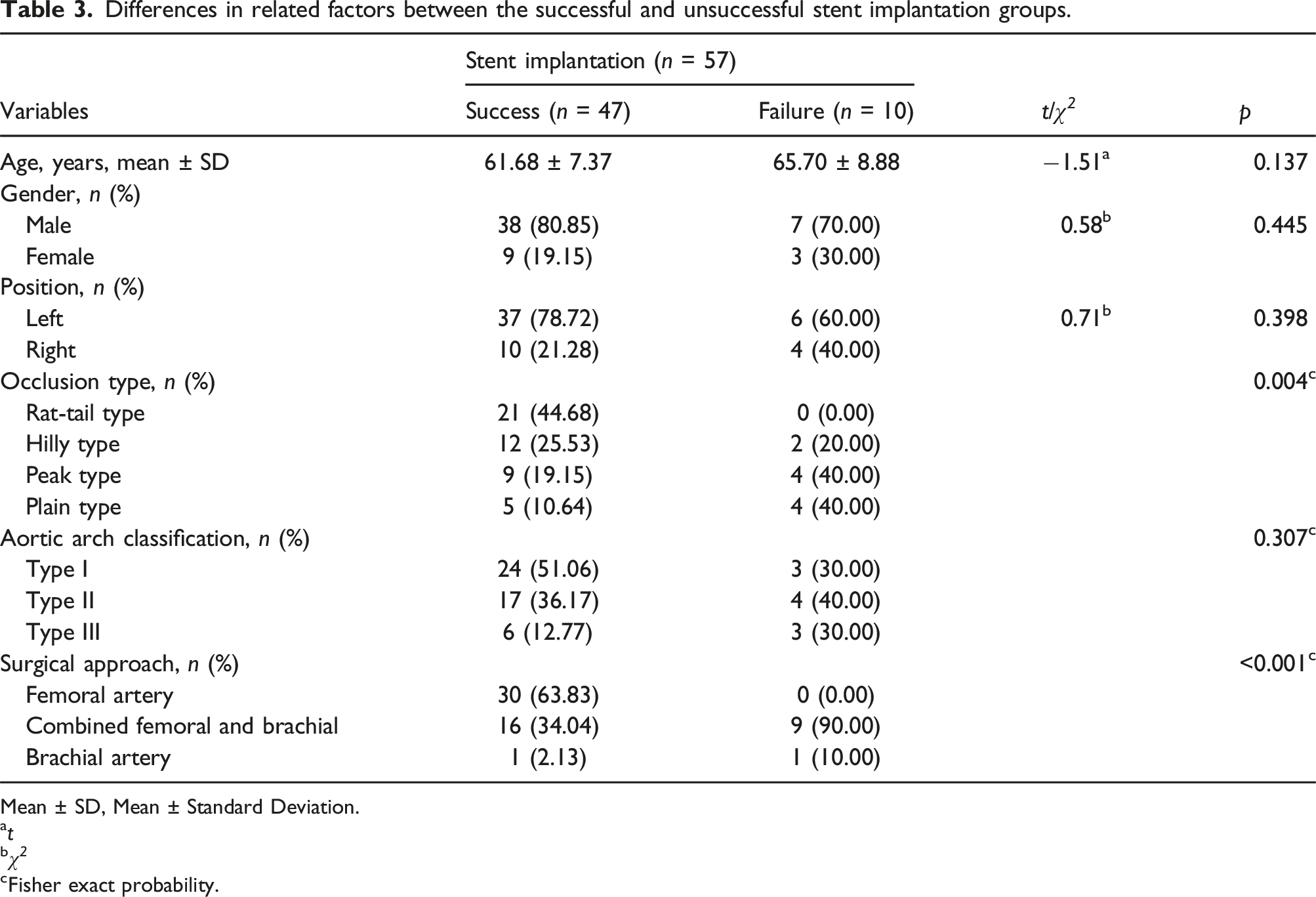
Univariate analysis for factors between the successful and unsuccessful stent implantation groups
Differences in related factors between the successful and unsuccessful stent implantation groups.
Mean ± SD, Mean ± Standard Deviation.
a t
b χ 2
cFisher exact probability.
Further evaluation of the effect of occlusion type and surgical approach on the outcome of stent implantation using PSM
The number of successful and unsuccessful stenting patients varies greatly, which may affect the stability of the results. PSM was used to perform a 1:2 matching control selection for patients with stent implantation failure (n = 10) to understand the occlusion type and surgical approach to stent implantation results. The match worked well (Figure 3). The matching factors for successful stenting patients included age, gender, occlusion site, and aortic arch classification. The results showed that the peak and plain types were risk factors for stent failure, while the combined femoral and brachial approach can improve the surgery success rate, which was consistent with the results of univariate analysis before unmatching (Table 4). Histograms of propensity scores before and after matching. Differences in the occlusion type and surgical approach mode distribution between successful and unsuccessful stent implantation groups after 1:2 matching.

All patients with stent implantation were followed up by outpatient visits or telephone after surgery, with a mean follow-up time of 34.6 ± 16.2 months. The cumulative stent patency rates at 1, 3, and 5 years in the calcified plaque group were higher than those in the non-calcified plaque group, with 95.5%, 86.4%, and 77.3% in the calcified plaque group and 92.0%, 76.0%, and 68.0% in the non-calcified plaque group, respectively (p < .05).
Discussion
SCA occlusion can cause SSS, which may lead to VBI and upper limb ischemia. Meanwhile, some patients may have no clinical symptoms. Therefore, the surgical indication is the key to treatment. Patients with SSS should be treated actively, and patients with good coronary collateral circulation and no steal can choose conservative treatment. 5 The early treatment of SSS syndrome is mainly open surgery, but the operation risk is high, with a mortality of as high as 10%, related morbidity of nearly 25%, and a mortality of 2%–5% even with chest technique.6,7 Both antegrade and retrograde approaches can be used for SCA with good clinical outcomes.8,9 SCA occlusion is usually caused by arteriosclerosis and inflammation, with a lower patency rate than SCA stenosis. 10 The plaques were divided into hard plaque, soft plaque, and mixed plaque. Hard plaques contained a large amount of calcification and collagen fibers, while soft plaques were mostly composed of cholesterol-bearing cells and foam cells with less collagen fibers and relatively heavy inflammation.11,12 The plaque was hard at the proximal end and soft at the distal end, making it relatively easy for the guidewire to pass through the stenosis.13,14 The surgical success rate of the calcified plaque group was higher than that of the non-calcified plaque group. The calcified plaque group tends to have gaps in which the guidewire could easily pass, but it was difficult to find these gaps. In the non-calcified plaque group, the plaque was hard, and the inflammation was severe, making it difficult for the guidewire to pass through the stenosis.
For some SCA occlusions involving the vertebral artery and covering the vertebral origin, balloon-expandable stents were not preferred because the balloon can push the atheromatous plaque into the vertebral artery. Intracranial embolism can occur during antegrade revascularization. Either a self-expanding stent or a small-diameter balloon relative to the blood vessel could be selected for pre-expansion. If there was plaque debris after vertebral artery expansion, the antegrade flow did not restore at this time, and retrograde flow flushed the debris into the upper extremity. As far as possible, avoid coverage of the vertebral artery origin for stenosis of the vertebral artery. The guide wire can be positioned in advance if necessary, and remedial measures can be taken in case of complications during balloon dilation or stent implantation.
The surgical success rates of rat-tail type, hilly type, peak type, and plain occlusions were 100% (21/21), 85.7% (12/14), 69.2% (9/13), and 55.6% (5/9), respectively. In univariate and multivariate analysis, there were no significant differences in age, gender, occlusion site (left and right), aortic arch classification, and other factors between patients with successful and failed stent implantation. The failure of stent implantation was mainly related to occlusion types and surgical approaches. The occlusion types of patients with endovascular treatment failure were mainly peak and plain types (40%), which were significantly higher than those in the successful implantation group (19.65%, 10.64%). The femoral artery or brachial artery approach was used in 31 patients with successful stent implantation. After the failure of the single approach, the combined femoral and brachial approach was used, and 16 patients were successfully implanted with stents. It can be seen that the combined femoral and brachial approach can improve success rate. When the SCA occlusion types are peak and plain, the catheters do not easily fix the stump, which cannot be opened through a single approach, and arterial dissection is easily formed by repeated operation. Therefore, the combined femoral and brachial approach can improve the success rate and reduce the incidence of arterial dissection.
The cumulative stent patency rates at 1, 3, and 5 years were 95.5%, 86.4%, and 77.3% in the calcified plaque group and 92.0%, 76.0%, and 68.0% in the non-calcified plaque group, respectively. The 3-year and 5-year patency rates in the calcified plaque group were higher than those in the non-calcified plaque group (p < .05). Mingoli reported that these approaches have been shown to have excellent long-term patency rates (range: 82% to 100%).15,16 A clinical report of 62 cases with SCA stenosis and occlusion reported by Onishi et al showed that the long-term follow-up patency rates were initially estimated to be 97.7% at 1 year, 97.7% at 3 years, 93.1% at 5 years, and 87.6% at 7 years, respectively.17,18 The follow-up results showed the 5-year stent patency rate of George et al. 19 was about 70%, which was generally consistent with the results of this study. The possible reasons were as followed: (1) intimal hyperplasia was observed after stent implantation in patients with atherosclerosis; (2) the SCA had high blood flow velocity at the neck outlet, and the stent as a foreign body repeatedly stimulated the blood vessels; (3) restenosis was more common in male smokers; (4) many patients with SCA occlusion do not have calcification but rather have inflammatory hyperplasia. Poor stent shape after implantation was prone to secondary thrombosis. Therefore, dual anti-platelet therapy (DAPT) was usually used for patients with SCA calcification. For patients with thrombosis and a high possibility of inflammatory hyperplasia, postoperative treatment with rivaroxaban or LMWH combined with DAPT was taken, which can improve the long-term patency rate.
Conclusion
Different occlusion types and surgical approaches can affect surgical success rate, and the combined femoral and brachial approach can improve the rate of recanalization of SCA occlusions. The patency rates at 3 and 5 years in the calcified plaque group were higher than those in the non-calcified plaque group. This single-center study supported the safety and efficacy of endovascular treatment for totally occlusive lesions of SCA in a limited patient cohort, thus emphasizing the need for larger-scale clinical trials to evaluate this approach.
Footnotes
Author contributions
All authors contributed to the study conception and design. Material preparation, data collection and analysis were performed by Shuangchao Liang, Andong Zhang, Fangkuan Li, Youchuan Xia, Guilin Feng, Bei Xu, and Haoran Wang. The first draft of the manuscript was written by Shuangchao Liang and all authors commented on previous versions of the manuscript. All authors read and approved the final manuscript.
Declaration of conflicting interests
The author(s) declared no potential conflicts of interest with respect to the research, authorship, and/or publication of this article.
Funding
The author(s) disclosed receipt of the following financial support for the research, authorship, and/or publication of this article: This work was supported by the Open Project of Key Laboratory of Anhui General Universities (Wannan Medical College) [grant numbers:RNA201905]; Funding agency: Key Laboratory of Anhui General Universities, Wannan Medical College. The Talent introduction project of The First Affiliated Hospital of Wannan Medical College (Yijishan Hospital of Wannan Medical College [grant number YR202211].
