



Abstract
Background
Chronic total occlusions with ambiguous proximal caps present a significant challenge in endovascular interventions of patients with Buerger’s disease.
Objective
We aimed to evaluate the effectiveness of transpedal retrograde wire just marker technique in patients with Buerger’s disease presenting proximal cap ambiguity and flush occlusions.
Methods
Seventeen patients with the diagnosis of Buerger’s disease who had below the knee artery chronic total occlusions with ambiguous proximal caps were enrolled. Procedural success, post-intervention Rutherford stage, wound scores, pedal loop scores, and amputation rates were recorded.
Results
Final study group consisted of 13 patients after exclusion of 4 patients due to pedal loop formation failure and severe vasospasm preventing equipment advancement. Post-intervention angiographic success rate was 100%. The post-intervention Rutherford stage showed excellent improvement (mean preprocedural Rutherford stage = 5 vs mean post-intervention Rutherford stage = 2; p = 0.003). Additionally, the average Saint Elian Wound Score System (SEWSS) decreased significantly (Preprocedural 14.9 ± 4.0 vs Postprocedural 11.3 ± 4.7, p < 0.001) after the interventions. Two patients had a major amputation during the follow-up indicating that higher post-intervention pedal loop scores are associated with higher amputation rates.
Conclusions
Transpedal retrograde wire just marker technique is an effective and practical method for revascularization of below the knee artery occlusions with ambiguous proximal caps. Including pedal loop angioplasty as a routine part of this technique can significantly increase blood supply to the pedal arch., thereby enhancing the likelihood of wound healing.
Introduction
Buerger’s disease, also known as thromboangiitis obliterans (TAO), is a non-atherosclerotic, segmental peripheral vascular disease impacting small- and medium-sized arteries and veins. Characterized by thrombus formation rich in inflammatory cells, Buerger’s disease causes vascular obstruction in extremities, leading to ischemic pain and ulcer formation. 1 Symptoms vary and include intermittent claudication, critical limb ischemia, and ulcers affecting both lower and upper extremities. In severe cases, chronic limb-threatening ischemia (CLTI) may necessitate amputation. Standard treatments often yield suboptimal outcomes. 2
Endovascular revascularization has become a promising approach for Buerger’s disease patients with CLTI, particularly those at high amputation risk. However, the small vessel size, extensive corkscrew collaterals, poor distal vessel bed run-off, and ambiguous proximal caps often complicate procedures and limit success. 3 Among alternative techniques, retrograde transpedal access shows promise, 4 though it may fail when proximal caps are ambiguous, 5 as attempts at blind puncture can result in procedural failure and severe complications, such as compartment syndrome, which can necessitate amputation.
To address these challenges, we have developed the “Transpedal Retrograde Wire Just Marker Technique,” an approach designed to improve antegrade puncture and navigation of ambiguous proximal caps. 6 This method employs a retrogradely advanced guidewire with a looped distal tip as a subintimal marker at the intersection of the proximal stump and main vessel, enabling precise antegrade penetration and effective revascularization. Here, we primarily evaluate the impact of this technique on procedural success and, secondarily, on outcomes in 17 patients with ambiguous proximal caps and flush occlusions.
Methods
Patient population
This study included 17 consecutive patients with below-the-knee artery occlusions who underwent endovascular intervention using the Transpedal Retrograde Wire Just Marker Technique between January 2021 and February 2024. Eligibility was based on angiographic evidence of below-the-knee artery occlusions with ambiguous proximal caps and unclear vessel courses. Buerger’s disease diagnosis followed Olin’s criteria, 2 confirmed through a comprehensive evaluation of clinical characteristics (age <45, current/recent smoking, claudication, rest pain, ischemic ulcers, or gangrene in distal extremities documented by non-invasive tests) and the exclusion of autoimmune diseases, hypercoagulable states, diabetes mellitus, and proximal emboli sources via echocardiography or arteriography, with confirmation of angiographic findings like corkscrew collaterals and extensive arterial occlusions.
Preprocedural evaluation
All patients underwent preprocedural assessment, including physical exams, ankle-brachial index (ABI) measurements, and imaging via duplex ultrasound and/or CT angiography. Patients received aspirin (81-100 mg/day) and clopidogrel (75 mg/day) at least 24 hours before the procedure. Baseline demographics, CLTI severity according to Rutherford classification wound scores using the Saint Elian Wound Score System (SEWSS), technical intervention details, and recovery or amputation rates were documented. 7 Follow-ups, including clinical assessment and Doppler ultrasound, were conducted at 1 and 6 months post-procedure. Informed consent was obtained, and the study was approved by the local ethics committee in adherence to the Declaration of Helsinki.
Endovascular procedures
An ipsilateral femoral antegrade approach was used in all cases, with 6-Fr x 10-cm introducer sheaths (Radifocus Introducer II, Terumo) inserted into the common femoral artery (CFA) under fluoroscopic guidance. Digital subtraction angiography of the affected limb, including below-the-knee and pedal arteries, identified target vessels per the angiosome concept, and distal run-off scores were calculated. 8 Patients received heparin (100 IU/kg) adjusted via activated clotting time (ACT), with additional doses as needed. Nitroglycerin (100–200 µg) was given through the arterial sheath to prevent vasospasm. Lesion crossing utilized intraluminal and subintimal methods; Controlled antegrade and retrograde tracking (CART), reverse CART, and Subintimal tracking and reentry (STAR) techniques.9–11 Following lesion predilation with transluminal angioplasty catheters, drug-eluting balloon catheters were used. At the procedure’s end, intra-arterial nitroglycerin (200–400 µg) was administered, and angiography was performed to assess the pedal arch. Hemostasis was achieved with manual compression at the puncture site, followed by sandbag compression.
Transpedal retrograde wire just marker technique
In cases with ambiguous proximal caps and unclear exit points below the knee artery, we employed the transpedal retrograde wire just marker technique. 6 Antegrade ipsilateral access was established via the common femoral artery, advancing a 0.018” guidewire (Asahi Gladius, Asahi Intecc) and a peripheral balloon catheter (Minerva SC, Brossmed Medical) through the superficial femoral and popliteal arteries. Balloon diameters ranged from 1.5 mm to 2.5 mm, with lengths from 40 mm to 150 mm, based on individual cases.
Access to the anterior tibial artery (ATA) or distal posterior tibial artery (PTA) was achieved with the Gladius wire, followed by completing the pedal loop with a 0.014” hydrophilic coated polymer guidewire (Hi-Torque Pilot 150, Abbott). The retrograde guidewire’s distal part was positioned at the intersection of the occluded vessel and its branching artery. Intraluminal placement was confirmed via contrast injection. The retrograde wire served as a marker for antegrade puncture, utilizing another 0.018” Asahi Gladius guidewire manipulated with a 4F Vert Catheter (Merit Medical). The catheter was directed towards the looped retrograde guidewire to facilitate antegrade access.
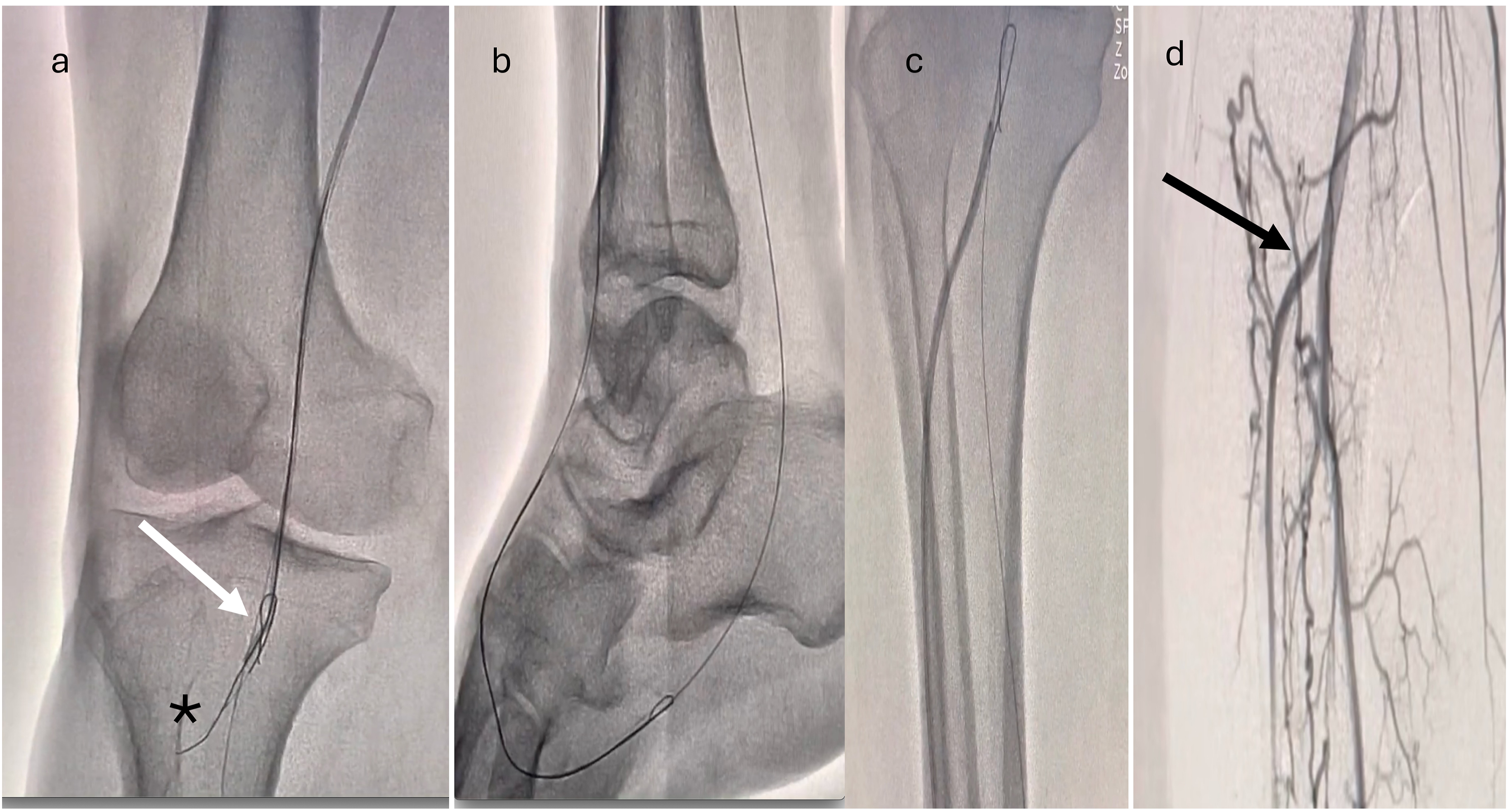
After puncturing the proximal cap and advancing the antegrade guidewire through the distal target artery, its intraluminal position was verified through contrast injections from two angiographic views. The 4 F Vert catheter and 0.018” guidewire were advanced toward the distal target artery and pedal loop, followed by balloon dilations using appropriately sized catheters. Final angiographic images confirmed the patency of the target vessels and pedal arch (Figures 1, 2 and 3). Angiographic images showing complete occlusion of the anterior tibial and peroneal arteries with ambiguous stumps, occlusion of the posterior tibial artery in the distal part just before the tarsal region and occlusion of the arteria dorsalis pedis. a. Distal posterior tibial artery (PTA) crossing and transpedal loop with a 0.018” guidewire b. Performing angioplasty to distal PDA and transpedal loop. c. Advancing the looped guidewire at the site of the anterior tibial artery (ATA) origin and tibioperoneal trunk intersection. White star indicates positioning of the looped guidewire as a marker for antegrade penetration point. a. White arrow indicates steering of angled vertebral catheter to the point where the transpedal retrograde wire loop was held as a marker for penetration. Black asterisk displays crossing of the 0.018’’ guidewire through the ambiguous proximal cap into the proximal anterior tibial artery b. Advancement of 0.018’’ guidewire through distal ATA, dorsalis pedis, and transpedal loop c. Balloon angioplasty of the osteal and proximal ATA. d. Black arrow indicating successful revascularization of the ATA with excellent distal runoff.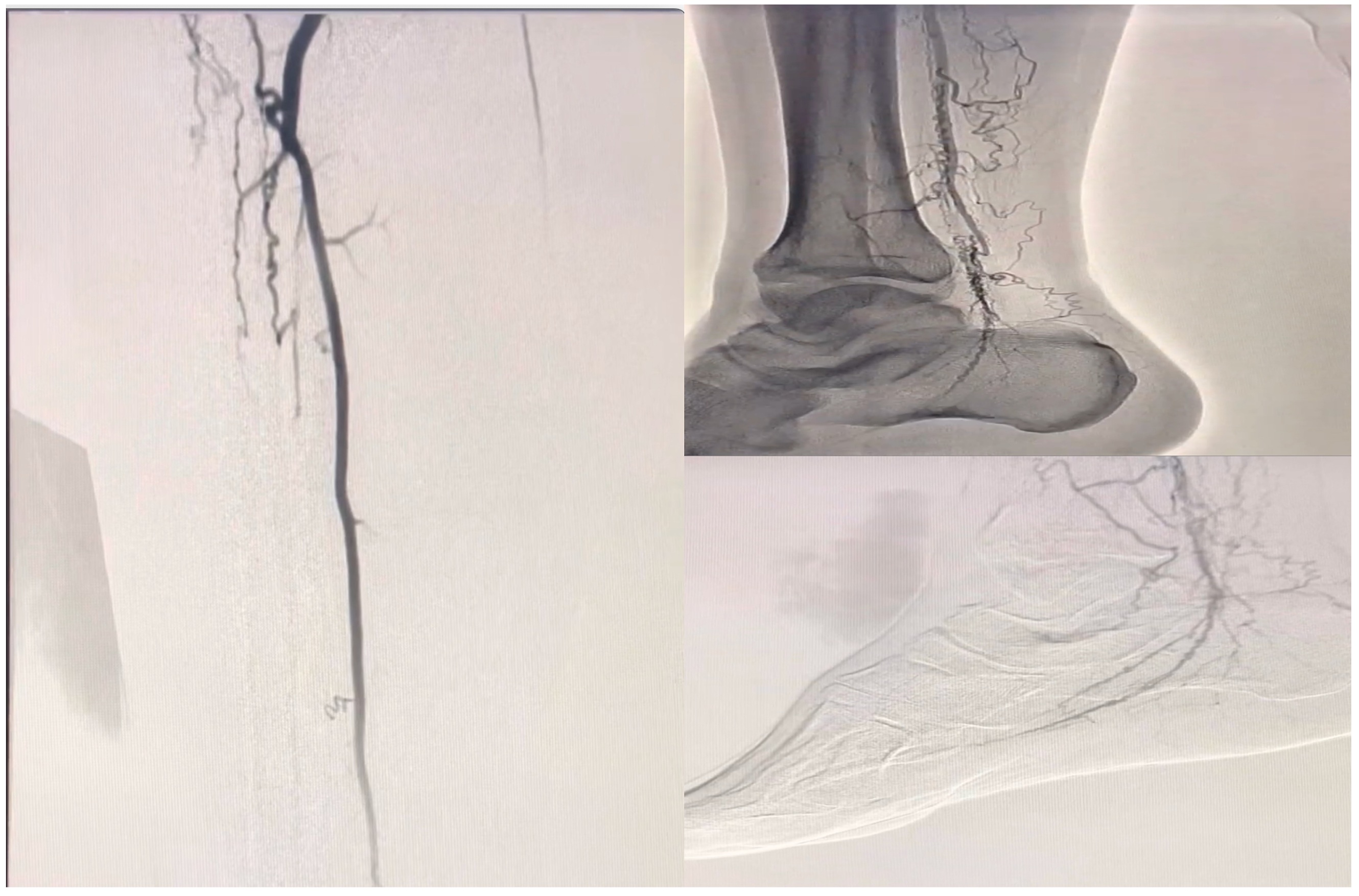

Study outcome measurements
The study assessed both primary and secondary outcomes. The primary outcome focused on the technical success or failure of the procedure, defined by post-intervention angiographic success as either residual stenosis <30% or no residual stenosis in the target vessel, with satisfactory distal run-off to the pedal arteries.
Secondary outcomes included post-intervention Rutherford class, SEWSS wound scores, and amputation rates during follow-up. Major amputations were classified as those above the ankle, including transtibial, transfemoral, knee, or hip disarticulations, while minor amputations referred to those involving the toes or below the ankle. Additionally, the pedal loop score was calculated immediately after the procedure, as described by Kawarada et al., 12 to predict post-intervention wound healing.
Statistical analysis
The Shapiro-Wilk test assessed the normality of numeric variables. Categorical variables were reported as frequencies and percentages, while continuous variables were presented as means and standard deviations or as medians and interquartile ranges (25th–75th percentiles), depending on their distribution. The Wilcoxon test was used for non-parametric variable comparisons between two periods, and the paired t test was applied for normally distributed variables. A two-tailed p-value of <0.05 was considered statistically significant.
Results
Baseline clinical characteristics
Baseline demographics and clinical characteristics.
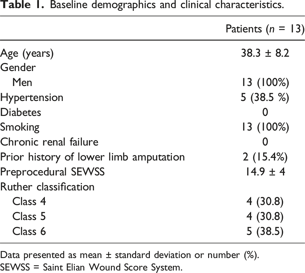
Data presented as mean ± standard deviation or number (%).
SEWSS = Saint Elian Wound Score System.
Procedural data
Procedural characteristics.
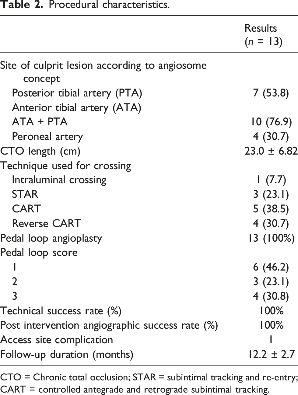
CTO = Chronic total occlusion; STAR = subintimal tracking and re-entry; CART = controlled antegrade and retrograde subintimal tracking.
Primary and secondary outcomes
The procedure failed in four of the initial 17 patients due to severe vasospasm, preventing pedal loop formation and the advancement of wires and balloons. Although the initial procedural success rate was 77%, the technical success rate reached 100% in the final group of 13 patients, where a pedal loop was successfully formed using the transpedal retrograde wire just marker technique. The post-angiographic success rate was also 100%, indicating effective luminal gain without significant residual stenosis in the target vessels. Access site vascular complications occurred in one patient, who developed a hematoma which was treated conservatively.
Secondary outcomes in patients with below the knee artery occlusions treated via transpedal retrograde wire just marker technique.

Data presented as mean ± standard deviation or number.
SEWSS = Saint Elian Wound Score System.

Preprocedural and postintervention SEWSS (a) and Rutherford Stages (b) indicating wound scores. (SEWSS = Saint Elian Wound Score System).

Relationship between follow-up amputation rates and postprocedural pedal loop scores.
Discussion
Buerger’s disease is a rare, non-atherosclerotic inflammatory disease affecting small- and medium-sized arteries and veins, leading to vascular obstructions, CLTI, and amputations if left untreated. Buerger’s disease predominantly affects young male smokers, usually under 45 years of age, and has limited treatment options. Smoking cessation remains the only proven strategy to prevent disease progression.1,2 While various treatments have shown limited success,13,14 endovascular revascularization still offers a promising alternative for Buerger’s disease despite all the drawbacks.15–18 However, performing endovascular treatment in Buerger’s disease can be technically challenging due to the distal location of lesions, small vessel size, extensive corkscrew collaterals, compromised distal run-off, and ambiguous proximal caps. 3
One of the key challenges is proximal cap ambiguity, which significantly complicates the revascularization of chronic total occlusions. 5 Target lesions in Buerger’s disease usually presents as below the knee artery chronic total occlusions with ambiguous proximal caps and unclear proximal vessel course of the occluded artery. Procedural failure and complication rates rise significantly in cases of flush occlusions where the artery’s exit point and ostium are not visible. Attempting to puncture and penetrate the ambiguous proximal cap blindly can result in perforations, which may lead to compartment syndrome. This can further deteriorate perfusion of the extremity and potentially necessitate amputation of the affected limb. Therefore, accurately identifying the vessel origin and proximal occlusion site is critical.
Up to our knowledge our study is the first to evaluate the “Transpedal Retrograde Wire Just Marker Technique” specifically in Buerger’s disease patients with chronic total occlusions and proximal cap ambiguities. This technique involves advancing a guidewire retrogradely through the transpedal loop and positioning it adjacent to the occlusion site. After confirming that the retrograde wire is intraluminal, it serves as a marker for antegrade puncture using a penetration guidewire with an angled catheter. We avoided forcing retrograde penetration and wiring through the ambiguous proximal cap with the guidewire already advanced to the distal cap retrogradely, as this could result in undesirable outcomes like major dissections and perforations in the branching main vessels. Instead, we opted for a safer technique by using the retrograde intraluminal guidewire not only as a marker for antegrade penetration and crossing but also as a landing zone for safe subintimal dissection and re-entry.
The cornerstone of the presented technique is retrograde advancement of a polymer coated guidewire to the occlusion site via pedal loop and ipsilateral antegrade femoral access. Previous studies have suggested that the transpedal retrograde approach is feasible and may be an effective first-line treatment option for limb salvage, especially when proximal caps are ambiguous. 4 Our technique not only facilitates antegrade crossing but also includes pedal loop angioplasty to enhance blood flow to the pedal arteries and promote wound healing. Previous studies reported that interangiosome connections can influence healing, regardless of the specific angiosome revascularized, and the quality of the pedal arch. was associated with healing outcomes.19,20 Hence, transpedal loop angioplasty which is routinely performed in our technique is of great importance in terms of restoring blood flow to the extremity and increased limb salvage rates.
While previous studies reported endovascular intervention success rates in Buerger’s disease patients ranging from 80% to 96%,3,15–18 our initial technical success rate of 77% highlights the challenges faced, particularly due to arterial vasospasm and pedal loop formation. Notably, our final study group achieved a 100% technical success rate with the implementation of our novel technique. Furthermore, clinical follow-up demonstrated a significant reduction in Rutherford stages and SEWSS scores, indicating potential clinical benefits from our interventions. Importantly, we identified a strong correlation between pedal loop scores and wound healing, reinforcing its role as a predictor of adverse outcomes following endovascular interventions, in alignment with Kawarada et al. and Kim et al.12,16
Study limitations
This study was limited by its retrospective, single-center design and a small patient cohort. A longer follow-up period would be beneficial for observing secondary outcomes, and a larger sample size would enable robust statistical analyses, including Kaplan-Meier survival curves for major and minor amputation-free survival rates. Additionally, our technique may have limitations in certain patients, as navigating the pedal loop can be challenging, particularly in cases of severe vasospasm. Lastly, ipsilateral antegrade femoral access is preferred over crossover access, as the balloon shafts currently used may not adequately advance through lesion sites in this context.
Conclusion
The Transpedal Retrograde Wire Just Marker Technique is a novel and practical method for treating chronic total occlusions of below-the-knee arteries with ambiguous proximal caps in patients with Buerger’s Disease. This technique, which routinely uses pedal loop angioplasty, not only facilitates the revascularization of the challenging target arteries with ambiguous stumps but also significantly increases blood flow to the pedal arch. promoting wound healing in the affected limb. While our results are promising, further studies with controlled designs are necessary to establish the routine applicability of this approach in clinical practice.
Footnotes
Declaration of conflicting interests
The author(s) declared no potential conflicts of interest with respect to the research, authorship, and/or publication of this article.
Funding
The author(s) received no financial support for the research, authorship, and/or publication of this article.
