



Abstract
Objectives
To investigate the effect of catheter-directed thrombolysis (CDT) combined with drug-coated balloon (DCB) dilatation in the treatment of femoral-popliteal thromboangiitis obliterans (TAO).
Materials and Methods
The clinical data of 26 patients with femoral-popliteal TAO who underwent CDT combined with DCB dilatation were collected from April 2020 to November 2023. The clinical efficacy at different time points (preoperative, 24 hours postoperatively. and every 6-month intervals after the procedure) in terms of the pain score, Rutherford classification, and ankle-brachial index (ABI) was compared. The primary patency was assessed at 6 months and 1 year postoperatively. Amputation-free survival and limb salvage rate were also evaluated.
Result
The femoral-popliteal artery occlusions were successfully recanalized in all 26 patients. No complications such as thrombosis, rupture of vessel, and distal embolism occurred during the operation. Thrombolytic-related hemorrhage occurred in one patient. The pain score and Rutherford grade were lower and ABI was higher at 24 hours, 6 months, and 1 year postoperatively (p < .05). The primary patency rate was 88.5% (23/26) at 6 months and 65.3% (17/26) at 1 year postoperatively. Amputation-free survival and limb salvage rate were both 92.3% at 1 year.
Conclusion
The efficacy of CDT therapy combined with DCB dilatation for femoral-popliteal thromboangiitis obliterans is acceptable at 6 months and 1 year, and the long-term effects need to be further observed.
Keywords
Introduction
Thromboangiitis obliterans (TAO), also known as Buerger’s disease, is a nonatherosclerotic, inflammatory vascular disease that affects the small- and medium-size arteries and veins mainly in the lower extremities. The pathological hallmark of this disease is the presence of a highly inflammatory thrombus in the affected arteries and veins. It is more prevalent in young males, with a significant proportion of these individuals having a history of smoking. Clinical symptoms mainly manifest as intermittent claudication and rest pain, and in severe cases, gangrene of the toes or even major amputation may occur.1,2 In recent years, endovascular minimally invasive techniques, represented by percutaneous transluminal angioplasty (PTA), have been widely used in the treatment of lower extremity arterial occlusive disease. However, patients with TAO are prone to restenosis or reocclusion shortly after endovascular treatment, making it difficult to maintain patency of the lumen. This is related to the inflammatory proliferative lesions present in these patients. 3 Currently, catheter-directed thrombolysis (CDT) has achieved good results in the treatment of arterial or venous thrombotic diseases.4–6 Moreover, drug-coated balloon (DCB) can inhibit intimal hyperplasia, thereby preventing the occurrence of restenosis after endovascular treatment.7–9 In this study, we investigated the efficacy of CDT combined with DCB dilation for the treatment of femoro-popliteal TAO.
Materials and methods
Eligible population
Clinical data were collected from patients with femoro-popliteal TAO who underwent CDT in combination with DCB dilation between April 2020 and November 2023. Inclusion criteria were a diagnosis of femoro-popliteal TAO according to the Shionoya criteria (Appendix 1) and angiographic findings compatible with TAO, Rutherford classification 4 or higher, and presence of one or more good infrapopliteal arterial outflow. Exclusion criteria were patients with a history of allergic reaction precluding angiography, with a history of hematological or renal dysfunction.
Technique
A 45 cm long sheath was inserted through a puncture in the contralateral common femoral artery. Digital subtraction angiography (DSA) was performed to clarify the lesion location, and the guidewire and catheter were then advanced to pass through the lesion segment. If the guidewire and catheter encountered significant difficulty in traversing the occluded vessel segment, an appropriate plain balloon was selected based on the vessel diameter for predilatation; if they passed through the lesion effortlessly, CDT was performed.
Based on the lesion scope and location indicated by DSA imaging, a thrombolytic catheter with appropriate working length (20–40 cm) was selected. The catheter was positioned along the guidewire, ensuring that its effective infusion segment completely covered the thrombus. The thrombolytic catheter and sheath were connected to micro-infusion pumps. The thrombolysis catheter was infused with urokinase, where 250,000 units of urokinase were dissolved in 0.9% sodium chloride and diluted to 60 mL and then continuously infused at a rate of 6–8 ml/h. The cumulative daily dosage ranged from 1 to 1.5 million units, adjusted according to body weight. The sheath was infused with unfractionated heparin, where 6250 units were mixed with 0.9% sodium chloride to a total volume of 60 mL and infused simultaneously with urokinase. During the treatment, activated partial thromboplastin time (APTT) and fibrinogen (Fib) levels were closely monitored, and the heparin dosage was adjusted to maintain APTT at 1.5–2.5 times of the normal reference value. Urokinase was discontinued when Fib was <1.0 g/L.
After maintaining CDT for 2 to 4 days, DSA imaging was performed again. If the lumen was satisfactorily opened, direct DCB dilatation was carried out. If there was residual stenosis or persistent occlusion, predilatation with a plain balloon was performed to fully open the occluded lumen and maintained for 1 to 2 minutes. If the dilatation was unsatisfactory, it was necessary to replace the plain balloon with a larger diameter for redilatation. Subsequently, an appropriate DCB was selected based on the target vessel diameter and lesion length for dilatation, lasting for 3 minutes. The DCB should completely cover the lesion area and extend 2 to 3 mm beyond each edge. If multiple DCBs were required for continuous dilatation, there should be a 10 mm overlap between the two balloons. The procedure was completed when the repeated DSA imaging showed that the residual stenosis of the target vessel was no more than 30%.
Follow-up
The clinical efficacy at different time points (preoperative, 24 hours postoperatively, and every 6-month intervals after the procedure) in terms of the pain score, Rutherford classification, and ABI was compared. Pain scores were assessed according to the World Health Organization’s pain guidelines. 10 At their follow-up visits, a hemodynamic evaluation was done by performing duplex ultrasound. Restenosis was defined as a decrease in ABI by 0.15, loss of palpable pulses, and a peak systolic velocity ratio of >2.4 on an ultrasonography scan. 11 Computed tomography angiography was performed in cases where recurrent stenosis was >50% as measured by ultrasound. Amputation-free survival and limb salvage rate were also evaluated.
Statistical analysis
SPSS 21.0 statistical software was used for the analysis. Normally distributed continuous variables were expressed as
Results
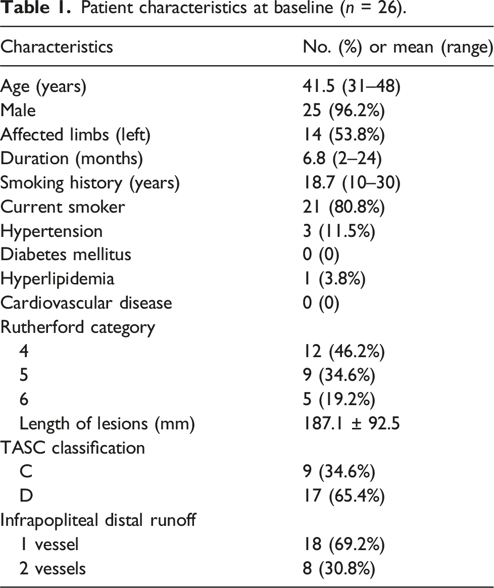
Patient characteristics at baseline (n = 26).
Comparison of clinical outcomes at different time points in 26 patients.

Compared with preoperative, *p < .05.
The primary patency rate was 88.5% (23/26) at 6 months postoperatively. No patients required target lesion revascularization at 6 months postoperatively. Three patients had a stenosis rate of ≥50%, but their clinical symptoms did not significantly worsen, and they continued with conservative treatment including medication and limb functional exercises. The primary patency rate was 65.3% (17/26) at 1 year postoperatively. Four patients underwent reintervention during the follow-up period because of relapsing of the symptoms and occlusion of the treated vessels. Two patients with technical failure underwent major amputation. There were no mortalities. Amputation-free survival and limb salvage rate were both 92.3% at 1 year.
Discussion
Thromboangiitis obliterans (TAO) remains a challenging arterial occlusive disease in vascular surgery. The underlying pathogenesis of TAO is not yet fully elucidated, yet both thrombosis and vasculitis play pivotal roles in its initiation and progression. The current surgical management of TAO encompasses a range of procedures, including vascular bypass, arteriovenous shunt, lumbar sympathectomy, and endovascular angioplasty. Endovascular angioplasty, in particular, has demonstrated favorable short-term outcomes in TAO treatment; however, its long-term efficacy still awaits further validation.12–15
In clinical practice, TAO typically manifests as extensive occlusion of the infrapopliteal arteries, whereas ilio-femoral and femoro-popliteal involvement is less common. PTA has shown limited success in the treatment of infrapopliteal artery lesions, but it shows acceptable efficacy in supragenicular lesions with adequate outflow tracts. Yuan et al. 16 characterized supragenicular TAO lesions as atypical TAO and in their report of nine patients with atypical TAO involving the external iliac to superficial femoral arteries, PTA treatment achieved good clinical outcomes, with restenosis occurring in only one patient during a mean follow-up of 20.9 months.
The main causes of restenosis after endovascular treatment of TAO include vascular elastic recoil, thrombosis, and flow-limiting dissection. Pure PTA cannot resolve these issues. Stenting has become a routine practice in the treatment of occlusive arteriosclerotic diseases, 17 but implanting stents in TAO lesions can lead to in-stent thrombosis or intimal hyperplasia, resulting in short-term in-stent occlusion. Therefore, stenting is not a recommended treatment for TAO lesions. In recent years, drug-coated balloons (DCBs) have shown good clinical efficacy in patients with occlusive arteriosclerotic diseases, especially in those with femoro-popliteal occlusive arteriosclerosis.18,19 They inhibit the proliferation of vascular smooth muscle cells through drugs such as paclitaxel, maintaining luminal patency and reducing the restenosis rate. 20 However, there are currently a few clinical studies on the use of DCB in the treatment of TAO. Therefore, this study applied CDT combined with DCB to treat TAO. CDT can dissolve thrombus, while DCB can inhibit intimal hyperplasia. The results showed that all 26 patients successfully opened their femoro-popliteal occluded arteries without complications such as thrombosis, vascular rupture, or distal embolism during the procedure. At 24 hours, 6 months, and 1 year postoperatively, pain scores and Rutherford grades were lower, and ABIs were higher than preoperative values, with statistically significant differences. The primary patency rate at 6 months and 1 year postoperatively was 88.5% and 65.3%, respectively. The preliminary study results confirmed satisfactory immediate and midterm effects.
In conclusion, the midterm efficacy of CDT combined with DCB in the treatment of patients with femoro-popliteal TAO is promising, but further confirmation through large-scale, prospective, comparative studies is required to substantiate these findings.
Footnotes
Acknowledgments
J.Z., L.W., and M.L. contributed equally to the article.
Declaration of conflicting interests
The author(s) declared no potential conflicts of interest with respect to the research, authorship, and/or publication of this article.
Funding
The author(s) disclosed receipt of the following financial support for the research, authorship, and/or publication of this article: This work was supported by the National Natural Science Foundation of China (grant nos. 81273522 and 81370441).
Informed Consent
Informed consent was obtained from all individual participants included in the study.
