




Abstract
Introduction
Ultrasound has traditionally been regarded as the first-line imaging modality for screening, diagnostic evaluation and monitoring treatment and disease progression of vascular pathology, including both the arterial and the venous branch of the vascular system. Albeit of its well-tolerated nature, wide availability and low cost, ultrasound above all, has the advantage of providing the clinician with clinically significant information related to both intraluminal irregularities and extravascular disease. Ultrasound has the potential not only to anatomically describe tissues but also to assess physiology by evaluating blood flow characteristics in real time.
Discussion
The already fundamental role of ultrasound has been even more expanded with the introduction of newer techniques like contrast-enhanced ultrasound, tissue-elastography and 3D ultrasound and the incorporation of research advances into clinical practice. The purpose of this review is to present and discuss some of the latest advances in the field of vascular ultrasound in attempt to illustrate how the adoption of multiparametric ultrasound into everyday clinical practice could address the patient's needs. Pathology and applications discussed include carotid and aortic disease, portal and peripheral venous abnormalities.
Conclusion
Widespread availability of modern technology in ultrasound devices has made the application of contrast-enhanced ultrasound, tissue elastography and 3D ultrasound feasible options, ready to contribute to the diagnostic performance of the ultrasonographic technique.
Introduction
From the first demonstration of Doppler ultrasound (US) for the detection of heart wall motion, in 1956, US has played an increasingly pivotal role in the diagnosis of vascular diseases, throughout the human body. US is reliably regarded as the first-line and often the only imaging modality required for screening, initial diagnosis or follow-up of virtually any vascular disease. This success primarily relies on the technique's inherent advantages including low cost, non-invasive nature, widespread availability and repeatability, as well as on the availability of a series of flow-detecting and analyzing techniques. Beyond visualizing blood flow, US has also the potential to investigate vessel wall-related pathology and abnormalities adjacent to the vessel; something not evident on the gold-standard imaging modality of conventional angiography. 1 Doppler technique with colour and spectral analysis offers the possibility to quantitatively analyze blood flow in healthy and abnormal vessels, being atherosclerotic arteries or thrombosed veins; information constituting the basis for diagnosis and grading of vascular disease. The umbrella term multiparametric ultrasound (MPUS) has been recently introduced in order to describe the series of existing and new ultrasonographic technologies available for applications in vascular imaging. These additional applications include contrast-enhanced ultrasound (CEUS), elastography and 3D US.2,3 The purpose of this review is to present insights into the recent developments and clinical applications of vascular US, regarding carotid atherosclerosis, abdominal aortic aneurysm (AAA), portal vein and peripheral veins thrombosis.
Carotid plaque
Conventional US
Atherosclerosis is the commonest arterial pathology affecting virtually every arterial system. A major manifestation of atherosclerosis is in the carotid artery, with significant morbidity related to cerebrovascular events. Risk stratification and the decision to treat carotid stenosis have been traditionally based on the degree of internal carotid artery (ICA) stenosis and the occurrence of neurologic symptoms.4,5 However, it is now well established that factors other than the degree of stenosis are equally, if not more important for the occurrence of stroke, especially elements that influence the mechanism of arterio-arterial embolization. Such plaque characteristics have led to the term “vulnerable” used for those plaques more prone to rupture. 6 The entity of “vulnerable” carotid plaque and its imaging identification plays a major role in modern research, and CEUS has contributed significantly to this field.
The detection of vulnerable plaque characteristics constitutes the hallmark of modern carotid imaging research and US is positioned to provide many significant developments. A series of imaging features have been unequivocally identified as markers of vulnerability, increasing a plaque's potential for rupture and acute stroke. Such features include plaque's lipid or necrotic content, intraplaque haemorrhage, surface irregularities, ulcerations, intraplaque neovascularization and inflammation. Each one of these factors can be evaluated using various qualitative or quantitative methods; for instance, a plaque's content can be assessed subjectively using the Gray-Weale's echogenicity classification or objectively using the Grey Scale Mean (GSM) technique.6–8 Similarly, a plaque's surface can be subjectively classified as smooth, irregular or ulcerated. 6 However, a quantitative method of analyzing surface irregularities would be more robust and reproducible. This was addressed by Kanber et al. 9 who conceived a potential index for the quantification of carotid plaque surface irregularities and concluded that plaques with ipsilateral hemispheric symptoms show significantly greater surface irregularities when compared to asymptomatic ones. Theoretically, the incorporation of multiple vulnerability features in a single risk classification scheme or arithmetic index would be helpful. The carotid plaque risk index, entirely US based, was proposed, taking into account the degree of stenosis, a quantitative index of surface irregularities and the GSM, suggesting that a multivariable vulnerability index increased both sensitivity and specificity for differentiating symptomatic from asymptomatic plaques compared to the degree of stenosis alone. Nevertheless, prospective studies are required for confirmation of these initial results. 10 Eyding et al. 8 have proposed an integrated multivariable system for classification of carotid plaque risk, incorporating clinical data, the degree of stenosis, GSM, surface irregularities and plaque neovascularization on CEUS. 8
CEUS
The introduction of ultrasound contrast agents (UCAs), microbubble based, and CEUS imaging has allowed superior visualization and insights into the pathophysiology of atherosclerotic plaques both in terms of surface morphology and internal structure.11–13 Superficial ulceration is thought to be a crucial contributor to plaque vulnerability, attributed to potential for arterio-arterial embolization of thrombotic material and aggregated platelets. Virtually any imaging modality investigating patients with carotid disease has the potential to visualize ulcerations with varying degrees of diagnostic accuracy.
11
Ten Kate et al.
14
demonstrated that CEUS was characterized by superior sensitivity, positive and negative predictive value for the diagnosis of ulcerated carotid plaque, as compared to colour Doppler technique, having computed tomographic angiography (CTA) as the reference method (Figure 1). In this study, it was observed that CEUS demonstrated ulcerations that were not seen by the reference method based on the superior inherent resolution of US, questioning whether CEUS outperforms CTA. The excellent spatial and temporal resolution in the near field when imaged with high-frequency transducers during a carotid examination would be supportive of this suggestion.
14
Hamada et al.
15
were able to confirm the superiority of CEUS over conventional US for the detection of carotid ulcerations using histology as the reference method, demonstrating that a cut-off value of only 1.3 mm for depth can be used to classify a concavity as an ulceration with a sensitivity exceeding 91%. Even if no UCA is administered, the introduction of new diagnostic criteria and improved image quality provided by modern US devices allows improved diagnostic accuracy for the detection of ulcerations. Explicitly, a concavity can be characterized on US as a true ulceration if its basal border echo is weaker compared to the adjacent plaque fibrous cap, discarding the previously reported criterion of size or internal flow reversal.
16
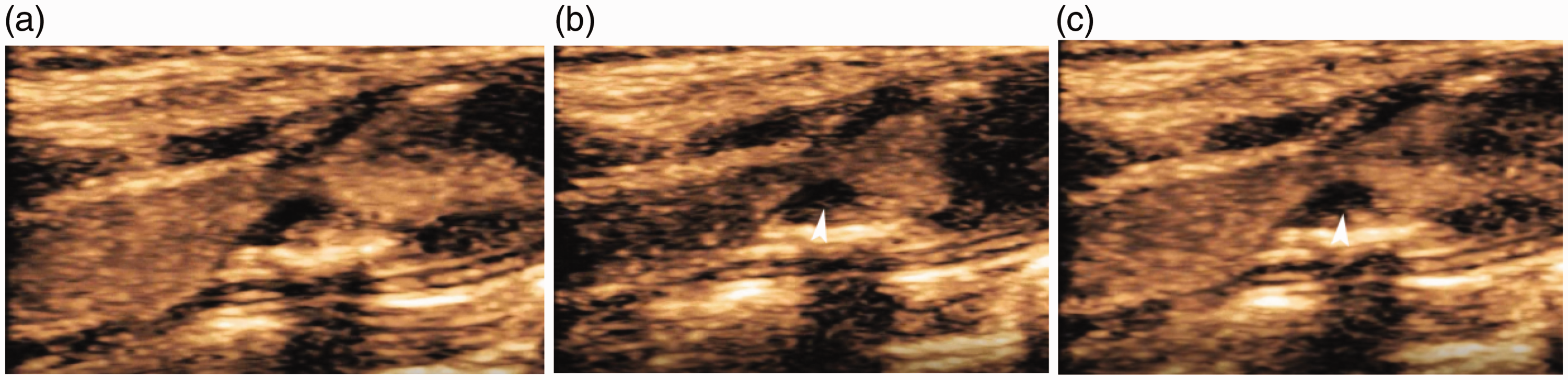
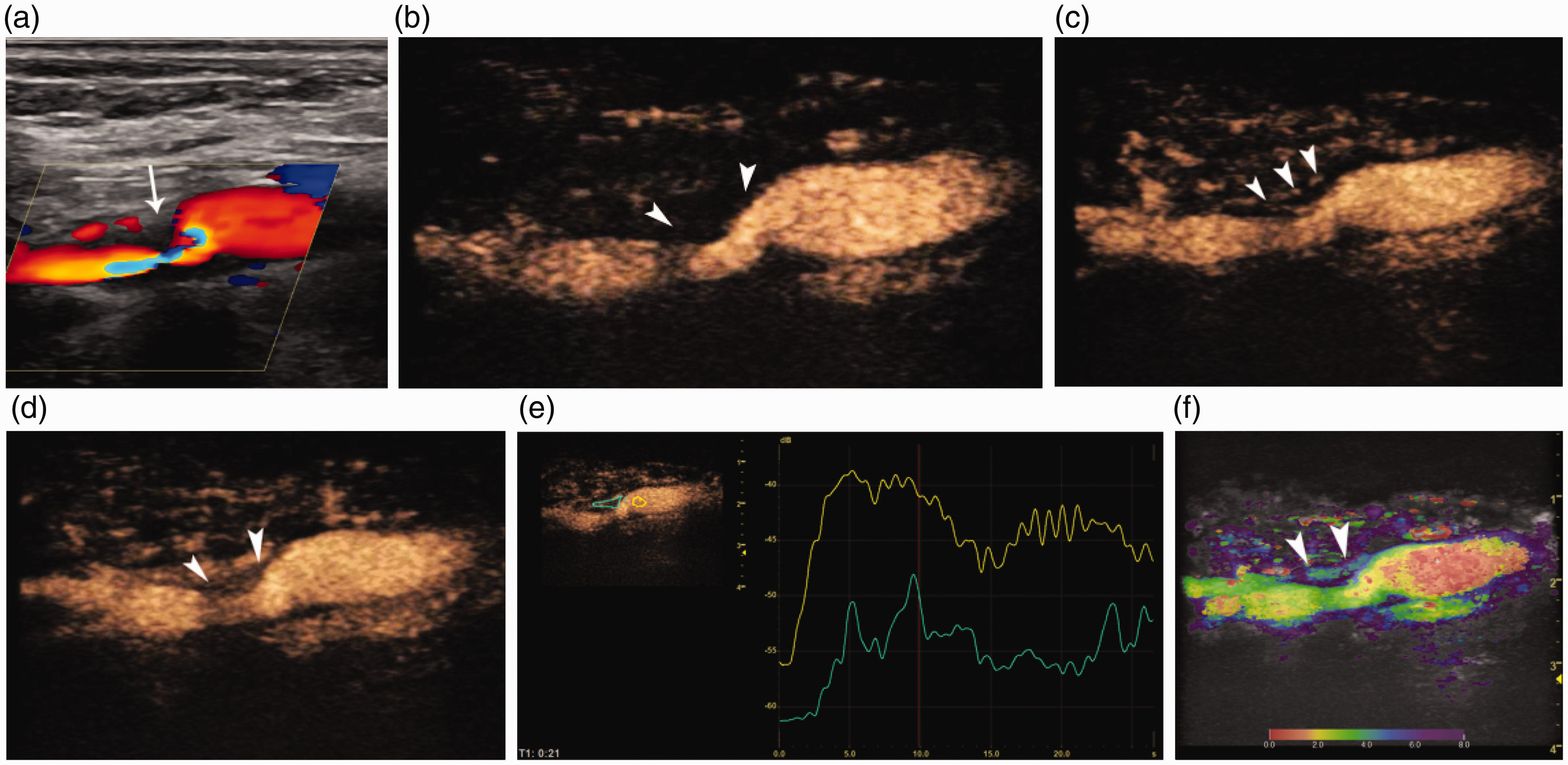
This echogenicity criterion is attributed to the lower acoustic impedance of the thrombus compared to that of the intact fibrous cap and reflects the improved ability of US for tissue characterization. With the evaluation of carotid plaque internal morphology, CEUS has offered new insights into the pathophysiology of atherosclerosis allowing both to observe in a qualitative way and quantify intraplaque neovascularization: – an emerging feature of plaque vulnerability (Figures 2 and 3). CEUS represents an excellent modality for intraplaque neovascularization as the inherent property of UCA to strictly remain within the vascular lumen allows UCA to act as an intravascular ‘tracer’; not possible for computed tomography (CT) and magnetic resonance (MR) imaging contrast agents. Plaque enhancement on CEUS, irrespective of the qualitative or quantitative assessment, has been correlated both with histologically detected neovessels and the occurrence of symptoms, with symptomatic plaques exhibiting higher enhancement than asymptomatic.12,13,17–20 Furthermore, increased enhancement on CEUS has also been associated with inflammatory cells and intraplaque haemorrhage, also essential features of plaque vulnerability.
17
Going forward, CEUS evaluation of carotid intraplaque neovascularization will need to be validated with technique standardization which will allow for risk stratification algorithms targeted to predict the occurrence of stroke in asymptomatic patients. Apart from the evaluation of the atherosclerotic plaque, CEUS can be used for differential diagnosis of carotid occlusion and near-occlusion: an ongoing dilemma with clinical significance as a near occlusion requires surgical intervention (Figure 4).
A 64-year-old male patient presenting with acute stroke and with an ipsilateral ulcerated ICA plaque. Long-axis colour Doppler image (a) shows a hypoechoic plaque causing severe stenosis (asterisk). Corresponding CEUS image (b) delineating a superficial ulceration (arrowhead) on this extremely stenotic plaque. Axial colour Doppler image (c) showing the stenotic ICA lumen surrounded by a hypoechoic plaque (asterisk). Corresponding CEUS image (d) confirming the presence of ulceration (arrowhead) (asterisk: ICA lumen; E: external carotid artery). CTA image (e) confirming CEUS findings, with contrast in the ulcerated plaque (arrowhead). A 60-year-old male patient with an acute stroke. Ipsilateral carotid artery US confirmed the presence of severe ICA stenosis. Consecutive CEUS images (a, b, c) demonstrate the presence of moving microbubbles (arrowheads) within the initially hypoechoic part of the plaque; findings consistent with mild intraplaque neovascularization. US and CEUS findings in a 57-year-old female patient with a right-sided stroke. Colour Doppler US (a) shows a hypoechoic plaque causing severe stenosis of the ipsilateral ICA (arrow). CEUS image immediately after the arrival of the microbubbles (b) demonstrates the plaque as uniformly hypoechoic and accurately delineates the plaque's border (arrowheads). Consecutive CEUS images (c, d) detected moving microbubbles (arrowheads) within the plaque, which gradually shows uniform enhancement (arrowheads); findings consistent with severe intraplaque neovascularization. A time-intensity curve (e) quantifies the plaque enhancement in comparison with the lumen enhancement. Parametric image (f) characterizing the plaque's enhancement based on the time of microbubble's arrival. It is assumed that the plaque enhances in a centrifugal pattern, probably due to the presence of adventitial neo-vessels (arrowheads pointing to the plaque). Imaging findings in a 74-year-old male patient with pre-occlusive stenosis of the ICA. Colour Doppler US (a) raised suspicion of ICA occlusion. However, CEUS (b) readily identified a thread-like opacified lumen establishing the diagnosis of near-occlusion (arrow), which was also confirmed with CTA (arrowheads in c).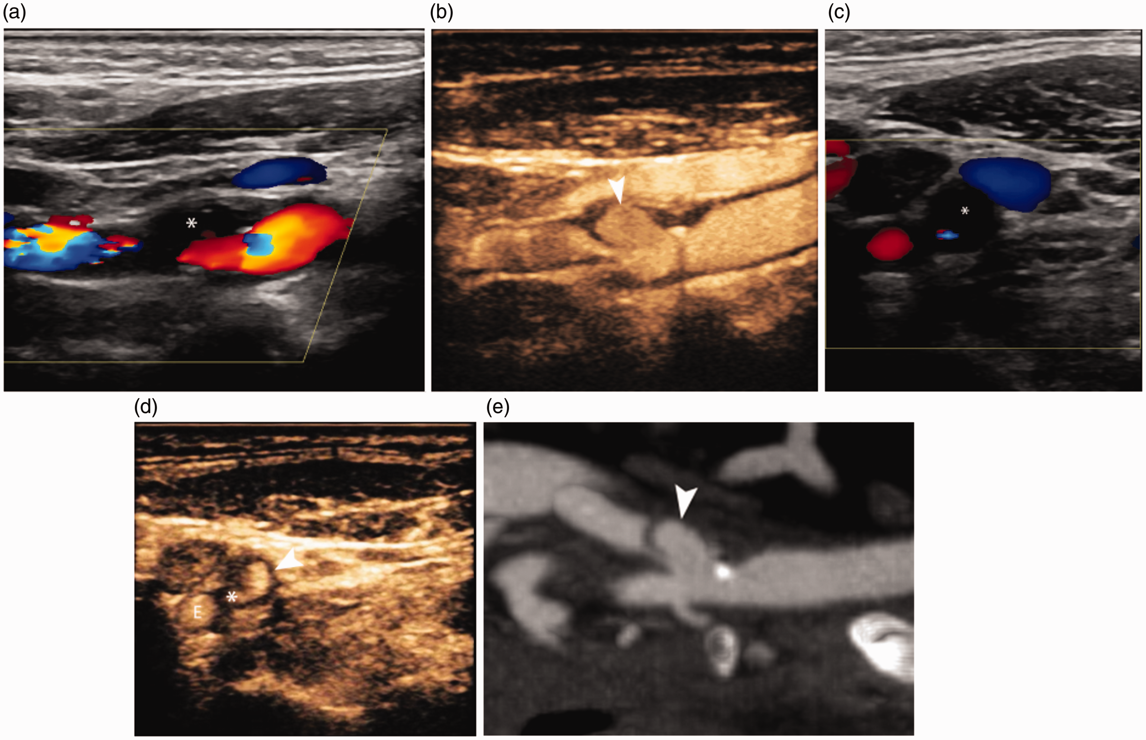

Tissue elastography
US elastography is a recent advance in carotid US imaging which can be used to evaluate plaque vulnerability and could be potentially incorporated into some type of multifactorial analysis.21,22 Ramnarine et al.
23
have used shear wave elastography to quantify plaque elasticity, concluding that symptomatic plaques had a significantly lower mean of Young's Modulus compared to asymptomatic plaques and that the incorporation of elastography to the degree of stenosis improved both sensitivity and specificity for the detection of vulnerable plaque. Lou et al.
24
have shown that the calculation of Young's modulus with shear wave elastography has excellent reproducibility and is significantly correlated with the Gray-Weale's echogenicity classification, importantly confirming that symptomatic plaques show a lower mean Young's Modulus and the combination of Young's modulus and degree of stenosis yields a higher sensitivity and specificity for the identification of symptomatic plaques.
24
When using histology as the reference method, it was found that the mean Young's modulus was significantly lower in unstable plaques as well as those with intraplaque haemorrhage, thrombus and increased number of foam cells.
25
In the light of these results, tissue elastography appears as a promising method in the risk stratification of carotid plaques, which could be used in the future for selection of plaques more prone to the occurrence of neurologic symptoms (Figure 5).
A 65-year-old male patient with stroke was diagnosed with an ipsilateral hypoechoic internal carotid artery plaque (colour Doppler US on a). Shear-wave elastography (b) has showed the plaque to be soft based on low elasticity values. A different 70-year-old asymptomatic patient was diagnosed with a heavily calcified internal carotid artery plaque which on shear-wave elastography was found to be hard based on high elasticity values.
Three-dimensional US
Three-dimensional US (3D US) has also been investigated in carotid imaging proving feasibility and a valuable technique to visualize the whole extent of the carotid system, course variations and stenotic segments. 26 Moreover, 3D US detects more ulcerations compared to conventional techniques and offers superior inter-observer agreement. 27 Kuk et al. 28 have used 3D US to prospectively follow-up patients with carotid stenosis and showed that a total volume of ulcerations > 5 mm3 was associated with a significantly higher risk of developing stroke or transient ischemic attack, myocardial infarction or death.
Even with initial promising results, further studies are needed to establish the role of all modern techniques including elastography, CEUS and 3D US in the evaluation of carotid vulnerable plaque. Furthermore, standardization of techniques and widespread consensus are essential before these techniques are implemented into routine clinical practice based. A number of technical factors have the potential to introduce technique variability. For CEUS, it is important to establish which microbubbles visualization technique is better (pulse inversion or amplitude modulation), define a default value for mechanical index (MI), the dose of contrast agent and pattern of injection as well as other US parameters like gain, frame rate or frequency. Similarly, it is essential to decide whether strain or shear-wave elastography would be more appropriate for the characterization of carotid plaque.
Aortic disease
Endovascular aortic repair
The aorta and major branches can be successfully investigated with US as a first-line imaging modality, although widespread availability of CT and MR imaging has altered the diagnostic approach of aortic diseases, replacing the traditional method of invasive angiography. Many patients are referred for AAA investigation, using US, which is the established imaging modality for the detection and monitoring of AAA. Currently, either a traditional surgical approach or an interventional approach (endovascular aneurysm repair, EVAR) is the course of treatment.
Even though EVAR has many advantages over the surgical technique, there is a need for lifelong imaging surveillance consequent to the risk for complications such as an endoleak or graft migration. The term endoleak refers to the presence of persistent blood flow within the aneurysm sac but outside the stent wall, with five different types of endoleaks described based on the origin and direction of blood flow. CTA is regarded as the best modality for the detection of endoleaks and is the primary technique used for follow-up of these patients. However, lifelong follow-up with CTA would entail an associated risk due to the exposure to ionizing radiation and nephrotoxic contrast agents. Colour Doppler US is also used for post-EVAR evaluation, although with varying degrees of sensitivity and specificity. CEUS has yielded superior diagnostic accuracy compared to conventional US and equivalent to CTA. CEUS inherent capability for real-time, dynamic and prolonged visualization of blood flow allows for observation of flow within a stented aorta; CTA does not have this capability, where the detection of an endoleak is limited by up to three instantaneous phase acquisitions. CEUS has a superior potential to characterize endoleaks and classify them based on the accurate detection of flow origin and direction.1,13,29–31 A meta-analysis of CEUS detection of endoleaks has shown that CEUS has a pooled sensitivity of 91.4% and a pooled specificity of 78.2%, although the latter may be limited by study heterogeneity.
32
Another systematic review has concluded that MRA and CEUS are superior to CTA for the detection of endoleaks and equivalent for endoleaks classification.
33
Four-dimensional CEUS is another recent development which was found equivalent to CTA in terms of accuracy for the assessment of aneurysm diameter, volume and endoleak detection.
34
Based on these encouraging results, CEUS can be used as a second imaging modality after initial screening for endoleaks with US, in order to increase the sensitivity and reduce the number of CTA examinations for patients after EVAR (Figure 6).
An 80-year-old-man, following EVAR, with a persistent enlarged aneurysmal sac, with renal impairment undergoing surveillance with follow-up ultrasound. B-mode ultrasound (a) in an axial view demonstrating the stented aorta (arrow) and surrounding echogenic thrombus within the aneurysmal sac. Colour Doppler US (b) suggests a leak with colour Doppler signal (arrow) identified within the aneurysmal sac. The CEUS image (c) demonstrates a clear type II endoleak (arrow).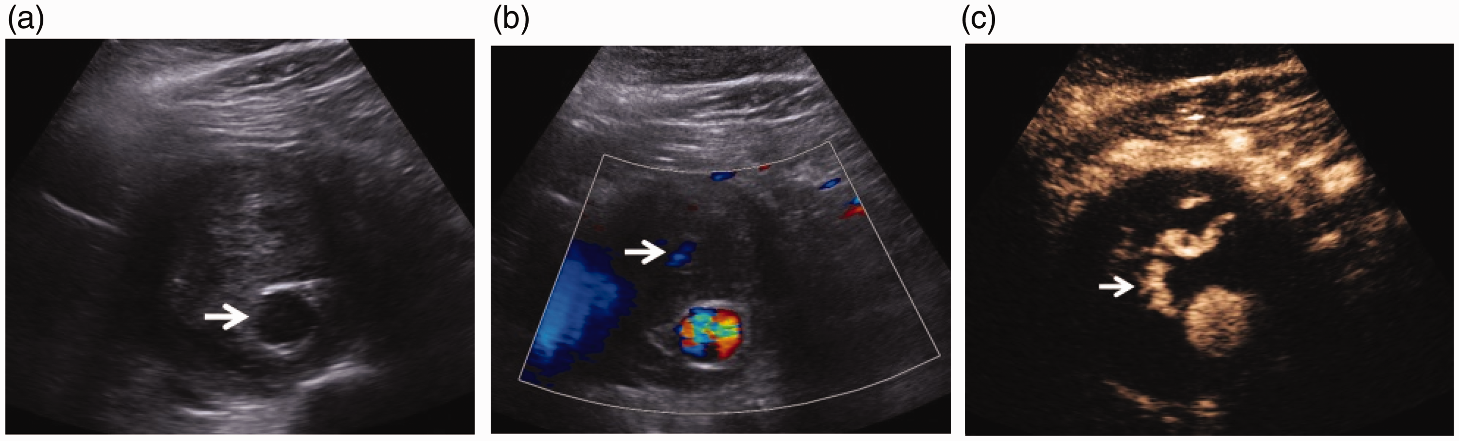
Aortic rupture
Beyond post-EVAR follow-up, CEUS has also been found superior to the unenhanced US for detection of AAA rupture, as it is possible to visualize active UCA extravasation and dependent pooling of the UCA, with excellent correlation with the reference method of CTA. 35 It is reported that aorto-caval fistulas, a complication of untreated AAA, can be visualized with CEUS. 36 CEUS has also been reported for evaluation of the microvasculature in the fibro-inflammatory reaction in chronic periaortitis. The technique was also able to document a reduction in the peri-aortic enhancement after steroid treatment. 37 Although such reports illustrate the superior spatial and temporal resolution provided by CEUS, these are isolated reports and do not yet substantiate routine use of CEUS.
Venous applications
Portal vein
Neoplastic thrombus constitutes a contraindication for liver transplantation. Imaging findings suggestive of a benign thrombus include gradual decrease in size or recanalization on colour Doppler US, whereas increase in size, disruption of the vascular wall and infiltration of the nearby tissues are suggestive of malignancy. The hallmark for the diagnosis of neoplastic thrombosis remains the demonstration of neovascularization within the thrombus. 38 Conventional B-mode and colour Doppler US can be used to detect portal vein thrombosis and raise suspicion of malignancy if blood flow signals are detected within the thrombus although they lack sensitivity for small thrombi and unfavorable body habitus due to the deep location of the vessel under examination.
Recent applications of CEUS include investigation of suspected portal vein thrombosis, where the important differential is distinguishing bland and neoplastic thrombus in patients with hepatocellular carcinoma or cirrhosis. CEUS was superior to the unenhanced US for the diagnosis of malignant portal vein thrombosis, demonstrating pulsatile enhancement of the thrombotic material during the arterial phase and earlier to the portal lumen opacification, as expected for malignant neovascularization (Figure 7). These findings account for the increased sensitivity and specificity of CEUS for the diagnosis of malignant portal vein thrombosis, which can be incorporated to the diagnostic pathway in order to avert fine-needle biopsy, the reference standard.38–40
Portal vein thrombus in a 27-year-old female patient. Colour Doppler US (a) demonstrates no flow within an expanded portal vein (arrow). CEUS image (b) demonstrates early re-canalization of the central aspect (arrow) of the occluded portal vein.
Peripheral veins
In the evaluation of peripheral veins, B-mode and colour Doppler US are usually sufficient for the thorough evaluation of abnormalities including acute or chronic thrombosis and venous insufficiency. An interesting application could be the use of elastography for accurate dating of thrombus, based on the notion that as thrombus becomes mature, it becomes stiffer. However, there is only limited evidence available for this application, stemming both from experimental and clinical studies and further clinical studies are required to confirm the validity of these findings.41,42 In another recently published paper, CEUS was used to evaluate perivascular perfusion in the setting of acute deep vein thrombosis. It was found that the peak enhancement and wash-in rate of a perivenous region are significantly higher in thrombosed veins and return to normal levels after three months, findings that can be attributed to inflammation associated with acute deep vein thrombosis. 43
Conclusion
With the many developments described, it can be concluded that US remains the cornerstone of vascular diagnosis, acting as the well-established first-line imaging modality. Cross-sectional modalities such as CTA and MRA can be set aside for further investigation of patients only after an initial assessment with US. The incorporation of CEUS and elastography in the ultrasonographic protocol of a single examination session could expand the value of US, already showing promising results and bringing implications for patient risk stratification in carotid disease or allowing for accurate diagnosis in endoleak detection or portal vein thrombus characterization. The widespread availability of high-end ultrasonographic devices has rendered the implementation of these technological advances feasible and it remains to be seen whether the future will lead to their adoption into clinical routine for the benefit of the patient.
Footnotes
Declaration of Conflicting Interests
Author VR has no conflicting interest regarding the publication of this manuscript.
Author PS has received lecture fees from Bracco, Siemens, Samsung, Philips and Hitachi.
Funding
The author(s) received no financial support for the research, authorship, and/or publication of this article.
Ethics approval
Ethical approval to publish this retrospective review was requested and waived by the hospital board. Written informed consent was obtained by all patients for publication of the submitted images.
Guarantor
PS.
Contributors
VR researched literature, VR and PS conceived the paper, wrote the paper and reviewed and approved its final version.