



Abstract
Real-time ultrasound imaging (US) to measure abdominal muscle dimensions has aided low back pain rehabilitation and research. Notwithstanding, ultrasound imaging measurement of transversus abdominis muscle activation in chronic low back pain populations has been characterized by variable and generally suboptimal intra-observer reliability. Methodological deficiencies of ‘freehand’ ultrasound imaging are uncontrolled probe–skin pressure, inclination and roll of the probe. Despite previous attempts to standardize these parameters, intra-observer reliability in chronic low back pain was poor to moderate (0.32–0.62). Therefore, a standardized method that controls and records probe force, inclination and roll during ultrasound imaging may optimize measurement reliability in chronic low back pain. This pilot study investigated utility, standardization and intra-observer reliability of ultrasound imaging transversus abdominis thickness measurement in chronic low back pain patients (n = 17). Transversus abdominis imaging over two separate measurement sessions was conducted using a novel method to standardize probe parameters. Resting and contracted transversus abdominis thickness, and transversus abdominis activation measurements were obtained from duplicate paired images (n = 68). Intra-class correlation coefficients were reported with 95% confidence intervals. Transversus abdominis thickness at rest (intra-class correlation coefficient = 0.97 confidence interval: 0.93, 0.99), when contracted (intra-class correlation coefficient = 0.99 confidence interval: 0.97, 0.99) and transversus abdominis activation (intra-class correlation coefficient = 0.93 confidence interval: 0.81, 0.97) measurements were highly reliable. Ultrasound imaging of transversus abdominis using the novel standardized ultrasound imaging method produced highly reliable intra-observer transversus abdominis measurements, superior to ‘freehand’ ultrasound imaging, despite the physical limitations typically associated with a chronic low back pain population. Unique standardizing ranges for ‘probe force device’ probe parameters were obtained. This novel standardized ultrasound imaging method may optimize transversus abdominis activation assessment in chronic low back pain and other populations, aiding future research.
Introduction
Real-time ultrasound imaging (US) has been used to measure abdominal muscle dimensions to aid rehabilitation and research in low back pain for many years. 1 The measurement of one specific abdominal muscle, the transversus abdominis (TrA), has become preeminent because of its role in spinal stabilization. 2 TrA thickness at rest (RTrA), and when fully contracted (CTrA), are US measurements used to calculate TrA activation (TrA-C). 3
Using US, previous studies have compared TrA-C in symptomatic and asymptomatic individuals with chronic low back pain (CLBP). Some studies have demonstrated TrA-C anomalies, 4 TrA morphological changes 5 and the efficacy of some therapeutic interventions to improve TrA-C. 6
Notwithstanding, US TrA-C measurement in CLBP populations has been characterized by widely variable and generally suboptimal intra-observer reliability. 7 Only two prior studies have reported intra-observer TrA-C measurement reliability in CLBP.3,8 Reliability was suboptimal (intra-class correlation coefficient (ICC) = 0.32–0.72). 7 Such variability has been considered solely dependent upon the failed application of rigorous application protocols, 9 yet a consensus regarding such protocols is currently lacking and is problematic given that US is a ‘freehand’ procedure. 7
Methodological deficiencies inherent with ‘freehand’ US are uncontrolled probe force (amount of pressure applied to the patients skin), inclination (side-to-side tilt of the probe perpendicular to the plane of the probe) and roll (forward and backward movement parallel to the scan plane of the probe). Maintenance of probe inclination perpendicular to the TrA during US is a prerequisite to identify the hyper-echoic TrA fascial borders between which TrA thickness is measured. 9 Moreover, variable probe-to-skin force may distort TrA dimensions. 9 Despite previous attempts to standardize these parameters by enclosing the probe in a foam housing secured to the participant with a belt, intra-observer reliability in CLBP was poor to moderate (0.32–0.62). 3 In addition, the raised body mass index (BMI) typical in CLBP 10 results in increased tissue adiposity and abdominal wall compliance, which thwarts the acquisition of clear measureable TrA images 11 and challenges control of probe force and orientation during ‘freehand’ US.
Probe force has been subjectively classified in ‘freehand’ US studies as ‘gentle’, ‘light’ or ‘minimal’.8,3,12 This is suboptimal, but a standardized method that controls and records probe force, inclination and roll during US, 13 may optimize TrA measurement reliability in challenging populations, such as CLBP. The objectives of this study were (1) to investigate the utility of a novel standardized US method in a CLBP population, (2) to apply this method for image acquisition and interpretation and (3) to calculate the intra-observer reliability using the standardized method.
Methods
Study design
This blinded pilot intra-observer reliability study was conducted in a multicultural specialist back pain clinic in a developed western country, with secondary day procedure facilities, as part of an existing registered prospective study (ACTRN: 12613000267752). Participants were referred to the clinic by either a primary healthcare physician or a healthcare specialist. Potential participants were consecutively approached as they attended the clinic between July and October 2015. Study approval was obtained from the human research ethics committee of the James Cook University, the Townsville Hospital and Mater Health Services (H4387/10QTH53/MHS20150512-07). An explanation and information sheet detailing aims and methods of the study was provided to participants, and written consent was gained prior to study commencement.
Inclusion and exclusion criteria
Participants were 18 years of age or older and presented with CLBP which adversely influenced their functional status. CLBP was defined as pain between the level of T12 and the buttock crease, with or without associated lower limb symptoms, 14 and present for longer than 12 weeks. 15 Participants were excluded if they had undergone previous surgery to the lumbo-sacral spine, were pregnant, unable to communicate, had psychiatric disorders that might interfere with the participant’s interpretation of instructions, systemic illness or infection, tumours, trauma/fractures, abdominal surgery in the previous year, current litigation, insurance or other compensation claims, medical conditions that resulted in CLBP, fibromyalgia, osteoporosis, presence of medical ‘red flags’ indicative of potentially serious medical conditions and progressive neurological disturbance.
Examiner
The primary investigator, a physiotherapist, with five years of experience at level one musculoskeletal US, screened potential participants for inclusion and exclusion criteria, recorded participant data and demographics, conducted all US and TrA measurement under blinded conditions.
Experimental equipment, outcome measures and procedure
Standard protocols were used to measure height and weight.
16
US was conducted in movie mode (GE Healthcare Venue 40 MSK; General Electric Company; Wauwatosa, WI, USA), using a 3.1 MHz, curved array abdominal probe 4C-SC model 5337596 (65 mm × 15 mm footprint), to capture real-time video images of the participants’ dominant side RTrA and CTrA over two separate measurement sessions (‘measurement 1’ and ‘measurement 2’). A probe force device (PFD)
13
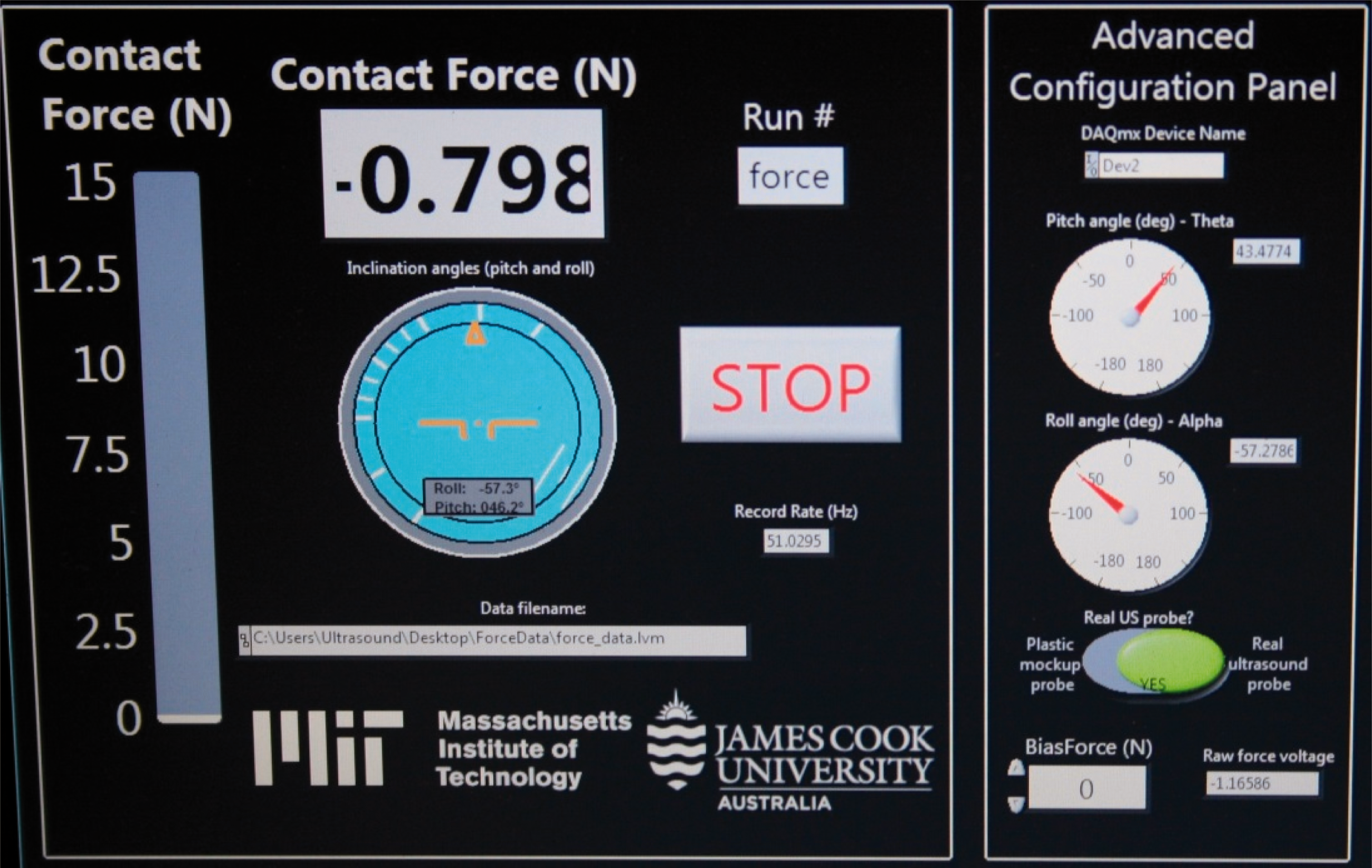
was attached to the US probe (Figure 1), and real-time on-screen display of probe force (N), inclination (°) and roll (°) was recorded at 60 Hz via a LabVIEW virtual instrument link on a laptop computer
13
and stored for later analysis (Figure 2). During imaging the US machine, PFD and the examiner were positioned to the right of participants who lay on a surgical procedure bed in supine ‘crook’ position, with no pillow head support.
17
Pelvic and lumbar position were standardized by palpation and auditory cueing.
18
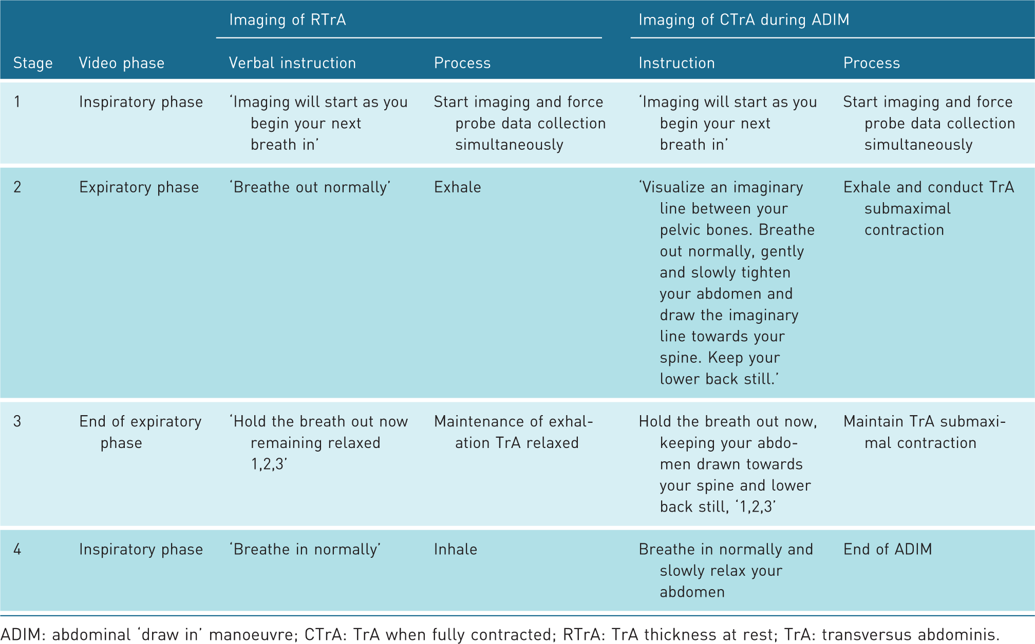
Goniometry was used to standardize hip and knee joint angles at 30° and 90°, respectively. A breathing cycle protocol was used for image capture of RTrA and CTrA (Table 1).
19
A familiarization session was conducted prior to US. For RTrA, participants were instructed to breathe normally and to then hold end expiratory phase for up to 3 seconds. For CTrA, participants were taught an abdominal ‘draw in’ manoeuvre (ADIM),
19
to selectively activate the TrA.
20
During familiarization, a method reported previously to standardize CTrA was applied.
21
Participants were instructed to conduct ADIMs at 50% maximum effort. Participants conducted several 50% maximum effort practice attempts, with repeatability assessed by the examiner using measurements taken with the US machines on-screen caliper.
PFD attached to real-time US probe and standardized position using adhesive template. PFD on-screen display. A single breathing cycle protocol during imaging of RTrA and CTrA ADIM: abdominal ‘draw in’ manoeuvre; CTrA: TrA when fully contracted; RTrA: TrA thickness at rest; TrA: transversus abdominis.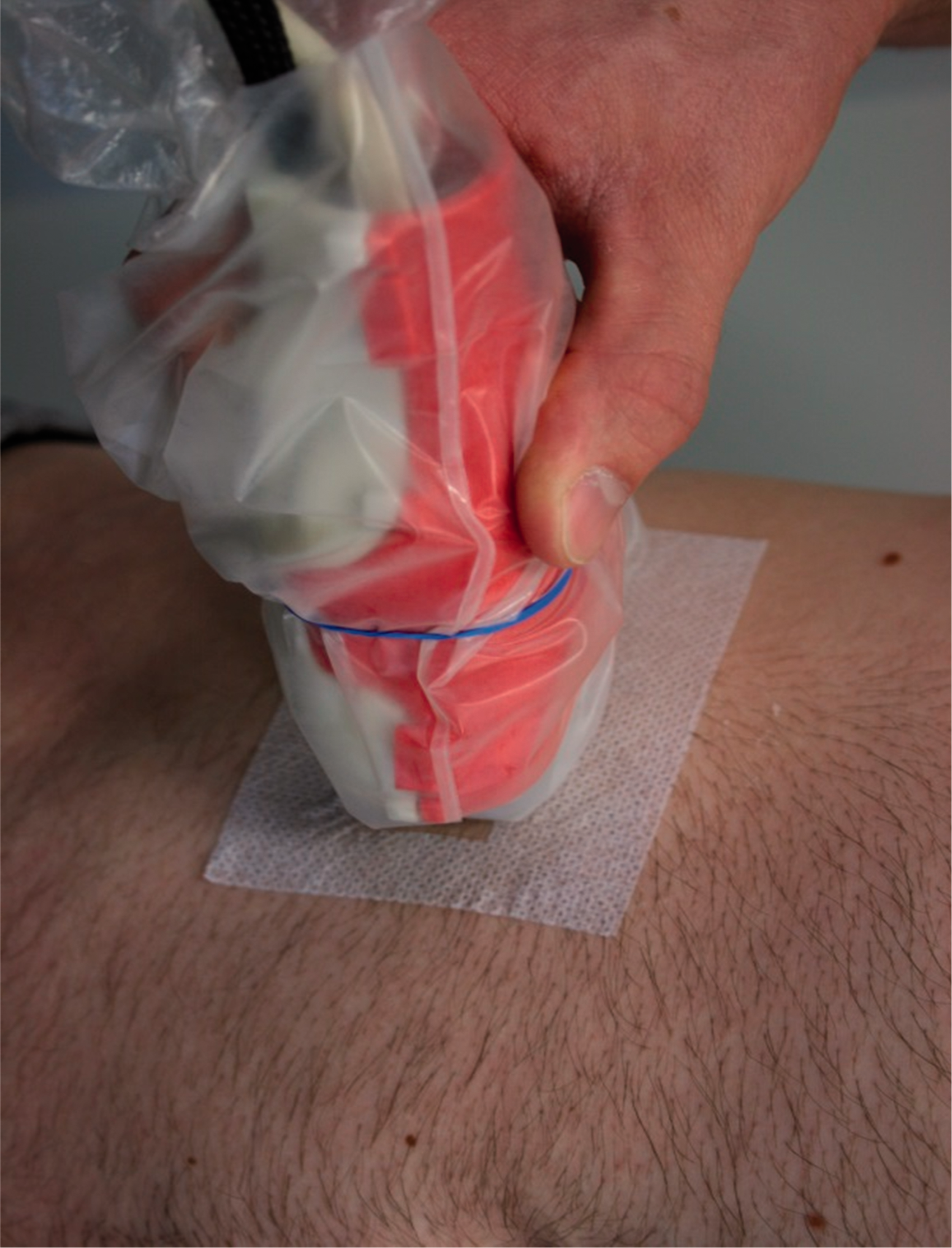
The US probe was clipped into the PFD shell and prepared for imaging (Figure 1). The laptop computer software and US machine were activated, probe cable ‘strain relieving’, and force bias ‘zeroing’ was conducted. 13 Coupling gel was applied and the probe was placed lateral to the umbilicus, midway between the iliac crest and the lower ribs. 8 Preliminary US established the probe position required to achieve optimal RTrA and CTrA views, and this position was marked with a Hypafix® (BSN medical Luxembourg Finance Holding S.à r.l.; Luxembourg) adhesive windowed template adhered to the participants’ skin (Figure 1). The window allowed a space for direct probe–skin contact, whilst maintaining repeatability of probe–skin placement.
Descriptive characteristics
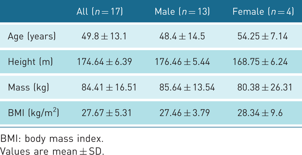
BMI: body mass index.
Values are mean ± SD.
To avoid ADIM ‘training bias’, participants were blinded to the US display using a screen between participants and the machine. US and probe force, inclination and roll data were automatically stored to a memory card for later processing and analysis.
Still image extraction
Data collection yielded one RTrA and one CTrA video for ‘measurement 1’ and for ‘measurement 2’ per participant (n = 4), from which cropped video sections of RTrA during end expiratory phase, and of CTrA during ADIM for each ‘measurement’ (stage 3 of the breathing cycle protocol) (Table 1), were extracted and stored (n = 68). Cropped videos for each participant were time matched to US probe data. Using Microsoft excel™, data were sorted to identify data time points, where probe force, inclination and roll at ‘measurement 1’ matched that of ‘measurement 2’. The matched data time points were identified on each video, and four corresponding still US images per participant were acquired (n = 68). Each still image was copied prior to image measurement, producing duplicate paired images.
Examiner blinding prior to image measurement was conducted to avoid measurement bias. An independent research assistant anonymized all images, which were measured by the examiner on a personal computer using ImageJ measurement software.
22
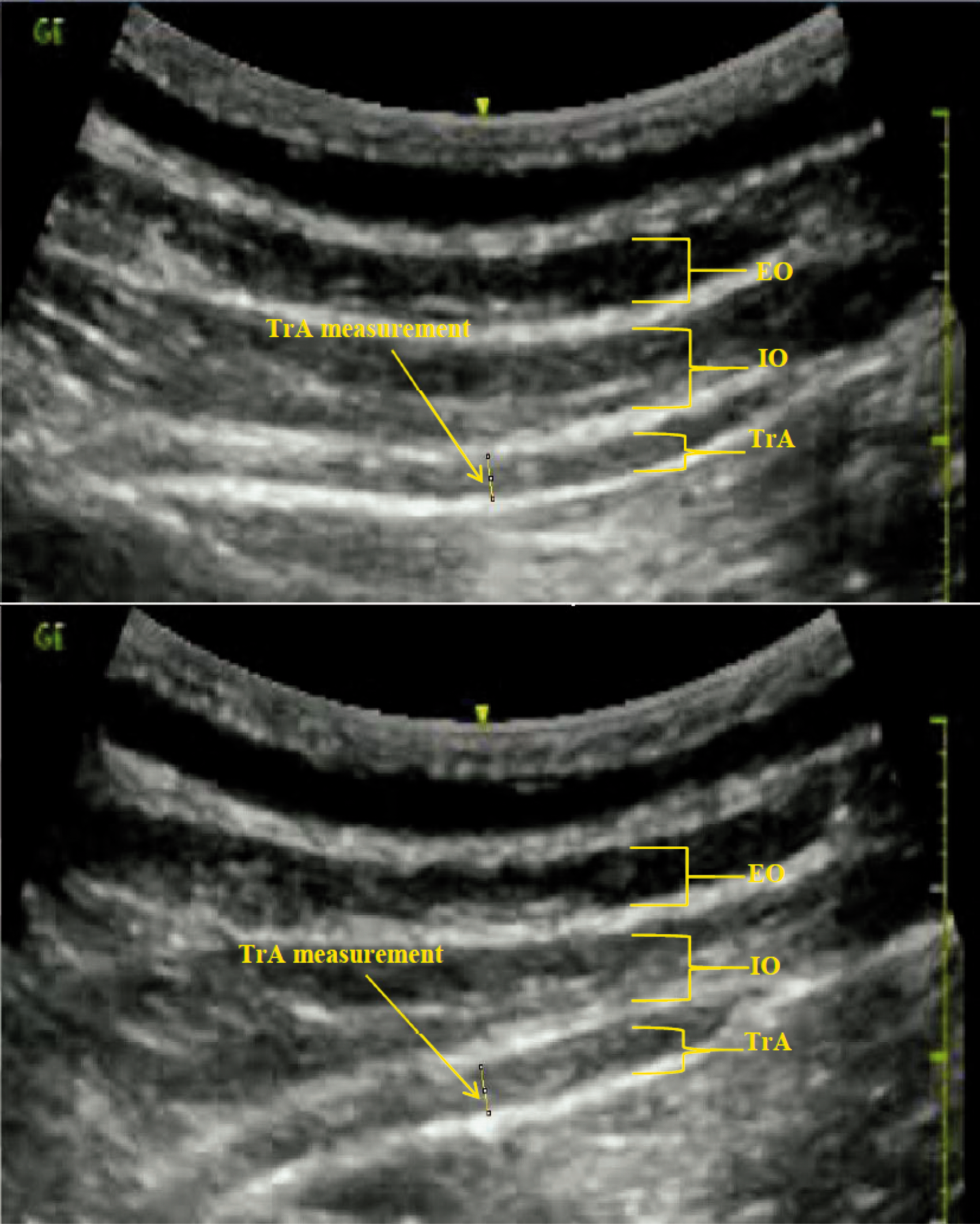
Image size was calibrated, and TrA thickness (mm) was measured as the perpendicular distance between the inside margin of its upper fascial borders, taken from a point 25 mm from the inside edge of the medial fascial joint (Figure 3). Following measurement the independent research assistant re-identified images prior to data input. For each participant duplicate paired measurements were averaged and reported as ‘RTrA’ or ‘CTrA’, and TrA-C was represented for each participant as their ‘RTrA’ to ‘CTrA’ percentage change using the formula
Images showing measurement of TrA during rest and abdominal draw-in manoeuvre. EO: external abdominal obliques; IO: internal abdominal obliques; TrA: transversus abdominis.
Data analysis
Statistical analysis was conducted in SPSS software, version 22 (SPSS Inc., Chicago, IL, USA). Descriptive statistics were calculated for gender, age and BMI as a measure of participant’s relative size, calculated from height and weight measurements (kg/m2). 23 The PFD has a six-axis force/torque measurement expressed in X, Y and Z axes, 13 thus negative and positive values of inclination and roll were represented using the right-hand rule for reporting of kinematic data. 23 Ranges of probe force, inclination and roll, and numerical variables for RTrA, CTrA and TrA-C were analysed for normality of distribution using the Shapiro–Wilk test, and reported using minima, maxima, mean and standard deviation (mean ± SD), or median with inter-quartile range as appropriate.
ICC3,2 with 95% confidence intervals (CI) were calculated for the two mean repeated imaging measurements. Measurement precision was reported as the standard error of measurement, calculated using the formula
Results
Descriptive statistics for the characteristics of the study population (n = 17) are summarized in Table 2. Two repeated US videos yielded a total of 68 extrapolated still images (17 participants × 1 RTrA and 1 CTrA × 2 trials). These were copied, resulting in 68 pairs of duplicate images, all of good quality and measureable.
Intra-examiner reliability

CI: confidence interval; CTrA: TrA when fully contracted; ICC: intra-class correlation coefficient; RTrA: TrA thickness at rest; SEM: standard error of measurement; TrA-C: TrA activation.
Discussion
This study investigated the utility of a novel standardized TrA US method to determine and report a single observer’s measurement reliability of RTrA, CTrA and TrA-C, in a CLBP population. Only two prior TrA US studies had reported observer reliability specifically in CLBP.3,8 ICCs reported in those studies, ranged from 0.32 to 0.72.
In this study, dominant-side TrA thickness measurements were highly reliable, for RTrA (ICC = 0.97 CI: 0.93–0.99) and CTrA (ICC = 0.99 CI: 0.97–0.99). TrA-C was also highly reliable (ICC = 0.93 CI: 0.81–0.97). Such results appear superior to those previously obtained.
In this study, blinding of participants to on-screen visual feedback of CTrA during US and examiner image measurement blinding was conducted. Such rigorous blinding methods have not been clearly stated in the previous CLBP studies.3,8 Therefore, results endorse the application of the methods used in this study using real-time feedback to ensure consistency in terms of probe force and orientation throughout TrA US.
Such consistency permitted the comparison of images obtained at two measurement sessions, each of RTrA and CTrA with probe force, inclination and roll matched across four images. Thus, probe parameters were controlled during TrA measurement. Such control cannot be reproduced in ‘freehand’ US. Even in one study where US transducers were secured within foam, and subsequently fixed to the patient by a belt, TrA-C ICC values of only 0.62 were obtained. 3
Notwithstanding, other factors could also have influenced our results. The training status of the examiner is important in any study of observer agreement. In this study, the examiner was a physiotherapist with five years’ experience in ‘Level-One’ musculo-skeletal US. Whilst examiner training level was mentioned in the TrA-C CLBP study by Costa et al., 8 this was not mentioned in that of Mannion et al. 3 Further, fundamental US methodology has varied between TrA-C CLBP studies. For example, in contrast to the current study, Costa et al. 8 utilized an involuntarily CTrA during isometric knee movement with a standardized force.
This is the first study to report intra-observer US TrA measurement reliability using this standardized method to control force, inclination and roll across repeated within participant measures in CLBP patients. The ranges observed in the current study reflect the residual variation still apparent in probe force and orientation despite the advantage of visual feedback associated with this novel standardized US method. Optimal ICCs for RTrA, CTrA and TrA-C were obtained from standardized ranges of probe force, inclination and roll in the current study. It is likely that greater ranges would be observed during ‘freehand’ US and that these would account for the lower ICCs observed in prior studies.
Limitations of this study include that only intra-observer reliability and dominant-side TrA examinations were reported, and males outnumbered females in this convenience pilot study of CLBP patients. Additionally, a potential source of measurement bias was associated with difficulties in standardizing participants’ 50% TrA-C effort. Thus, future studies are required to assess both intra- and inter-observer reliability and standardize TrA-C effort.
Conclusion
This novel study established the utility of a new and novel standardized method for TrA US and achieved highly reliable intra-observer measurement in CLBP patients. Results obtained were superior to prior reports using ‘freehand’ US in comparable populations. Unique standardizing ranges, with mean values, for probe force, inclination and roll were obtained to aid future studies and research. This novel standardized US method may optimize TrA activation assessment in CLBP and other populations, aiding future research.
Footnotes
Acknowledgements
The authors wish to thank the nursing and administration staff of the Townsville, and Mater Hospitals, for their assistance and encouragement during this project, and independent research assistant Mr Paul Gabriel for conducting image blinding.
Declaration of Conflicting Interests
The author(s) declared no potential conflicts of interest with respect to the research, authorship, and/or publication of this article.
Funding
The author(s) received no financial support for the research, authorship, and/or publication of this article.
Ethics approval
Study approval was obtained from the Human Research Ethics Committee of the James Cook University, the Townsville Hospital and Mater Health Services (JCUH4387/HREC10QTHS53/MHS20150512-07).
Guarantor
CAF
Contributors
CAF and SJG researched literature and conceived the study. CAF, SJG and LGM designed the methodology. CAF and LGM conducted data collection. CAF conducted the data analysis. CAF wrote the first draft of the manuscript. CAF, LGM and SJG reviewed and approved the final version of the manuscript.