



Abstract
Introduction
Cerebral vasculopathy, elevated transcranial Doppler velocities and stroke are linked to excessive intravascular haemolysis in sickle cell anaemia. This study determined the prevalence and pattern of abnormal blood flow velocities in children with sickle cell anaemia from Northern Nigeria using transcranial Doppler and to correlate transcranial Doppler velocities with haematological and biochemical markers of haemolysis.
Methods
Full blood count, reticulocyte count, fetal haemoglobin and some selected biochemical markers of haemolysis of 100 children with sickle cell anaemia were determined at steady state. The time-averaged mean of maximal velocities in four major intracranial blood vessels was measured using non-imaging transcranial Doppler, which were then classified according to the stroke prevention in sickle cell disease trial protocol. A simple linear correlation between time-averaged mean of maximal velocities in the four major intracranial vessels as the dependent variable and haematological and biochemical markers of haemolysis as independent variables was also determined at the significance level of 0.05.
Results
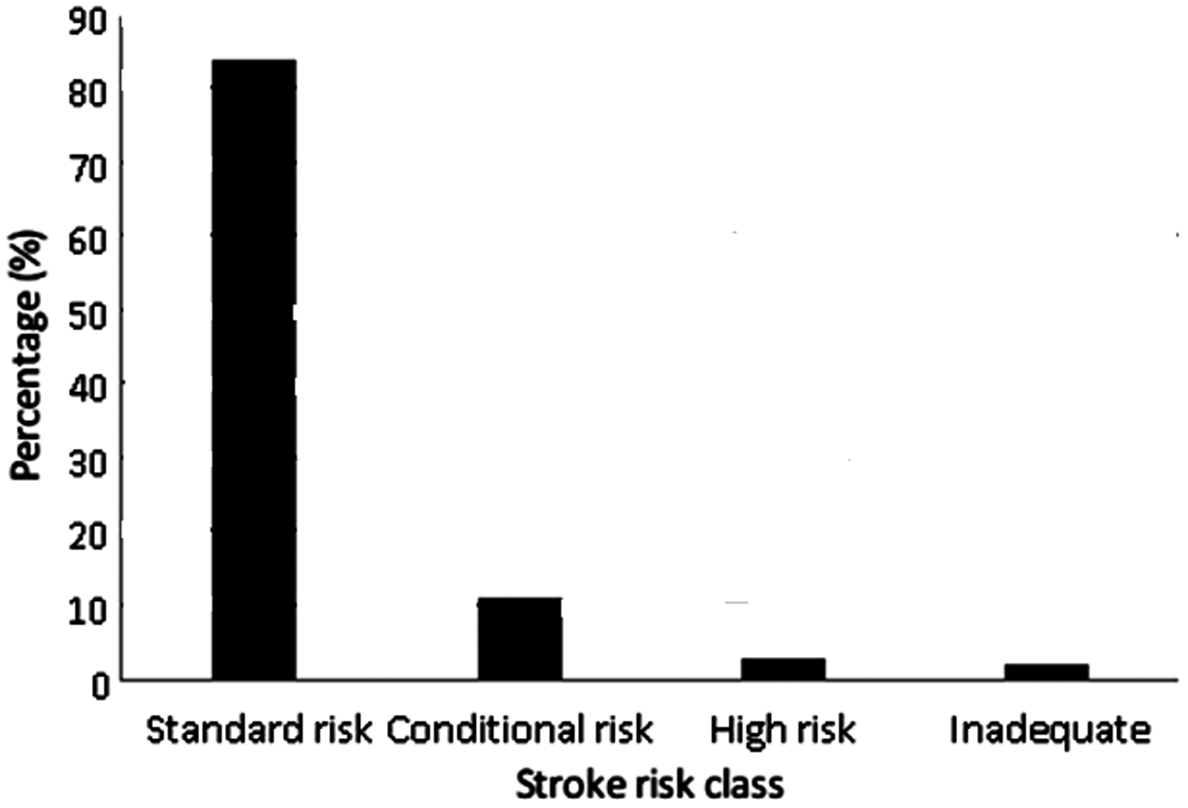
Abnormal transcranial Doppler velocities, classified as high risk, were found in 3% of the studied patients; 11% had intermediate risk while 84% had standard risk. Most high velocities were detected in the middle cerebral artery. The time-averaged mean of maximal velocities of middle cerebral artery positively correlated with total white blood cell count, absolute neutrophil count, platelet count, reticulocyte count, serum lactate dehydrogenase and total bilirubin, while it was negatively correlated with haematocrit and fetal haemoglobin levels.
Conclusion
Our study showed low prevalence of abnormal transcranial Doppler velocities and low prevalence is unrelated to low markers of haemolysis in our study population.
Keywords
Introduction
Sickle cell disease (SCD) is the most common single gene disorder worldwide. 1 The World Health Organisation (WHO) estimates an annual global birth of over 300,000 babies with severe forms of haemoglobinopathies, including sickle cell anaemia (SCA). 2 Sub-Saharan Africa bears the greatest burden of SCA worldwide, with over 75% of the affected children. 3 About 50–80% of these patients will die before adulthood. 4 The WHO estimated that 70% of SCA deaths in Africa are preventable with simple, cost-effective interventions. 2 In Nigeria, the prevalence of homozygous haemoglobin S (HbSS) disease is about 1–3% of the over 160 million population, and results in significant morbidity and mortality. 5
Stroke, especially cerebral infarction, occurs 5% to 10% of patients with SCA before they reach adulthood. 6 This complication usually results from an occlusive vasculopathy involving the arteries of the circle of Willis. 7 The risk of stroke increases with an increase in blood flow velocity in selected cerebral arteries as measured by transcranial Doppler (TCD) ultrasound. 8 The arterial process that leads to stroke develops over a period of months to years before cerebral symptoms develop. 9 This creates a “window of opportunity” to detect the abnormality and institute a preventive intervention before cerebral infarction occurs. Primary prevention of stroke by prophylactic transfusion therapy is feasible if the patients at risk are identified early. TCD ultrasonography can detect arterial stenosis and is ideally suited for screening large vessel disease in patients with SCA because it is safe, non-invasive, of relatively lower cost and is well-tolerated by children.
Previous studies have linked the development of vasculopathy in the large cerebral blood vessels, and hence stroke, with increased markers of haemolysis such as serum lactate dehydrogenase (LDH) and reticulocyte counts in SCA.10–12 Furthermore, reduction of intravascular haemolysis through blood transfusion or hydroxyurea therapy has been shown to increase cerebrovascular vasodilatory capacity and reduce both TCD velocities and stroke risk in SCA.12,13 Moreover, lower prevalence of elevated blood flow velocities in cerebral blood vessels as measured using TCD has previously been reported in children with SCA from in Nigeria.14,15 This is in spite of a higher prevalence of stroke in these children compared to those in the developed countries.14,16 This observation portrays TCD velocities as less predictive of stroke in our environment than ordinarily expected. Given the role of haemolysis in the pathogenesis of cerebral vasculopathy in SCA, we hypothesise that at least part of the reasons for the observation of low prevalence of elevated TCD velocities in our environment is due to low levels of haemolytic markers in our patients compared to others. To the authors' knowledge, no study to date directly compared cerebral blood flow velocities with markers of haemolysis in SCA patients living in the African setting. Therefore, we aimed at determining the prevalence of elevated blood flow velocities in children with SCA from Northern Nigeria using TCD. Other objectives were to investigate if low flow observed in Northern Nigerian children with SCA is due to low levels of haemolysis; and finally to correlate TCD velocities with haematological and biochemical markers of haemolysis.
Methods
One hundred male and female children with SCA between the ages of 2 and 16 were enrolled in the study. The study participants were recruited from the paediatric sickle cell clinic of Aminu Kano Teaching Hospital (AKTH) Kano, Nigeria. Approval was obtained from the hospital's Research Ethics Committee. Informed written consent and assent were obtained from the parents/caregivers before enrolment into the study. All procedures in the study were conducted in accordance with the Helsinki Declaration.
Inclusion criteria
Patients aged 2 to 16 years were consecutively enrolled in the study during routine visits to the clinics. These include all eligible patients who were previously diagnosed SCA as homozygous haemoglobin S disease (HbSS) using cellulose acetate electrophoresis at alkaline pH. This diagnosis was confirmed by Hb quantitation using high-performance liquid chromatography (HPLC). All patients were in steady state, defined as the absence of an acute illness (pain crisis, fever, acute chest or other SCA-related acute complications) or transfusion in the preceding four weeks.17,18
Exclusion criteria
Patients with acute illness such as fever, central nervous infection, previous stroke or seizures requiring anticonvulsant therapy were excluded from this study. However, headache alone was not considered grounds for exclusion unless associated with loss of consciousness or focal neurological symptoms. Other exclusions include children under the age of two, those receiving hydroxyurea and recipients of blood transfusion in the preceding three months. Additionally, patients with haemoglobinopathies other than HbSS, such as haemoglobin SC (HbSC) disease, were excluded because the risk of stroke has been shown to be low in these patients. 19
Data collection
Basic bio-demographic data
A structured questionnaire was used to obtain basic bio-demographic data such as age, sex, past medical, neurological and blood transfusion history.
Haematological studies
Venous blood was drawn by venepuncture aseptically into tubes containing ethylenediaminetetraacetic acid (EDTA) anticoagulant. All haematological tests were conducted as described by Dacie and Lewis. 20 Full blood count (FBC) (i.e. measurement of haemoglobin (Hb) level, haematocrit (HCT), total white blood cell (TWBC), absolute neutrophil count (ANC) and platelet count (PLT)) was performed by the impedance method 20 using automated haematology analyser (Alpha-Swelab®). Reticulocyte count was estimated manually from a peripheral blood smear stained with new methylene blue and expressed as a percentage of the total red cell count. Fetal haemoglobin (HbF) level was quantified by HPLC method 21 using HbGold® analyser (Drew Scientific Ltd., UK).
Biochemical analyses
Serum LDH was measured by quantitative in vitro determination in serum using the ultraviolet (UV) method described by Wroblewski and LaDue. 22 Serum total bilirubin was estimated using the colourimetric method described by Jendrassik and Groff. 23
Non-imaging TCD
Procedure
The procedure was performed by a medical doctor who has received specific training on and has years of experience in conducting TCD examinations in children with SCA. The examination was done with the patients lying in a comfortable supine position with head slightly elevated. The TCD procedure was explained to the patients in a way they would understand and they were asked to remain calm and not to sleep. The investigator sat at the head end of the patient with arms supported in order to maintain the stability of the hand with the transducer. A generous amount of coupling gel was applied over the temporal region (slightly above the zygomatic arch) and subsequent insonation performed through the temporal acoustic window. The middle cerebral artery (MCA), anterior cerebral artery (ACA), posterior cerebral artery (PCA) and terminal portion of the internal carotid arteries (tICA) were examined using a 2 MHz transducer of the Sonora/Tek (Intertek 81300, China) portable system with Viasys™ Healthcare software.
Haematological parameters
ANC: absolute neutrophil count; HbF: fetal haemoglobin.
The envelope tracing on the machine was set to encase the waveform over the entire strip automatically. The TCD recordings were post-processed by adjusting the gain to maximise the velocity readings while maintaining a tight envelope around the waveform. The highest spectral tracing was then obtained as judged from the visual observation and listening of audible sounds of the tracings. The freeze button was then activated to allow for velocity recording and the average of the highest recorded mean velocities over time for each artery was selected and saved. This was taken to be the most representative and was recorded as TAMMV. In order to standardise the technique and minimise inter-individual variations, the same examiner using the same machine obtained all measurements.
The highest TAMMV value thus obtained in any of the above-mentioned four vessels was then used to classify patients according to the stroke prevention in sickle cell disease (STOP) trial protocol
25
as follows:
Normal velocities (Standard risk) < 170 cm/s Conditional velocity (Intermediate risk) 170–199 cm/s Abnormal velocities (High risk) > 200 cm/s Inadequate scan was regarded as all scans where there was a poor sonic window, or the patient was uncooperative.
In line with the STOP trial protocol referral criteria, those with abnormal TCD were offered urgent referral for either commencement of chronic blood transfusion or hydroxyurea at the paediatric haematology unit of AKTH. Patients with conditional TCD were asked to return after two weeks to repeat the scan; patients whose second TCD scan was abnormal were treated as in the abnormal group above. The STOP protocol was chosen because the STOP trial is both the largest prospective application of TCD and the most widely used protocol in stroke prediction in both research and clinical practice settings. 25 This will ease direct comparison with other similar studies. Furthermore, the STOP study produced the velocities used to classify patients based on stroke risk mentioned above. Time-averaged mean of the maximum velocity (TAMMV) is one of the quantitative measures of blood flow using Doppler sonography and high TAMMV correlates well (more consistently compared to pea systolic velocity) with increasing risk of stroke in sickle cell anemia and vice versa.24–26
Data analysis
The data generated were analysed using the STATA® software v15.1, 2018 (College Station, TX, USA). For variables with normal distribution, means and standard deviations were computed and reported as (mean ± standard deviation). Categorical variables were reported as numbers and percentages. Correlation between haematological parameters and TCD velocities was determined using simple linear univariate regression analysis. A p-value of less than 0.05 was considered statistically significant.
Results
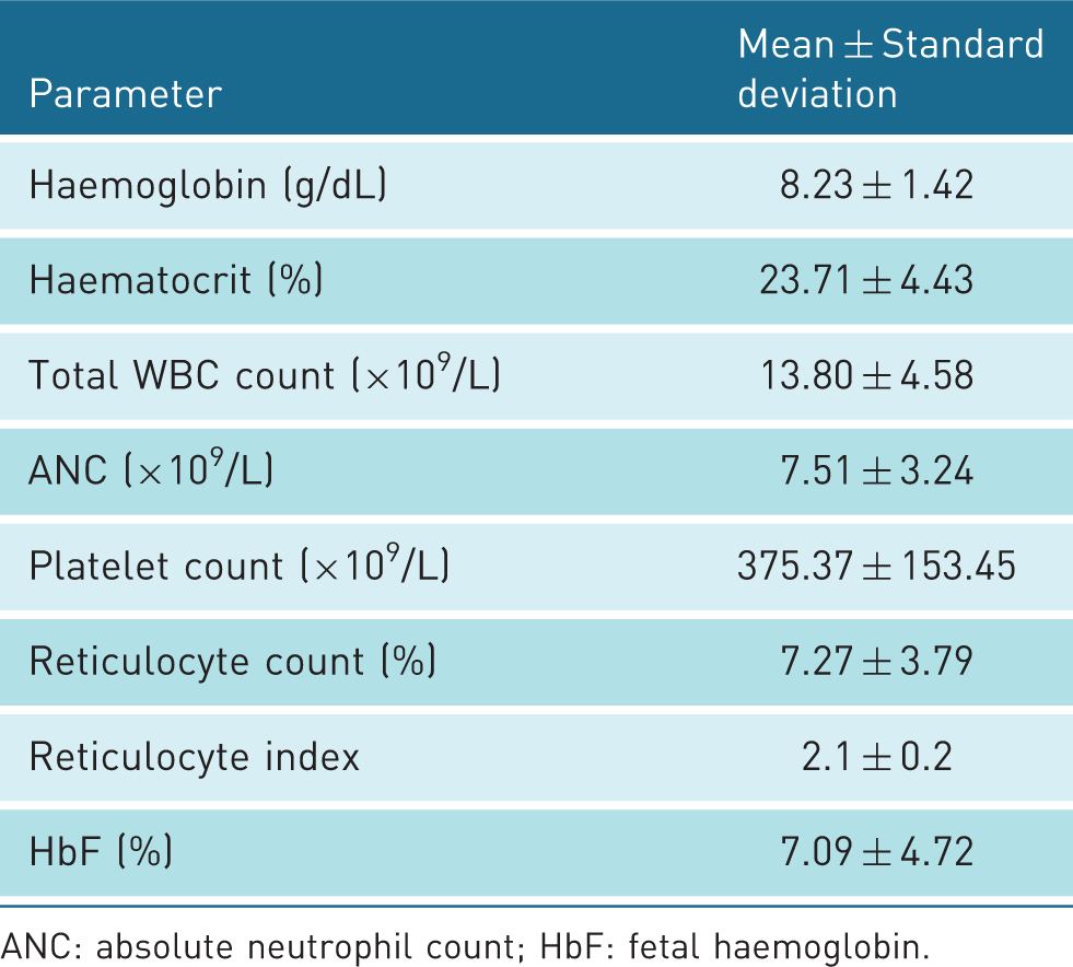
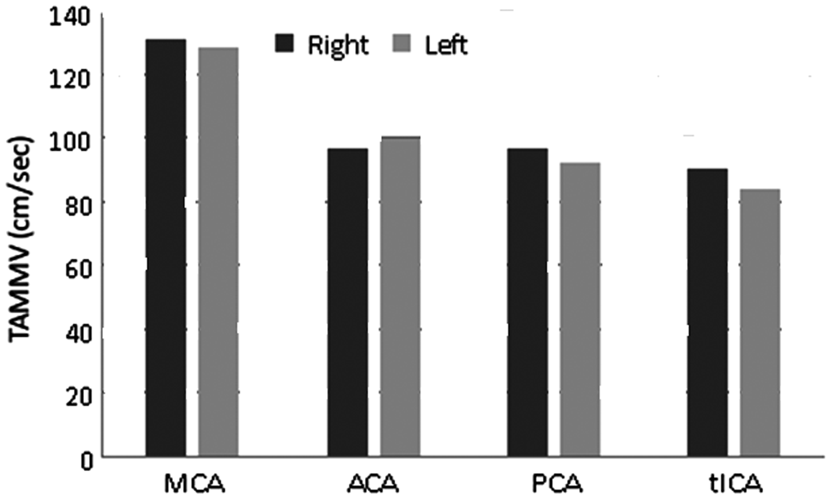
There was slightly more female (54%) than male (46%) patients in the study. Figure 1 shows the age distribution of the studied patients while Table 1 summarises their baseline haematological parameters. The TAMMV in the four intracranial blood vessels is shown in Figure 2. The highest values of TAMMV were found on the right MCA. The highest TAMMV in any of the examined vessels was used in the risk classification and the distribution of participants based on stroke risk is shown in Figure 3.
A bar chart, showing age group distribution of the study subjects. A bar chart, showing territorial distribution of blood flow velocities in cm/s in the arterial branches of the cerebral circulation, comparing right and left sides. A bar chart, showing classification of the blood flow velocities of the study subjects based on the criteria of STOP protocol.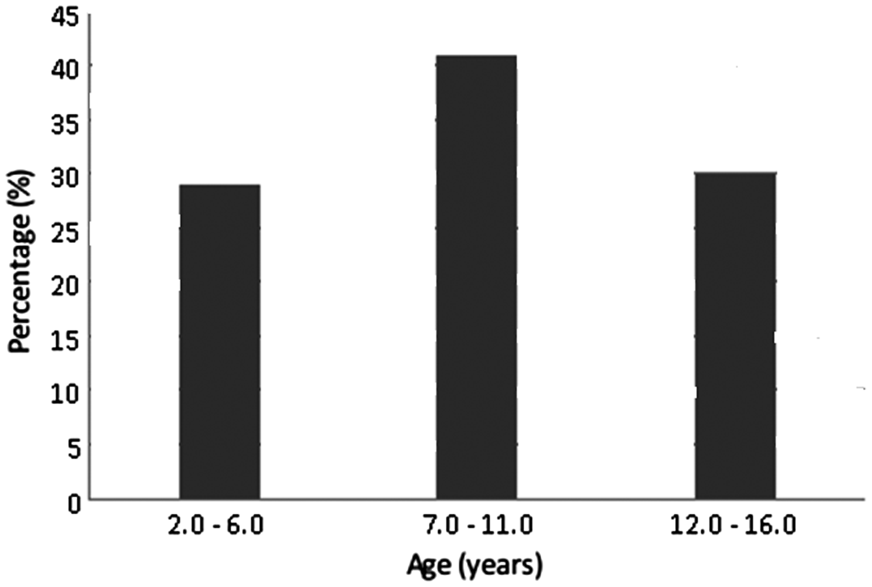
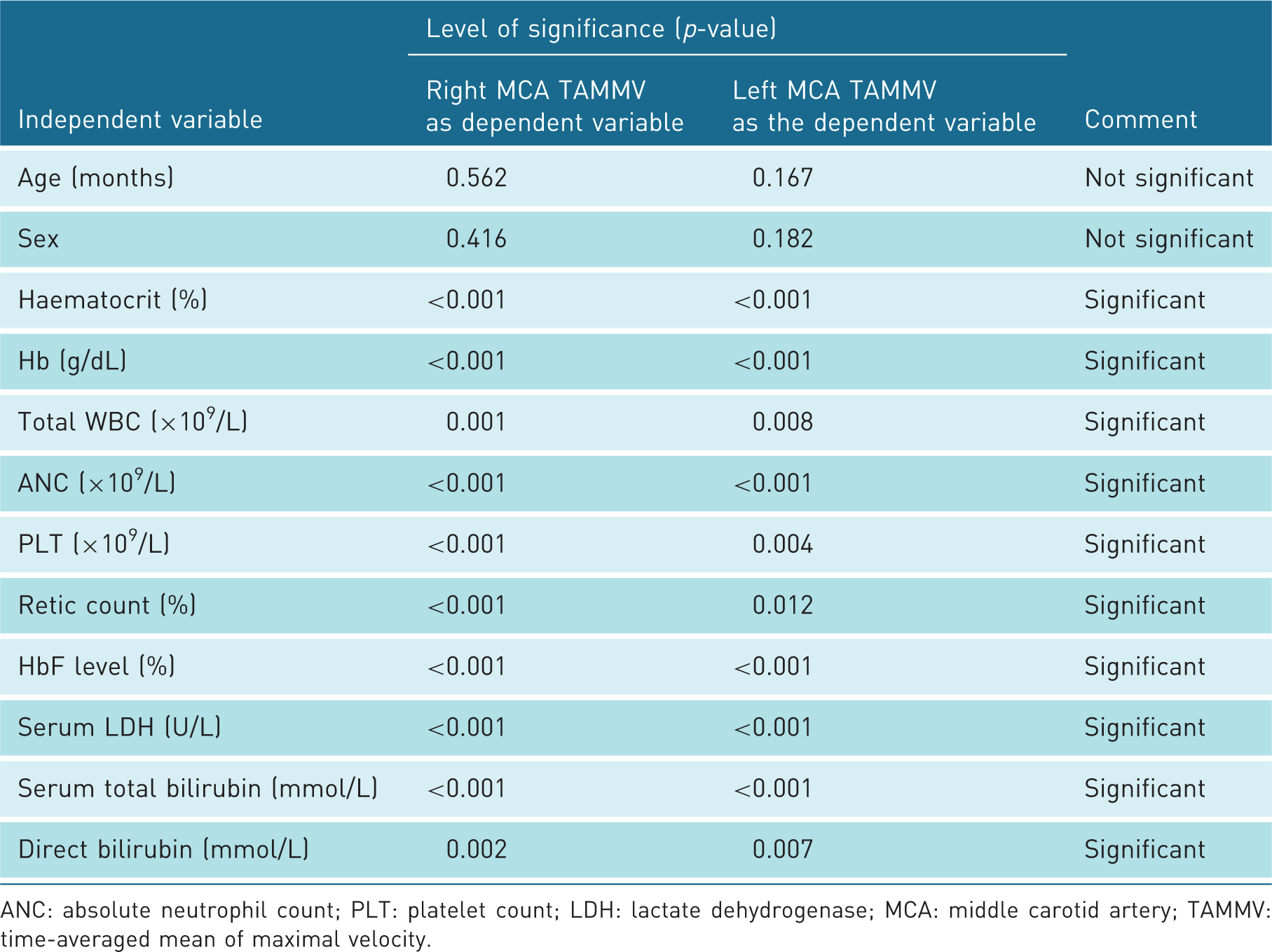
In order to determine the correlation between haematological and biochemical parameters and TCD velocities, a simple linear univariate regression analysis was made with the haematological parameters as independent variables and the TAMMV in the left and right MCAs. The results of univariate analyses are depicted in Table 2. Figure 4(a) to (g) shows the curve-fits for the univariate regression for the TAMMV in the right MCA as the dependent variable and selected haematological and biochemical parameters as independent variables.
(a) A scatter plot, showing a relationship between the blood flow velocity within the right middle cerebral artery and absolute neutrophil count. (b) A scatter plot, showing a relationship between the blood flow velocity within the right middle cerebral artery and proportion of fetal haemoglobin (HbF). (c) A scatter plot, showing a relationship between the blood flow velocity within the right middle cerebral artery and blood level of lactate dehydrogenase (LDH). (d) A scatter plot, showing a relationship between the blood flow velocity within the right middle cerebral artery and packed cells volume in percentage. (e) A scatter plot, showing a relationship between the blood flow velocity within the right middle cerebral artery and platelets count. (f) A scatter plot, showing a relationship between the blood flow velocity within the right middle cerebral artery and reticulocyte count. (g) A scatter plot, showing a relationship between the blood flow velocity within the right middle cerebral artery and total white blood cell count. Summary of results of univariate regression with haematological parameters as independent variables and TAMMV as dependent variable ANC: absolute neutrophil count; PLT: platelet count; LDH: lactate dehydrogenase; MCA: middle carotid artery; TAMMV: time-averaged mean of maximal velocity.
The linear regression equation for the univariate model is as follows

Discussion
TCD has emerged as a powerful tool for assessing stroke risk in patients with SCA. It is flexible, non-invasive and uses relatively simple, inexpensive equipment. With the appropriate training, it is also relatively easy to perform. Primary prevention of stroke in children with SCA became possible when TCD examinations became part of routine clinical practice. 25 This clinical utility has become the cornerstone in reducing the burden of one of the most feared and debilitating consequences of the disease, with a potential for improving the quality of life. 26 The search for predictors of stroke risk among SCA patients living in Africa is still ongoing. To our knowledge, this is the first descriptive report that correlated TCD velocities with haematological and biochemical markers of haemolysis in children with SCA from northern Nigeria.
This study evaluated the TCD characteristics of the most vulnerable group for stroke in SCA, age group 2 to 16 years. Ischaemic stroke due to SCA has been found to be uncommon before the age of 2, while the incidence diminishes after 16 years. 19 The cerebral vasculopathy that results in ischaemia and infarction is thought to develop gradually over time and appears to manifest as a stroke from the third year of life. 19 This partly explains the rarity of strokes in SCA before the age of two. Elevated HbF levels may offer additional protection against severe manifestations in infancy. 19
The main haematological findings in this study largely depict moderate anaemia, neutrophilia with absolute leucocytosis, reticulocytosis and platelet counts on the upper limits of normal. Our findings are similar to what Abdullahi et al. 27 reported earlier in children with SCA within the same population with mean steady-state values for Hb, WBC, PLT and reticulocyte counts of 7.31 ± 1.26 g/dL, 14.57 ± 6.63 × 10⁹/L, 350.36 ± 120.96 × 10⁹/L and 3.90 ± 1.96%, respectively. In comparison, non-SCA children with haemoglobin AA genotypes have mean values of 11.03 ± 1.10 g/dL, 7.25 ± 1.96 × 10⁹/L, 322.00 ± 68.77 × 10⁹/L and 0.83 ± 0.43% for Hb, WBC, PLT and reticulocyte counts, respectively. 27 Omoti in Benin City, 28 southern Nigeria, found similar steady-state values in patients with SCA. Mechanistically, chronic haemolysis, autosplenectomy and chronic inflammation provide the unifying pathophysiological bases for the above haematological findings in SCA.12,29,30 Several clinical studies have also linked severe anaemia, reticulocytosis, neutrophilia and thrombocytosis to adverse clinical events in SCA including acute chest syndrome, vasoocclusive crisis (VOC), hospitalisations, silent cerebral infarcts, overt stroke and death.28,31–35
The mean HbF level found in this study is similar to 7.1 ± 4.5% reported in patients with Benin haplotype of SCA, which is the predominant sickle cell haplotype encountered in Nigerian patients.36,37 Sickle cell patients with this phenotype are known to have intermediate clinical severity, being more severe than the Saudi Arabian haplotype but generally milder than the Bantu and the Central African Republic (CAR) haplotypes. 37 An elevated level of HbF has been associated with milder clinical phenotype in SCA characterised by fewer VOCs, fewer hospitalisations and longer survival in patients with SCA.38,39 High HbF has also been speculated to reduce the occurrence of both silent cerebral infarctions and overt strokes in SCA.38,39 Hence, increasing the HbF level is currently the main target of several established or experimental disease-modifying therapies in SCA including hydroxyurea. 13
Recent insight into disease mechanisms of SCA is increasingly recognising the role of chronic intravascular haemolysis in the pathogenesis of some of its complications. Serum LDH concentration has now emerged as an important marker of haemolysis in SCA. 40 The mean steady-state LDH concentration found in this study is significantly higher than 356 U/L found in Saudi Arabia 41 and 875 U/L found in the UK. 42 This is also significantly higher than both 610.6 U/L reported in the American National Institutes of Health (NIH) and 680.8 U/L in the CSSCD cohorts. 40 These findings suggest that patients in our environment have significantly higher steady-state haemolytic rates than their counterparts elsewhere. Thus, our findings disprove our earlier hypothesis that a lower haemolytic rate in our patients is the likely reason for the observed lower prevalence of elevated TCD velocities from previous studies. The exact reasons for the high haemolytic rates in these patients are not clear. However, a high prevalence of some acquired or inherited haemolytic anaemias such as due to malaria and G6PD deficiency seen in our environment might explain this.43,44 Our study did not test for malaria and G6PD deficiency; nevertheless, these factors can potentially increase steady-state haemolysis in SCA. Future studies need to consider these factors in order to control for their potential confounding effects on the steady-state haemolytic markers in SCA.
Two-sample t-test with unequal variances (left vs. right – interhemispheric)
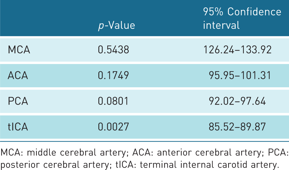
MCA: middle cerebral artery; ACA: anterior cerebral artery; PCA: posterior cerebral artery; tICA: terminal internal carotid artery.
The current study used a non-imaging TCD protocol to measure TAMMV in cerebral blood vessels. In comparison, the duplex imaging TCD technique has been reported to give velocity readings of up to 10% lower than those acquired using the non-imaging protocol. 47 This apparent difference has led to suggestions that a lower threshold of velocities is assigned to stroke risk categories. Going by this, McCarville et al. 47 have proposed a modification to the STOP trial criteria, by applying 10% correction to readings obtained with the imaging technique. They suggested that 180 cm/s or more to be considered abnormal velocity with the duplex (imaging) procedure, 153–179 cm/s as conditional, while <153 cm/s as standard risk. Padayachee et al. 48 have, however, pointed out the danger in this approach, suggesting that since the two techniques closely correlate with each other with little overall systematic bias, routinely applying this correction to the STOP trial velocities would only increase the number of patients categorized as abnormal or conditional. This would lead to the inappropriate selection of patients for chronic transfusion programs. They further suggested that the observed differences between the two techniques are largely attributable to physiological variations such as changes in pCO2 level. 48 As the present study used similar protocol as the STOP trial, there was no need to apply any correction for comparison with similar studies.
This study has also further confirmed a lower prevalence of abnormal TCD velocities in African children with SCA compared with their counterparts in North America, 49 with a finding of 3% prevalence of TAMMV ≥ 200 cm/s in the present study. This is similar to the 4.7% found in Ibadan, southwestern Nigeria. 14 Oniyangi et al. 15 found a prevalence of abnormal TCD velocities of 6.9% in SCA children in Abuja, central Nigeria using the imaging TCD technique. Similarly, a Kenyan study 50 of 105 children with SCA found both lower mean TAMMVs and 0% prevalence of high-risk TCD velocities. These findings are lower than 9.3% found in STOP trial. 25 Although the findings in the present study are similar to those of the American BABY-HUG study where a prevalence of 2% high TCD velocities was found, 51 the study subjects in the BABY-HUG were much younger (less than 2 years of age) while our sample size is much smaller. In contrast, Silva et al. 52 found up to 31.8% of children with SCA to have high TCD velocities in Portugal. The reason for this low prevalence of high TCD velocities in African children with SCA is not clear and is intriguing especially given that we have a higher prevalence of stroke in Africa than in America. 50 Lagunju et al. 14 had earlier speculated that it is quite possible that Nigerian patients with SCA may have relatively lower thresholds of TCD velocities at which they develop stroke compared to Americans. Another proposed possibility is that stroke might have already occurred in many patients with abnormal TCD velocities in this environment before the patients are screened. 14 Although these hypotheses are quite plausible, appropriately and specifically designed long-term prospective studies are needed to verify the propositions.
This study found a positive correlation between selected haematological and biochemical markers of haemolysis and blood flow velocities as measured by TCD. It also found a negative correlation with HbF, which is a known protective marker for high blood flow velocities and stroke in SCA. Our findings are in keeping with those reported in SCA children elsewhere. 44 The implication of these findings is that other hitherto undiscovered factors, unrelated to haemolytic markers, may be responsible for the low prevalence of elevated TCD velocities observed in this population. Our study further brought to fore the apparent paradox of low prevalence of high TCD velocities in a setting with high stroke burden among African SCA children. Given this paradox and the fact that elevated markers of haemolysis found in this study logically parallel the high burden of stroke in African children with SCA, our data suggest that blood-borne haematological and biochemical markers of haemolysis are potentially more predictive of stroke risk than TCD velocities in these patients. Alternatively, as the TCD velocity thresholds for stroke risk determination in SCA were set based on studies conducted in the developed world, it is possible that these thresholds are not appropriate in children living in Africa, where the vast majority of SCA children reside. If this happens to be the case, then many SCA children at high risk of stroke are missed in Africa in spite of TCD screening and risk categorisation that is hinged on extraneously determined cut-offset points. Hence, further studies are needed to determine the appropriate TCD velocity thresholds for proper risk stratification and their associated risk factors in children with SCA living in Africa.
Because of a lack of appropriate equipment, this study could not measure pCO2, which together with blood pressure, are important limitations of this study need to be acknowledged here. The study did not take measurements of pCO2 and blood pressure (BP), both important variables that can impact on cerebral perfusion. 53 It has been shown that for every mmHg change in arterial pCO2 level, the TAMMV can vary by up to 4%. 44 T, and together with age and hematocrit, pCO2 can account for 15% of variations in subsequent TAMMV measurements. 53 Measured blood flow velocity can also be higher with higher systemic blood pressures despite an intact autoregulatory system. 54 This relationship is particularly important in patients with subarachnoid haemorrhage (SAH) who are monitored for cerebrovascular vasospasm as manifested by elevated cerebral blood flow velocity and who may simultaneously be undergoing induced hypertension to treat vasospasm. The analysis of velocity measurements is particularly challenging in these patients. 55
Conclusion
This study of sickle cell disease patients from northern Nigeria confirmed a low prevalence of elevated TCD velocities, compared to what obtains in North America, which is intriguing given the higher prevalence of stroke in our patients. A significant positive correlation was also found between TAMMV in MCA and TWBC count, ANC, platelet count, reticulocyte count, serum LDH and total and direct bilirubin in these patients, while HCT and HbF levels were negatively correlated with the TAMMV. Levels of haemolytic markers do not appear to be the reason for the observed low prevalence elevated TCD velocities in Nigerian children with SCA. There is a need for larger prospective studies to determine the appropriate reference ranges for TCD velocities and stroke risk thresholds in African children with SCA.
Footnotes
Declaration of Conflicting Interests
The author(s) declared no potential conflicts of interest with respect to the research, authorship, and/or publication of this article.
Funding
The author(s) declared receipt of the following financial support for the research, authorship, and/or publication of this article: The management of Aminu Kano Teaching Hospital, Kano has provided part funding, which was useful to provide laboratory consumables.
Ethics Approval
Research and ethics committee of Aminu Kano Teaching Hospital, Kano, Nigeria has approved the conduct of this research. The reference number of the approval is: AKTH/AMC/SUB/12A/P-3/VI/1058.
Guarantor
Anas Ismail
Contributors
Anas Ismail, Aminu Abba Yusuf, Aisha Kuliya-Gwarzo, Sagir Gumel Ahmed, Abdulkadir Musa Tabari, Shehi Ali Abubakar.
Acknowledgements
The authors thank Dr Shehu Abdullahi of Paediatrics Department and Dr Abubakar Habib for their assistance during patient recruitment, Mr Fahad and Mrs Khadija for organizing the TCD sessions, Mr Ado Dakata, Mr Muhammad Bello and Dr Imoru Momodu for samples analysis, Drs Musa M Bello and Rayyan M for data analysis.