




Abstract
Isolated congenital mitral ring is a very rare subtype of congenital mitral valve malformation, which accounts for about one-third of congenital cardiac anomalies associated with left ventricular inflow obstruction. A mitral ring may be easily missed unless the disease is suspected. The mitral valve repair should be considered in symptomatic patients with mitral stenosis. We report a rare case of a 43-year-old woman with an intramitral ring who experienced previous embolic stroke with left hemiplegia. However, stroke happened several years ago and it does not completely prove causality.
Keywords
Introduction
Congenital mitral valve (MV) malformations as scarce diseases affect 5/100,000 of general population. 1 Isolated congenital mitral ring is a very rare subtype of congenital MV disorder. 2 It is usually associated with some cardiac anomalies, such as Shone’s Complex, subvalvular aortic stenosis and aortic coarctation. 3 Fischer in 1902 reported the disease for the first time. There is no exact data about incidence or sex predilection. However, it accounts for about one-third of congenital heart anomalies associated with left ventricular (LV) inflow obstruction. 4 The clinical presentation can vary significantly, depending on the severity of obstruction, the degree of mitral regurgitation, the presence of pulmonary arterial hypertension and the existence of concurrent anomalies. 5 Here, we report a rare case of a 43-year-old female patient with an intramitral ring who experienced previous embolic stroke with left hemiplegia.
Case report
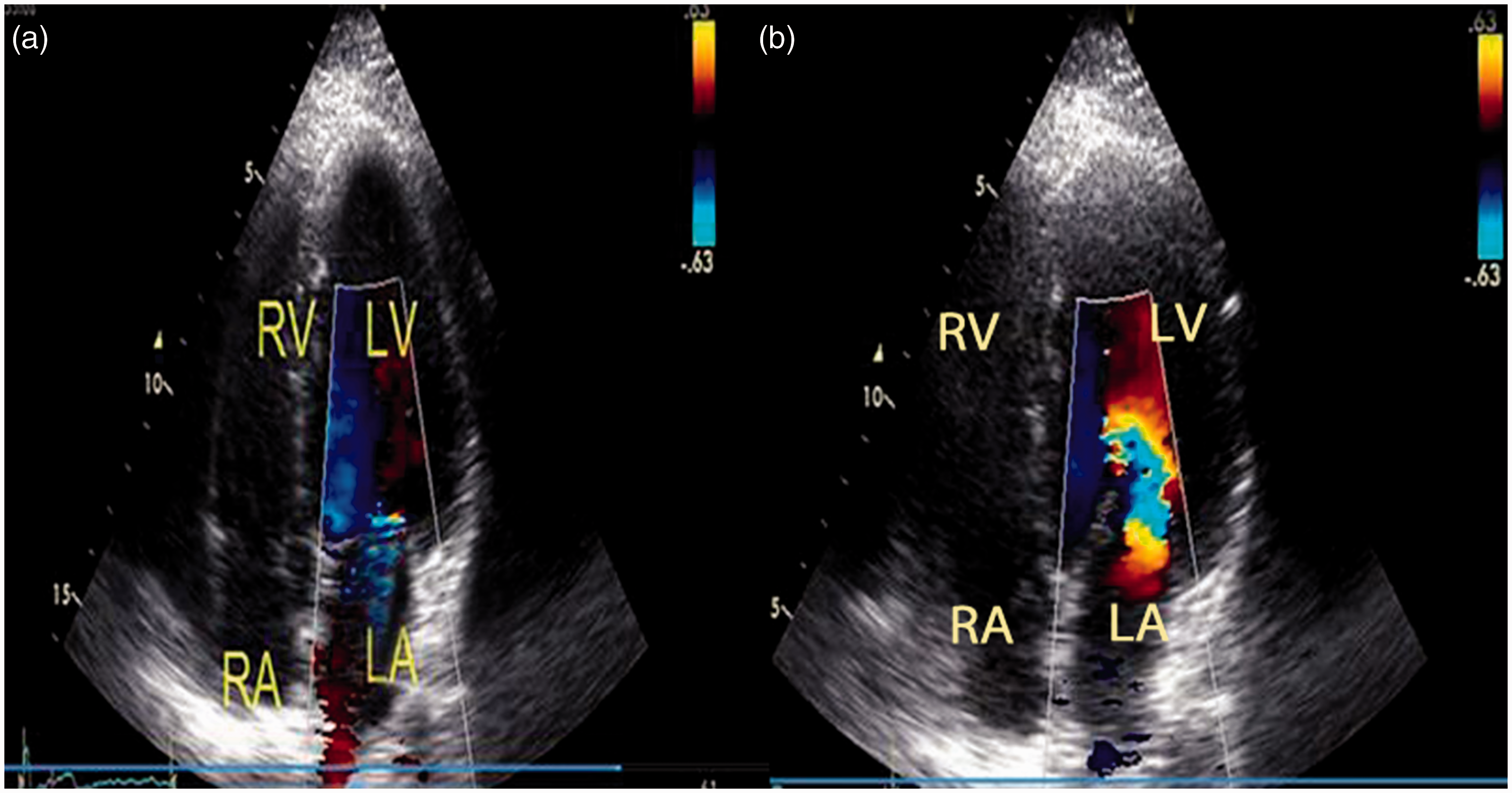
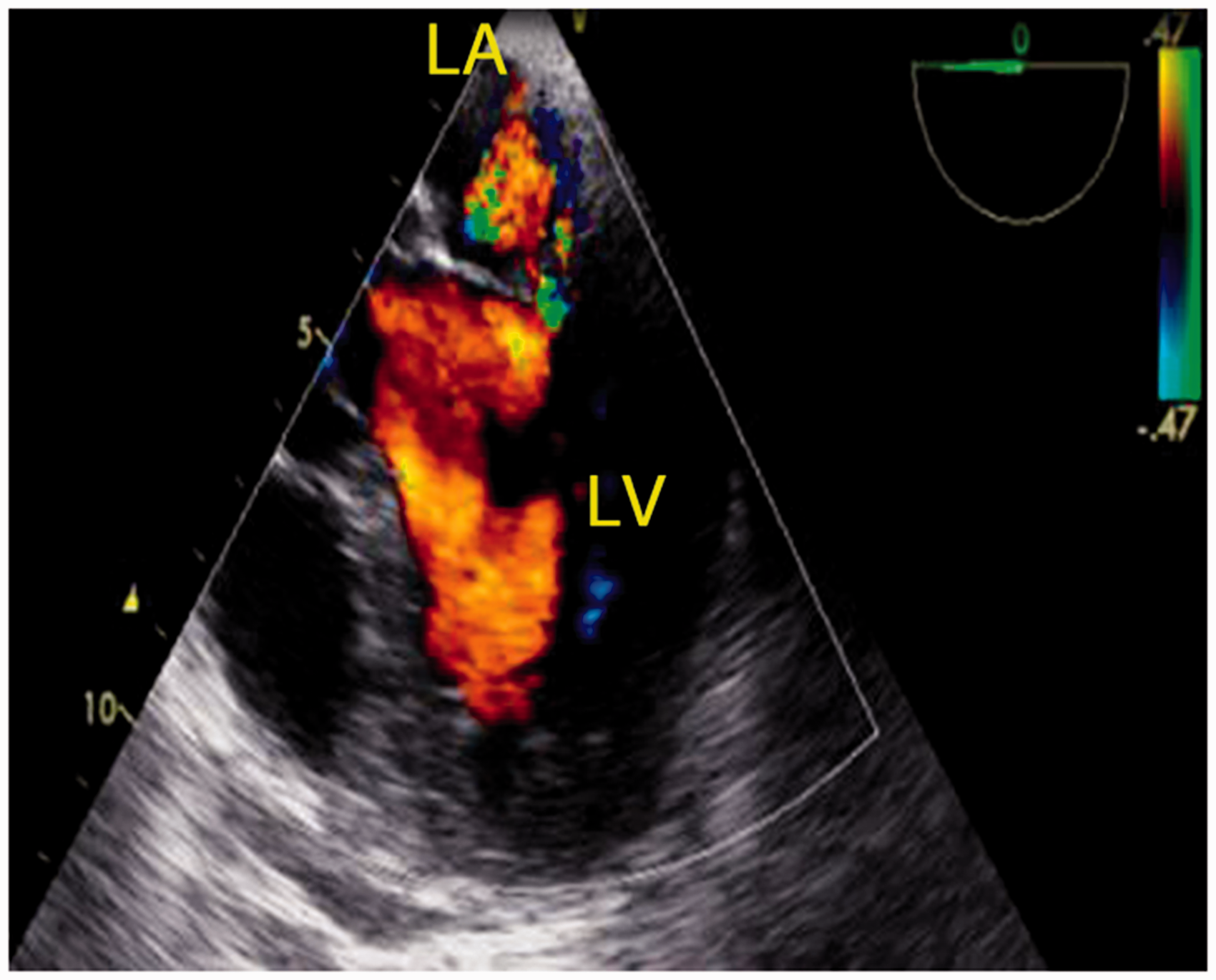
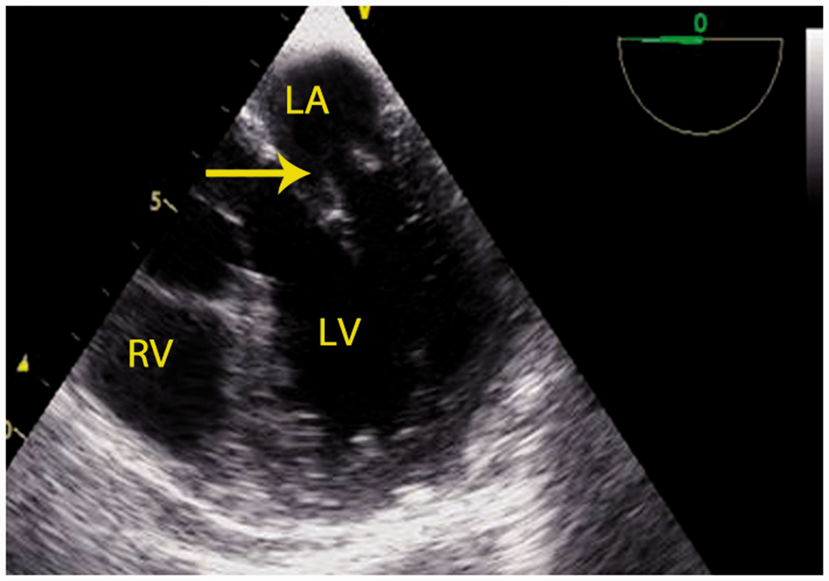
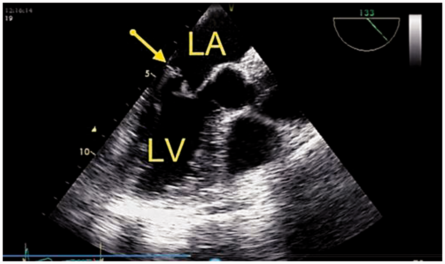
A 43-year-old woman was referred to our clinic because of an episode of extreme weakness and fatigue a week ago, whose symptoms were completely resolved within a few minutes. In medical history, she experienced an embolic stroke 20 years ago, which affected the right hemisphere of the brain and caused left hemiplegia. At the same time, full medical examinations, including transthoracic echocardiography, hypercoagulability testing and carotid Doppler ultrasonography, showed no obvious abnormalities as the causes of embolic stroke, and the patient reported no use of any drugs or history of rheumatic fever. Recent physical examination revealed pansystolic murmur grade II/VI at the cardiac apex. The electrocardiogram and chest X-ray exhibited normal appearance. Transthoracic echocardiogram showed normal LV size, systolic function and moderate MR due to restricted posterior MV leaflet motion with a normal pulmonary arterial pressure and no other cardiac abnormality, except for flow acceleration above the MV annulus (Figures 1 and 2, Supplementary Videos 1 and 2). However, the mean gradient across the MV was not elevated, and Doppler mitral inflow patterns showed nearly normal view (tracing of deceleration slope of the diastolic mitral flow showed a pressure half time of 106 ms and a calculated MV area of 2.1 cm2) (Figure 3). Transthoracic echocardiography (TTE) was followed by transesophageal echocardiography (TEE) to determine the etiology and severity of the MR, which confirmed the moderate MR (Figure 4, Supplementary Video 3). Further probe advancement revealed some semi-mobile rudimentary tissues attached to the atrial side of MV leaflet (Figure 5, Supplementary Video 4). The probe was then flexed posteriorly, and rotated about 120° that exhibited the rudimentary tissues were originated from an intramitral ring attached to the mid portion of the leaflets without restricting inflow across the MV (Figures 6 and 7, Supplementary Videos 5 and 6). The patient again underwent laboratory workup for systemic lupus erythematosus, anti-phospholipid antibody, inflammatory markers, erythrocyte sedimentation rate and blood cultures, which showed no abnormality. The patient was requested for anticoagulant therapy because of previous history of cerebrovascular event and semi-mobile rudimentary tissues on intramitral ring. She did not agree to perform further examination.
Two-dimensional parasternal long-axis view represented a restricted posterior MV leaflet motion. Two-dimensional apical four-chamber view with color flow Doppler represented a moderate MR (a, systole) and flow acceleration above the MV annulus (b, diastole). Continuous wave Doppler echocardiography with cursor placed at the tip of mitral leaflets shows that the mean gradient across the MV was not elevated. Transesophageal two-dimensional echocardiography and color Doppler flow velocity mapping in the midesophageal for chamber view represent a moderate MR. Transesophageal two-dimensional echocardiography in the midesophageal four-chamber view represents some rudimentary semi-mobile tissues (arrow) attached to the atrial side of MV leaflet. Transesophageal two-dimensional echocardiography in the midesophageal long-axis view (120°) represents an intramitral ring (arrow) attached to the mid portion of the leaflets. Transesophageal two-dimensional echocardiography in the midesophageal two-chamber view (84°) represents some rudimentary semi-mobile tissues (arrow) attached to the atrial side of MV leaflet.


Discussion
The mitral ring is a rare subtype of congenital mitral stenosis, ranging from a thin membrane to a thick fibrous ridge that encircles the mitral orifice partially or totally, and adheres to the mitral leaflet. There are two types of mitral ring: supra and intramitral variants. A supra mitral ring is usually associated with normal MV apparatus and may be concurrent with other congenital cardiac anomalies. The intramitral ring is often associated with Shone complex and abnormal subvalvular apparatus. These two types of congenital mitral ring seem to have different embryologic origins. Supramitral ring probably represents a form of left atrial outflow obstruction, whereas intramitral ring may be due to an intrinsic MV pathology. This anomaly has not been reported in the fetus, thereby suggesting postnatal development of the ring during early infancy. 6 Lack of prenatal detection may also be contributed to low resolution of mitral ring in a fetus. Our patient had intramitral subtype presented as a thin membrane originated from the mid portion of atrial side of mitral leaflets. Echocardiography allows high-resolution images of the MV apparatus. However, a mitral ring may be easily missed unless the disease is suspected. 1 Serra et al. in 2005 reported a 36-year-old male with progressive dyspnea. TTE showed LV enlargement, severe MR, moderate aortic regurgitation and suspected MV masses. TEE revealed a supramitral ring restricting the leaflets movement and impairing their opening with resultant mild diastolic gradient and severe MR. The patient underwent membrane excision, and the mitral and aortic valves replacement. 7 Baharestani et al. in 2012 reported a 17-year-old male with syncope undergoing a cardiac operation for ventricular septal defect closure and MV repair 15 years ago. TTE, TEE, and finally cardiac catheterization showed a neglected supravalvular mitral ring, which was resected in a second operation. 8 Similar to our patients, a diagnosis had not been confirmed until recently. TEE assessment can help to obtain precise visualization of the MV morphology. The ring may not be visible by two-dimensional TTE, and the most characteristic finding is the Doppler flow acceleration just above the MV annulus, as seen in our patient. TEE may enhance visualization of the membrane that appears as a fibrous structure adhering to the atrial side of the MV leaflets. 2 In our patient, the membrane was visualized by TEE that revealed a thin membrane with some attached semi-mobile rudimentary tissues. The soft mobile masses on both MV leaflets are highly suspicious for infective endocarditis. 9 However, it was excluded based on clinical findings, blood cultures and laboratory tests. Another differential diagnosis is marantic or nonbacterial thrombotic endocarditis, which is associated with mucin-producing adenocarcinomas and coagulopathies. These vegetations are usually small and irregularly shaped and are left sided and bilateral. 10 Our patient had no evidence related to this entity. Although rare on the MV, papillary fibroelastoma can also affect MV. However, it is often a solitary mass with a characteristic frond-like appearance. 9 This anomaly should also be differentiated from a cortriatriatum sinistrum, where the fibromascular membrane divides the left atrial cavity into a pulmonary venous chamber and a left atrial chamber. 2 The patients with supramitral ring represent a subgroup of patients with congenital mitral stenosis who have a nearly good prognosis. 6 However, the outcome is also determined by the presence of simple or complex associations. 4 The MV repair, including ring removal, should be considered in symptomatic patients with mitral stenosis. 1 To the best of our knowledge, such case of asymptomatic woman with an intramitral ring and semi-mobile rudimentary tissues attached to the membrane who experienced an embolic stroke in adulthood has not been reported previously. Embolic stroke in our patient might have been due to embolization of the rudimentary tissues from congenital intramitral ring. However, the stroke happened several years ago and does not prove causality.
Footnotes
Supplemental material
Supplemental material for this article is available online.
Acknowledgements
None.
Contributors
All the authors contributed for the publication of this article.
Declaration of Conflicting Interests
The author(s) declared no potential conflicts of interest with respect to the research, authorship, and/or publication of this article.
Ethics approval
Approval was obtained from the Ethics Committee of the Fatemeh Zahra teaching hospital.
Funding
The author(s) received no financial support for the research, authorship, and/or publication of this article.
Guarantor
MN.
Permissions
Written permission was obtained from the subject for publication of this case report.
References
Supplementary Material
Please find the following supplemental material available below.
For Open Access articles published under a Creative Commons License, all supplemental material carries the same license as the article it is associated with.
For non-Open Access articles published, all supplemental material carries a non-exclusive license, and permission requests for re-use of supplemental material or any part of supplemental material shall be sent directly to the copyright owner as specified in the copyright notice associated with the article.
