



Abstract
Introduction:
Undifferentiated paediatric patients can be difficult to correctly classify with the correct condition. This may be, in part, due to the unique differences in how symptoms present as well as the challenges clinicians face in undertaking a complete examination where cooperation may be hindered due to pain, anxiety or limited understanding. Point-of-care ultrasound is now recognised as a valuable tool to assist acute paediatricians in their decision-making and diagnosis when incorporated into their clinical assessment.
Conclusion:
We describe six cases which presented to the Paediatric Emergency Department, where point-of-care ultrasound played a crucial role in uncovering malignant or invasive lesions and subsequently expedited further investigations, leading to much quicker diagnosis.
Introduction
Undifferentiated paediatric patients can be difficult to correctly classify with the correct condition 1 owing to their unique differences in how symptoms present as well as the challenges clinicians face in undertaking a complete examination where cooperation may be hindered due to pain, anxiety or limited understanding. 2 Point-of-care ultrasound (POCUS) is now recognised as a valuable tool to assist acute paediatricians in their decision-making and diagnosis when incorporated into their clinical assessment. The role of paediatric POCUS continues to expand, allowing users to answer more advanced questions such as ‘does this child have evidence of raised intracranial pressure?’ or ‘does this child have a skull fracture?’.3,4
On average, there are approximately 1645 new cancer diagnoses in children and young people (0–14 years) in the United Kingdom each year, with 54% of new cancer diagnoses being made in the Paediatric Emergency Department (PED). 5 The importance of timely identification and recognition of signs and symptoms which may indicate malignancy or invasive disease cannot be overstated. We describe six cases that presented to the PED in which POCUS played a crucial role in uncovering malignant or invasive lesions and subsequently expedited further investigations, leading to much quicker diagnosis.
Case 1
A previously healthy 3-year-old girl was referred to the PED after her parents had noted mild swelling of her right upper eyelid for the past week. This had been treated using warm compresses and antibiotic eye drops provided by the pharmacy, with no improvement. On the day of presentation, her mother had noticed her right eye appeared subtly larger than the left (see Figure 1). She had been systemically well, with no recent fevers, headaches or dizziness. On review by ophthalmology, she was noted to have reduced visual acuity in the affected eye (Right Snellen: 6/12, Left Snellen: 6/6). There was no inflammation of the conjunctiva, and her optic discs appeared normal bilaterally. On examination, she had mild upper eyelid swelling, a full range of eye movements and no pupillary defects. The eye was painless, with no erythema or discharge. A POCUS was undertaken by one of the authors, which demonstrated a large heterogeneous retro-orbital mass which appeared to be impinging on the optic nerve and leading to papilloedema (see Figure 2). Due to the POCUS findings, the case was made for an urgent magnetic resonance imaging (MRI) with contrast (see Figure 3). This demonstrated a 28 mm × 21 mm × 18 mm mass within the right orbit, which demonstrated mostly iso-intense T1 signal and slightly hyper-intense T2 signal to grey matter. The lesion was closely related to the optic nerve and right medial rectus muscle, which showed no intralesional fat or calcification and enhanced homogenously following contrast administration. It extended up to, but not beyond, the optic foramen. After discussion with the tertiary neurosurgical and oncology teams, she was transferred for further investigations. The patient underwent a biopsy the following day, which showed an embryonal fusion-negative rhabdomyosarcoma (RMS). Staging investigations showed no evidence of metastatic spread. The patient has almost completed her initial chemotherapy regimen and commenced proton beam therapy. The patient has maintained her sight, and her oncologists are confident of achieving remission.

Subtle asymmetry between the right (affected) and left unaffected eye.
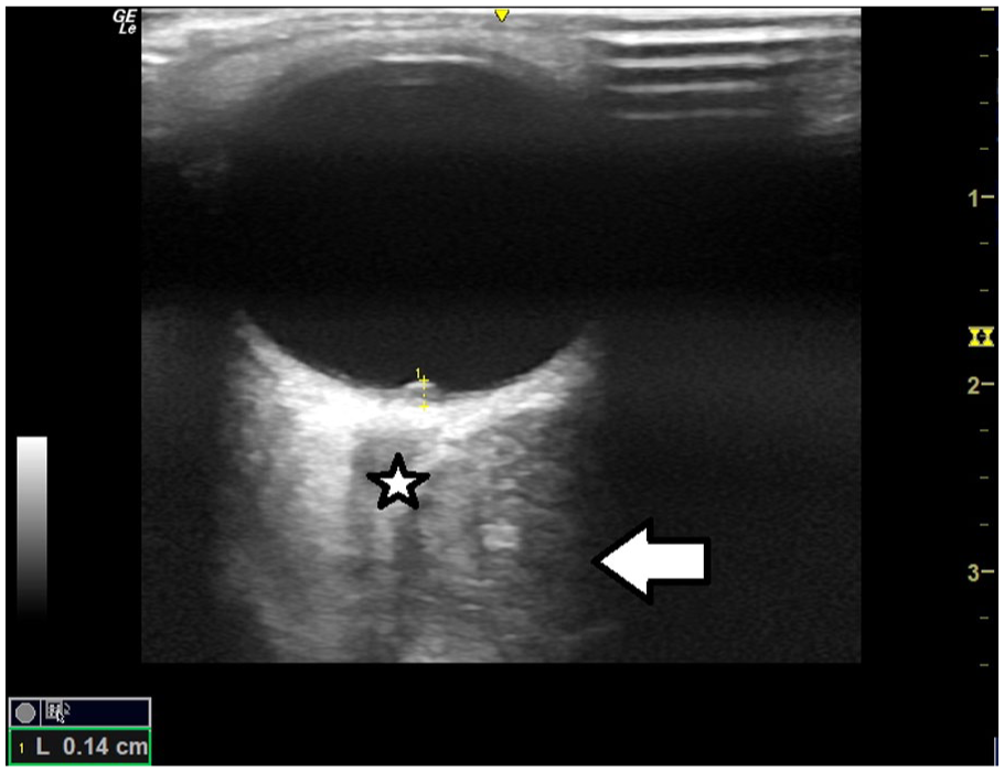
Ocular POCUS of the right eye using a high-frequency 12 MHz linear probe demonstrating a retro-orbital heterogeneous mass (white arrow). This is medial to and impinging on the optic nerve (star). There is also evidence of papilloedema denoted by the elevation of the optic disc to 1.4 mm.

MRI images showing a mass within the right orbit, closely related to the optic nerve and right medial rectus muscle.
RMS is a rare paediatric malignancy, with an incidence of 4.6 cases per million children. However, it is the most common orbital malignancy in childhood.6,7 Arising from undifferentiated mesenchymal cells, RMS has been divided into four major histopathological sites: embryonal, pleomorphic, alveolar and botryoid. 8 Embryonal is the most common form and carries the best prognosis, with a 5-year survival rate of 94%. Conversely, alveolar RMS is less common but carries a worse prognosis, with a 5-year survival of 74%. 9 Orbital RMS typically presents with proptosis, which occurs more rapidly in newborns and infants than in older children. Additional symptoms may include blepharoptosis and eyelid swelling. 10 The current treatment of orbital RMS includes a biopsy, either incisional or by complete surgical removal, followed by chemotherapy and irradiation. 11
Case 2
A 7-month-old boy born at full term was brought to the PED due to concerns about significant weight faltering. Initially born weighing 3.6 kg just below the 50th centile, he had steadily fallen to below the 0.9th centile. On assessment, he was quiet and slightly pale, but his initial observations were normal. On examination, his abdomen appeared slightly full with a palpable left-sided mass. A POCUS was carried out, which showed a large heterogeneous mass in the left side of the abdomen, obscuring the spleen and most of the left kidney (see Figure 4). Differential diagnoses considered included a Wilms tumour, renal cell carcinoma or neuroblastoma. An urgent computed tomography (CT) chest and abdomen was arranged, which showed a very large mass arising from the left kidney measuring 11.1 cm × 11.4 cm × 11.5 cm extending across the midline with associated displacement of the aorta and inferior vena cava. The appearances were suggestive of Wilms tumour (WT) (see Figure 5). The infant was transferred to the tertiary oncology centre, where a diagnosis of WT was made. He was commenced on an initial chemotherapy regimen, followed by delayed nephrectomy 4 weeks later. He remains in remission 2 years later.
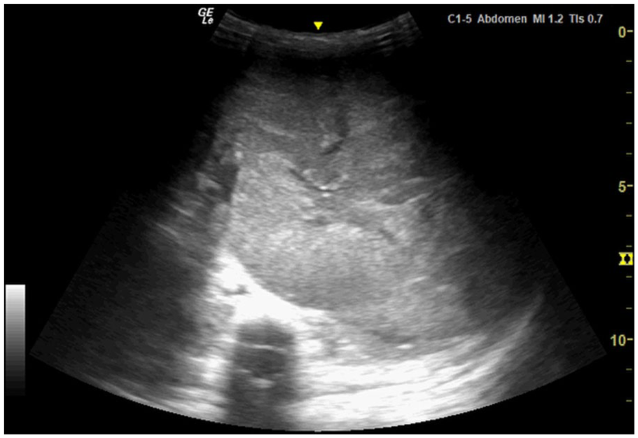
POCUS of the left upper quadrant using a 5 MHz curvilinear probe demonstrating a large heterogeneous mass obscuring the spleen and left kidney.
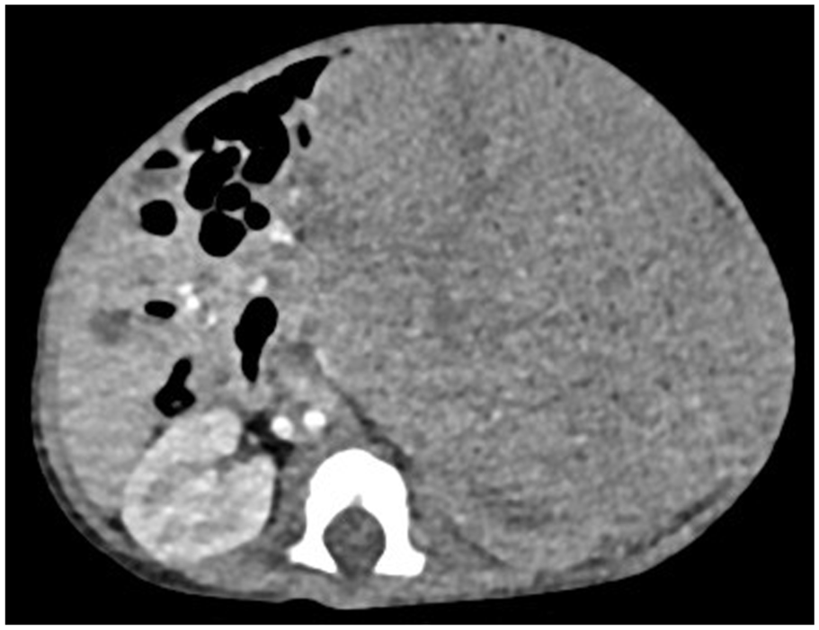
CT scan of the abdomen demonstrating a very large mass measuring 11.1 cm × 11.4 cm × 11.5 cm arising proof from the left kidney.
Renal cancer represents 6% of paediatric malignancies in the United Kingdom; WT, which is also known as nephroblastoma, is the most common type and accounts for approximately 95% of renal tumours diagnosed in children under 15 years.6,12 The overall survival rate of WT is now over 90% with modern advances in treatment. 13 The prognosis is largely dependent on the stage at diagnosis, where children with low-stage WT have been found to have the best prognosis. 14
WT is thought to be caused by abnormal embryonic development and has been associated with the presence of nephrogenic nests, which are clusters of persistent metanephric cells in a fully developed kidney. 15 Following biopsy, the histological features can have a large degree of differentiation, containing different proportions of epithelial, blastemal and stromal components. 16 While most cases are sporadic, as many as 8% are associated with a congenital abnormality. 17 WT typically presents with a non-tender abdominal mass, often first noticed by a parent. Other presenting symptoms may include abdominal pain, haematuria or hypertension. 15 Management involves a combination of chemotherapy, surgical excision and radiotherapy.
Case 3
A previously fit and well 14-year-old girl was sent to the PED out of hours after her general practitioner had undertaken blood tests, which highlighted ‘abnormalities to her renal function and calcium’. She described a 3-month history of dull ache in her upper chest and back, which had started to wake her at night. On enquiry, she described unintentional weight loss of 10 kg in this time and described nausea and early satiety. She also described fatigue resulting in an inability to attend school. On examination, she was slightly pale but had no palpable lymphadenopathy. Her abdomen was soft, non-distended, with no palpable masses. When asked to identify the site of her pain, she pointed to the right upper arm. One of the authors undertook a POCUS of this area (see Figures 6 and 7), which revealed irregular appearances of the bony cortex at the humeral head. These appearances were concerning for a bony malignant process in view of the history. POCUS of her abdomen revealed multiple hypoechoic lesions within the liver and kidney, suspicious of metastatic lesions (see Figures 8 and 9). In view of the POCUS findings, an urgent CT of the shoulder, chest, abdomen and pelvis was arranged. This demonstrated a large malignant mass infiltrating the mediastinum with multiple metastatic deposits to the liver, kidneys and bones (see Figures 10 and 11). Blood results showed a normal haemoglobin, white cell count and platelets, but showed the following abnormalities: urea 8.7 mmol/L, creatinine 122 µmol/L, uric acid 563 µmol/L, adjusted calcium 3.91 mmol/L, lactate dehydrogenase 555 U/L, PTH 0.6 µmol/L (NR 1.1–6.9). She was commenced on hyperhydration and transferred to the regional tertiary oncology centre. She was diagnosed with stage IV (mediastinum, skeletal, liver, renal and bone marrow) primary mediastinal large B-cell lymphoma. She had pathological fractures of the right humerus, manubrium and T5 vertebra. Subsequent positron emission tomography (PET) scan demonstrated avid disease spread, including multifocal markedly avid bony involvement at the skull base, spine, pelvis, ribs, sternum, bilateral humeri, scapulae, femurs, tibias and fibulae (see Figure 12).

POCUS of the right humerus using a high-frequency 12 MHz linear probe in longitudinal orientation demonstrating a bony cortical irregularity (white arrow) suspicious for bony malignancy.
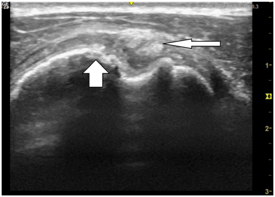
POCUS of the humeral head using a high-frequency 12 MHz linear probe in the transverse orientation, which shows degradation of the cortical surface (thick arrow) along with a hyperechoic irregular lesion superficial to this (thin arrow).
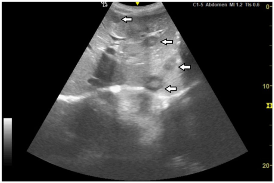
POCUS of the right upper quadrant using a 5 MHz curvilinear probe demonstrating multiple rounded, well-defined lesions throughout the liver (arrows) with the characteristic hypoechoic halo sign.

POCUS of the right upper quadrant using a 5 MHz curvilinear probe showing multiple large proof hypoechoic lesions within the right kidney (arrows).
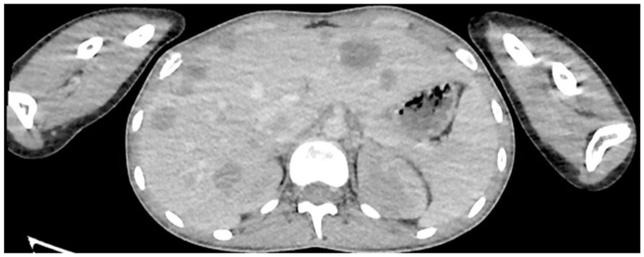
CT scan of the abdomen showing multiple metastases in the liver.
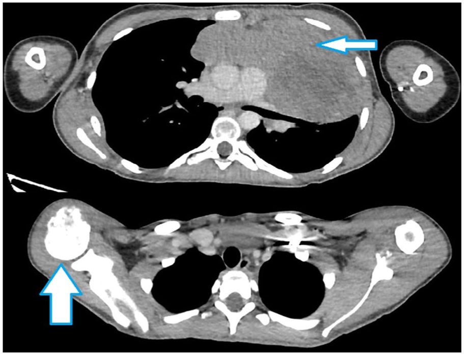
CT scan of the thorax demonstrating a very large mediastinal mass (thin arrow) and a lesion within the right humerus suspicious of a bony metastasis (thick arrow).
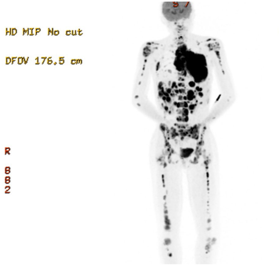
PET scan showing avid disease, including an extremely large anterior mediastinal mass with large volume bone, liver and renal involvement.
Primary mediastinal large B cell lymphoma (PMBL) is an aggressive B cell lymphoma that arises from transformed thymic B cells in the thymic medulla. 18 PMBL is a type of non-Hodgkin lymphoma (NHL), accounting for 2.4% of NHL cases. 19 Though there is a female predominance, the median age of diagnosis is 35 years, and the annual incidence is 0.4 per million, making this a rare diagnosis. 20 In contrast to adult cases, where NHLs are usually low-grade, paediatric NHL cases exhibit high-grade pathology and aggressive clinical behaviour. 21 Presentation commonly includes symptoms of cough, shortness of breath, neck swelling and raised lactate dehydrogenase. B symptoms are present in a small proportion of patients. 22 PMBL can also present with emergency complications, most commonly superior vena cava syndrome. Further emergency complications include tumour lysis syndrome and pericardial/pleural effusions. 20 Though research is limited in quantity, current research into treatment is promising, showing rituximab/chemotherapy treatment with 5-year event-free and overall survival rates of 93.2% and 100%, respectively. 23
Case 4
A 2-year-old girl with no significant past medical history was brought to the PED by her parents after they noted that she had a lump on the left side of her head. She was initially reviewed at an urgent care centre, where her swelling was thought to be a haematoma sustained following a minor head injury when she ran into the edge of a door 11 days ago. However, due to progressive enlargement, she sought a second opinion. On assessment, she was alert and systemically well with no neurological symptoms. Examination revealed a 5 cm × 5 cm × 2 cm boggy, tender swelling over the left parietal skull. POCUS revealed a haematoma with a large depression and discontinuity of the skull cortex beneath this (see Figure 13). Although a depressed skull fracture was considered as a differential diagnosis, the appearances were unlike other skull fractures previously identified using POCUS, with a much larger defect evident in the bony cortex. A CT head was undertaken, which demonstrated multiple lytic skull lesions with associated soft tissue components, including a 20 mm × 14 mm lesion in the left parietal region with underlying subcutaneous haematoma. Smaller lytic lesions were scattered throughout the right parietal bone, calvarium, orbital bones, petrous bones and skull base. No intracranial mass or haemorrhage was seen (see Figure 14). The differential diagnoses were either of neuroblastoma metastasis or eosinophilic granuloma. The child was urgently referred to paediatric oncology, where her pattern of disease was felt to be consistent with Langerhans cell histiocytosis (LCH), which was confirmed on biopsy 4 days later.
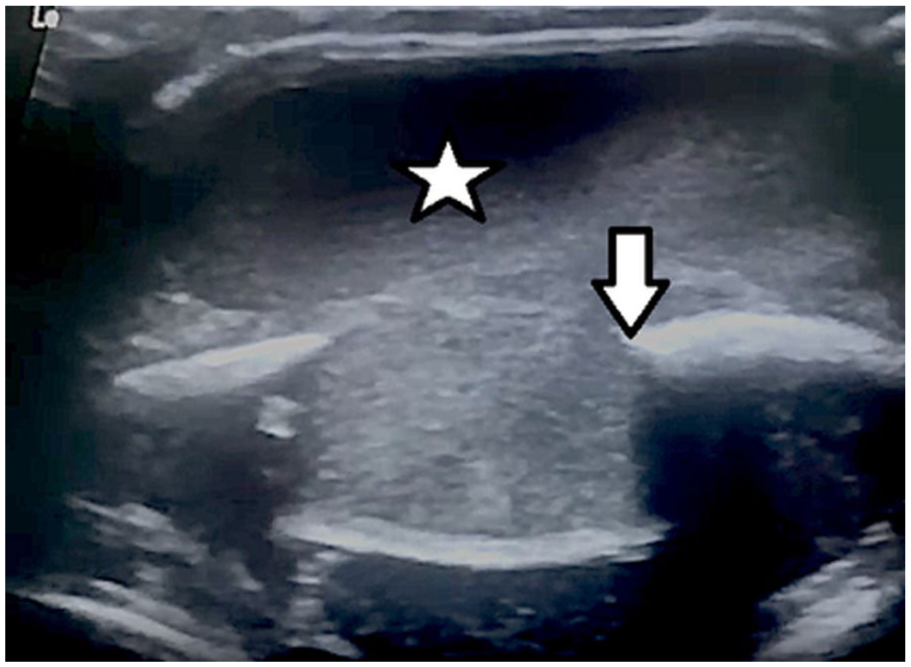
POCUS of the left parietal skull using a high-frequency 12 MHz linear probe in the transverse orientation, showing a large subcutaneous haematoma (star) overlying a depression and discontinuity of the bony cortex (arrow).

CT scan of the skull, which shows a large lesion in the left parietal bone with an underlying subcutaneous haematoma.
LCH is an inflammatory myeloid neoplasm, named due to the appearance of these cells resembling dendritic Langerhans cells, but relating to myeloid dendritic cells. LCH is more common in children than in adults, and more common in males than in females. Incidence is estimated at 5–10 cases per million children, most commonly in those aged 1–3 years old.24,25 In children, this most commonly affects bones and skin, but can also involve the bone marrow, liver, spleen, lungs, central nervous system and other organs, leading to diverse symptomatology. 26 Patients may be asymptomatic and a diagnosis made incidentally, or they may present with localised bone pain with a raised, soft, tender lesion evident. LCH can be classified into single-system single-site (SS-s), single-system multi-site (SS-m) and a multi-system type (MS), which impacts management and outcomes. 27 Treatment varies depending on the site involved, ranging from curettage for local bone lesions to vinblastine-prednisolone chemotherapy for multi-system LCH.25,28
Case 5
A 5-year-old boy presented to the PED after his mother noticed a firm lump on the left side of his neck 1 day earlier. The child was entirely well in himself with no other symptoms and no temperatures. His appetite was good, and he was gaining weight along the 25th centile. On examination, he had a large firm lump approximately 2.5 cm × 3 cm in the left mid-jugular region. There was no warmth, tenderness, erythema or any overlying skin changes. POCUS was carried out, which demonstrated a 1.2 cm × 2.2 cm well-defined, but very heterogeneous lesion with an echogenic internal component, with a degree of clean shadowing deep to this. Part of the lesion appeared to be within the sternomastoid muscle (see Figure 15). These appearances did not appear consistent with reactive lymphadenopathy or infective lymphadenitis. A radiology performed ultrasound was obtained, which was unable to fully characterise the appearances. Differential diagnoses included a traumatic tear in the sternocleidomastoid muscle with subsequent haematoma formation or a possible sarcoma. Four days later, an MRI scan was conducted, which revealed a heterogeneous lesion in the left anterior triangle anterior to the origin of the sternomastoid muscle. The impression was that this could be an inflammatory nodule representing an atypical infection or could be more sinister. The lesion began to develop overlying violaceous skin changes in the following 2 weeks, and the child was reviewed in an ENT outpatient clinic, where a diagnosis of non-tuberculous mycobacterial (NTM) lymphadenitis was made. The family were counselled regarding potential management options, including a watchful wait approach or surgical excision of the lesion. Four weeks later, the lesion had begun to spontaneously discharge; therefore, a decision was made to manage conservatively.
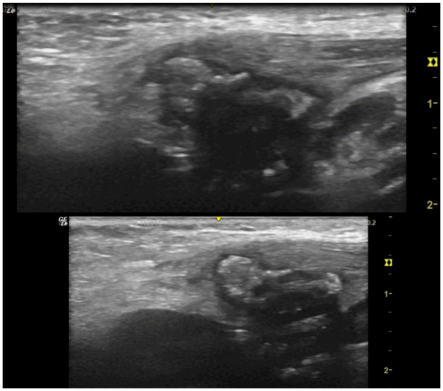
POCUS of the left neck level II using a high-frequency 12 MHz linear probe in the transverse orientation, which shows a 1.2 cm × 2.2 cm well-defined, but heterogeneous lesion with a strongly echogenic internal component, with a degree of post-acoustic shadowing.
NTM lymphadenitis is the most common manifestation of NTM disease in immunocompetent children, typically affecting those aged 1–5 years with an estimated incidence of 0.8–3.1 per 100,000 children. The disease is caused by environmental mycobacteria, most commonly the Mycobacterium avium complex (MAC). 29 It is a chronic, localised infection of the lymph nodes, most often in the cervicofacial region. Affected children typically present with a slowly enlarging, painless lymph node, often unilateral and located in the submandibular or preauricular area. Over time, the node may become fluctuant or develop overlying skin changes and sinus tract formation. Systemic symptoms are usually absent. 30 Sonographic features suggestive of NTM lymphadenitis are hypoechoic lymph nodes in the initial stages, followed by liquefaction with intranodal cystic necrosis and adjacent soft-tissue oedema. 31 Complete surgical excision of the affected lymph node is considered the gold standard treatment and is associated with high cure rates and low recurrence. Non-surgical management, either with a ‘watchful wait’ approach or with macrolide-based antibiotics, is an alternative but typically requires prolonged treatment. 32
Case 6
A 15-year-old boy attended the PED due to a painful lump on the left side of his upper chest. This had been gradually increasing over the preceding 3 weeks to the point where it was becoming painful and difficult for him to sleep. The child had no relevant past medical history and no family history of malignancy. On examination, he had a very firm, fixed lump just below the left clavicle near the sternum, approximately 2.5 cm × 2.5 cm. There were no overlying skin changes, and the lump was not warm or tender. The rest of his examination was unremarkable, with no evidence of organomegaly or lymphadenopathy. X-rays of the left clavicle were undertaken by the clinician who initially assessed him (see Figure 16). This did not reveal any abnormalities, so one of the authors was asked to undertake a POCUS. This revealed an unusual structure comprised of hypoechoic regions surrounded by a hyperechoic mesh-like network. There was no enhancement using colour Doppler within each hypoechoic region (see Figure 17). The appearances were not familiar to the user; however, differentials included a type of bony malformation, given the hyperechoic surrounding structure or possibly a lymphangioma. The appearances on POCUS were described to the on-call radiologist, and it was recommended that a CT scan would be the best modality to provide further detail. The orthopaedic team reviewed the CT and concluded that this could represent a sarcoma, or more likely a giant cell tumour. A subsequent MRI scan demonstrated an expansile lytic lesion involving the manubrium sterni with sclerotic margins most consistent with an aneurysmal bone cyst or giant cell tumour (see Figure 18). An incisional manubrial biopsy confirmed osteoclastic giant cell-rich tumour of bone. An initial resection was undertaken 6 weeks later with a plan to undertake further sclerotherapy of the lesion if required.

X-rays of the left clavicle, which do not show any abnormalities, were reported as normal.
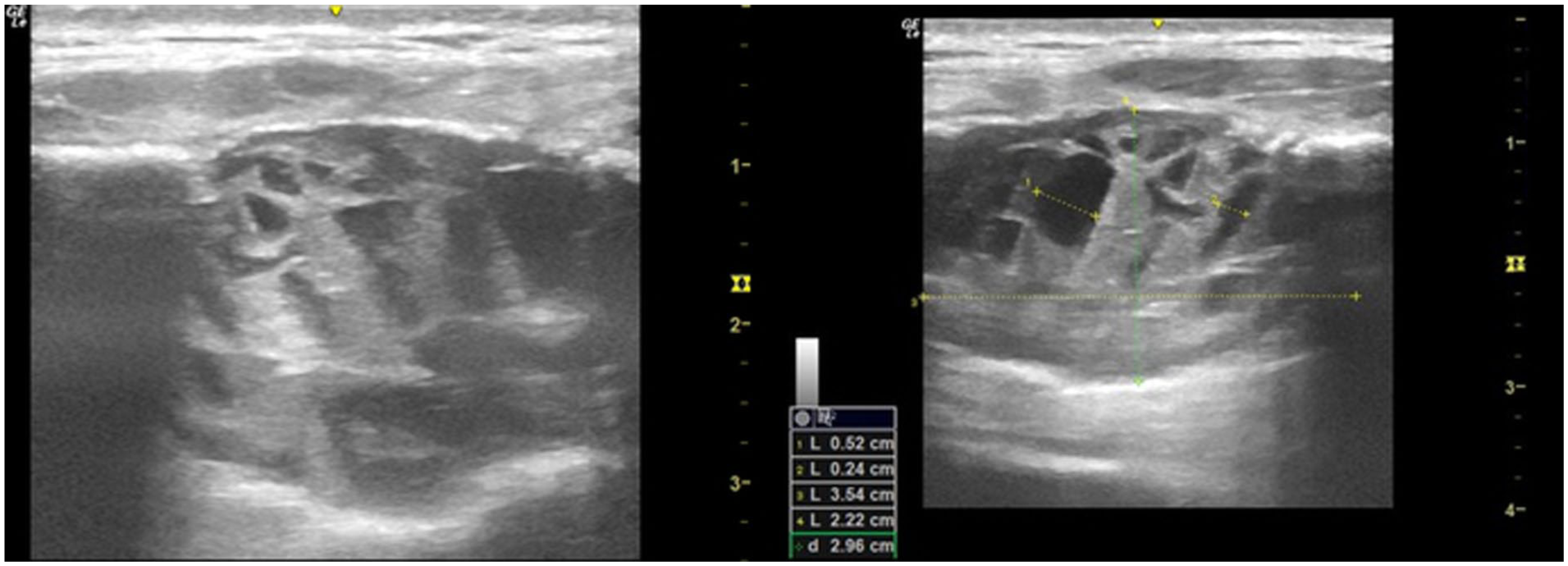
POCUS of the left sternoclavicular region using a high-frequency 12 MHz linear probe in the transverse orientation. This demonstrates a lesion more than 3.5 cm in diameter composed of a hyperechoic mesh-like network with multiple small hypoechoic regions within it.
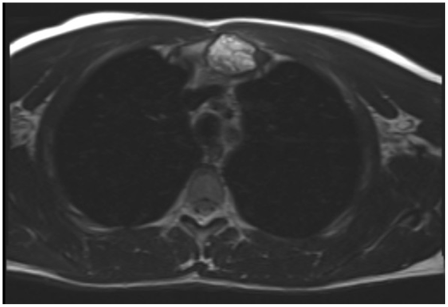
An MRI scan of the thorax, which shows a lesion involving the manubrium sterni with sclerotic margins most consistent with an aneurysmal bone cyst or giant cell tumour.
Giant cell tumour (GCT) of bone is a rare, generally benign but locally aggressive neoplasm characterised histologically by multinucleated giant cells. They account for approximately 5% of all primary bone tumours and 20% of benign bone tumours. 33 GCTs are uncommon in children and occur more frequently in older adolescents and post-pubertal individuals. The most common sites include the distal femur, proximal tibia and distal radius. 34 GCT originating from the ribs is extremely rare, with only six paediatric cases reported in the literature, with all but one originating from the posterior rib. 35 Children typically present with localised pain and swelling, which can lead to developing pathological fractures in advanced stages of disease. Surgical excision is the mainstay of treatment, and prognosis is generally favourable, though there is a risk of local recurrence and metastatic disease. 36
Discussion
Diagnosing cancer in children can be challenging as it is an uncommon diagnosis and because symptoms frequently mimic those of common benign conditions of childhood, or because the assessing clinician is less familiar with presentations of paediatric malignancy.37,38 Furthermore, clinical examination alone is not always sufficient in determining the origin of a child’s problem.39,40 Prompt diagnosis of cancer is the goal because it improves the prospect of cure, may lower treatment morbidity and help maintain better quality of life.38,41 In the United Kingdom, half of children and young people wait more than 4 weeks to be diagnosed with cancer from their initial presentation, with certain age groups, such as late teenagers, waiting on average almost 9 weeks. Prolonged diagnostic intervals such as these can increase the risk of cancer up-staging, disability and even sudden death. 42 Ultrasound is recommended as the most appropriate first-line investigation in suspected intrabdominal malignancy in children 40 ; however, access to those with sufficient paediatric expertise remains a challenge in the United Kingdom. 43 This is despite a 2010 report by the National Imaging Board, which recommended that a paediatric radiology service should be part of an integrated pathway for the management of site-specific cancers and integrated in the care pathways of Children’s Cancer Centres. 44
POCUS, utilised as an extension of clinical examination, has the potential to bridge some of these gaps by adding an additional layer of detail to the overall clinical assessment of the paediatric patient. The role of POCUS is not to attempt to provide a final diagnosis but to help detect occult lesions and to safely and efficiently expedite definitive imaging, thereby positively impacting the patient journey. POCUS protocols for use in existing cancer patients have been devised, such as The Focused Assessment with Sonography in Cancer (FASC), which is used in order to detect pleural effusion, pericardial effusion and ascites in patients with known malignancy. 45 There have also been other reports of paediatric emergency medicine (PEM) performed POCUS detecting paediatric malignancy very early in their presentation.46,47
The decision to use POCUS in each of these cases was guided by specific clinical cues and the need to achieve greater diagnostic information promptly. The users were faced with the situation where definitive imaging was either less readily available or required justification before it could occur, which POCUS was able to provide. These examples illustrate how POCUS can be integrated into existing triage pathways, offering a readily available method of imaging that can expedite and guide the sequencing of further imaging. By highlighting this, our aim is to show how POCUS supports real-world clinical decision-making, particularly to those unfamiliar with its application.
These cases represent the value POCUS can offer as a quick, well-tolerated, low-risk clinical imaging tool to aid in the early detection of serious paediatric conditions. Despite its obvious advantages, it is important to be aware of the limitations of this imaging modality. The main challenge of ultrasound lies in its operator dependence. This is of particular relevance in relation to POCUS, where training pathways are less well established and understanding the background experience of its users is of paramount importance when integrating their findings into patient care. Several studies highlight the risk of misinterpretation of artefacts or disease, both in POCUS and formal radiology imaging.48,49
All scans in this case series were undertaken by one of two PEM consultants, both of whom have completed postgraduate studies in POCUS and have over 7 years of experience. Despite being experienced, both users encountered findings that were unfamiliar and worked closely alongside their imaging departments to ensure the necessary definitive imaging studies were carried out.
Pitfalls also exist, specifically, within the challenges of differentiating very complex lesions using ultrasound, for example, in soft tissues, where an abscess or haematoma can resemble a tumour and vice versa, potentially leading to misdiagnosis or delay in management. 50 A further limitation of ultrasound is reflected in Case 3, in which bony cortical irregularity was detected using POCUS. As ultrasound cannot visualise beyond the bony cortex owing to the acoustic impedance mismatch between soft tissue and bone, it can only be relied upon to provide part of the clinical assessment and alternative imaging modalities are needed to image beyond the bony cortex and provide a more detailed assessment.51,52
This case series highlights the importance of utilising POCUS as an extension of clinical assessment in order to achieve a more detailed evaluation than standard examination alone allows, thus providing the clinician with greater certainty when ensuring the necessary definitive imaging investigations are obtained and in a prompt timescale. The importance of cultivating and maintaining close working relationships with experienced clinicians within hospital imaging departments cannot be overstated and was key to ensuring successful patient journeys in each of our cases. This is an important factor to consider for the POCUS user who is considering expanding their scanning repertoire.
Conclusion
POCUS provides important additional information when integrated into the clinical assessment of the undifferentiated paediatric patient. We describe six cases of children and young people presenting with malignant or locally invasive lesions in which POCUS allowed the PEM physician to promptly and efficiently expedite definitive imaging, thus leading to much quicker diagnosis and an improved patient journey. Close working relationships with experienced clinicians within hospital imaging departments are essential to achieving positive outcomes such as these.
Footnotes
Acknowledgements
None.
Contributors
None in addition to the listed authors.
Declaration of Conflicting Interests
The author(s) declared no potential conflicts of interest with respect to the research, authorship and/or publication of this article.
Funding
The author(s) received no financial support for the research, authorship and/or publication of this article.
Ethics Approval
Informed consent was provided by those who are authorised to do so for all images used. No patient identifiable information was visible. No specific approval from the NHS Research Ethics Committee was deemed to be required for the publication of this article, as it related to routine patient care only.
Permission From Patient(s) or Subject(s) Obtained in Writing for Publishing Their Case Report
YES consent was obtained for the use of images in this manuscript, by those who were authorised to provide it; in this case, given that the subjects were minors, this was from those with parental responsibility.
Permission Obtained in Writing From Patient or any Person Whose Photo Is Included for Publishing Their Photographs and Images
YES.
Confirm That You Are Aware That Permission From a Previous Publisher for Reproducing any Previously Published Material Will be Required Should Your Article be Accepted for Publication and That You Will be Responsible for Obtaining That Permission
n/a.
Guarantor
n/a.
