
Abstract
Numerous studies have demonstrated improved performance results for sport skills through attentional focus instructions. However, few studies have examined the effects of cueing on expert kinematic performance, and this is the first to assess cueing’s kinematic effects on expert soccer players performing .common techniques This study assessed the lower extremity kinematics of an inside of the foot soccer trap via motion capture cameras under different attentional focus cues to analyze how the cues affect the movement pattern among expert performers. 10 NCAA Division 1 soccer players trapped a launched ball on a scoring mat after receiving coaching cues inducing an internal, external, holistic, or control focus of attention. Statistical analysis revealed movement differences between the internal and external cue conditions for the ankle and knee joints during the movement. The difference at the knee was a steeper flexion followed by a correction and, at the ankle, a steeper flexion, both during the internal cue condition. Attentional focus cues inducing a conscious control of the movement may disrupt the movement patterns of highly skilled expert performers. Coaches should be acutely aware of the language they use in instruction, even among expert performers.
Introduction
Central to effective coaching is facilitating motor skill acquisition and facilitating peak performance in players. Particularly in soccer training, verbal instructions are an important means by which coaches are able to guide performers to the most effective and efficient movement patterns to achieve a desired outcome. 1 Research has indicated that the focus of attention which verbal instructions induce, either internal or external, has an “important impact on the effectiveness and efficiency of motor performance.” 2
An internal focus of attention directs the performer’s conscious thought toward their body movements with verbal cues that explicitly reference one or more of their body parts. 3 Contrarily, an external focus of attention directs the performer’s conscious thought toward the effect or outcome of their movement, with verbal cues that reference implements or apparatuses as well as outcome goals. 3
Beyond the two aforementioned focus types, Gucciardi and Dimmock 4 have proposed a third type of attentional focus cue, a global focus, which consists of a single word cue that metaphorically summarizes the desired quality of the movement outcome without referring to outcome nor coordination patterns. “Smooth” during a golf putt is an example from their work.
Becker, Georges, and Aiken 5 further developed this paradigm into what they termed a “holistic cue” and suggested it as a solution to activities where determining an effective external verbal cue is nonobvious. An example is from Lawrence, Gottwald, Hardy, and Khan’s 6 study of skilled gymnasts that correlated a performance decline when performing a novel gymnastic routine with an external focus. As a remedy, Becker et al., 5 found performance improvements with their holistic cue condition in standing long jump distance, expanding cuing language for when an external cue may be suboptimal.
This research has link the holistic cue condition to previous research has indicated that external cues enable higher performance outcomes and movement efficiency relative to internal focus in running, volleyball, ski simulation, dart throwing, and a wide variety of other skilled activities.7–9 To expand upon the research of cueing effects on performance outcomes, the present study aims to examine how these three attentional focus coaching cues affect the kinematic performance of an inside of the foot soccer trap of expert soccer players.
Among expert performers who have a highly developed and specialized procedural memory for the task, there are several theoretical frameworks which aim to explain the detrimental effect of an internal focus of attention, of which three will be considered here. First, Prinz’s common-coding theory of perception and action asserts that conscious thought of the movement interferes with the procedural memory of the movement. 10 Secondly, Wulf, proposed the constrained-action hypothesis that with an internal focus, less efficient conscious control overrides an automatic, more efficient, mode of movement control. 11 Finally, Russell 12 explained this effect within the framework of Bernstein’s 13 synergy theory, arguing that the conscious control of one segment during a complex, multi-segment movement disrupts efficient synergistic coordination patterns.
For an examination of these hypotheses, researchers have conducted a variety of experiments which analyze performance outcomes, qualitative analyses, kinetics and EMG activity with different attentional focus cues during a variety of movements and activities as follows. Wulf 2 asked novice participants during a golf chip shot to either focus on their arm swing (internal focus) or the club swing (external focus). The results indicated that the group with the external focused cue, club swing, scored significantly higher than the group who received the internal focused cue, arm swing. Additionally, qualitative analyses have been conducted as Wulf et al. 11 had experienced volleyball coaches observe the serve of novice and advanced players who had received either an internal focus or external focus cue. The coaches evaluated the movement and scored the external focus group higher than the internal focus group. Furthermore, in the same paper, Wulf 2 investigated the accuracy of soccer kicks with one set of instructions mentioning the participant’s foot and the other set mentioning the ball. This analysis demonstrated higher performance outcome and retention in the group with instructions mentioning the ball. For muscle activation, Marchant et al. 14 uncovered a decrease in EMG activity paired with increased peak joint torque when a bicep curl was performed with an external focus of attention. Finally, Gucciardi and Dimmock 4 and Becker et al. 5 revealed increased performance during golf putting and a standing long jump respectively when participants employed a single global or holistic word cue. The range of studies in the domain of attentional focus measure primarily outcomes, conduct qualitative analyses, EMG, and kinetics, yet a notable absence is an analysis of the underlying kinematic effects due to different attentional focus cues. It is in this manner that the present study builds upon the scant presence in the literature of kinematic examination of attentional focus cues.
One of the only studies to connect attentional focus and complex movement kinematics was a 2009 investigation of novice jugglers. Zentgraf and Munzert 15 measured the upper body kinematics and ball trajectory of three groups of learning jugglers: one who received ball-related cues; one who received body-related cues; and a no cue group. While all three groups improved in a retention test, there were significant differences in upper body kinematics and ball trajectories during the juggling task between the internal and external group. There was a lack of significant differences between the no cue group and the external cue group. This study focused on learners and examining the effects on expert performers was beyond the scope of the investigation.
However, Ford, et al. 16 examined the lower extremity of expert soccer players when the participants wore visual occlusion spectacles which gave full vision or no ball trajectory vision, ostensibly, as Wulf 2 interprets, inducing an external or internal focus of attention. The analysis examined hip-knee and knee-ankle kinematic variability, finding no significant differences between the conditions for both measures, but indicated a trend of less variability when the ball trajectory information was absent (internally focused) for both joints. Their analysis was based upon intrinsic feedback from the movement rather than instructional cues; however, it was an important advance in the application of attentional focus to soccer-specific movements as it uncovered that attentional focus may alter movement kinematics. This study is a further step to examine the link between attentional focus and kinematics in expert soccer players.
Described as the “defining action of soccer,” the biomechanical analyses of soccer techniques have focused primarily on the kick. 17 Numerous previous studies have assessed, quantified, and analyzed a wide variety of common soccer kicking techniques from the standpoint of kinematics, kinetics, EMG, and motor control. 17 Perhaps equally as important as kicking is the technique of controlling or “trapping” a soccer ball. Renowned coach and soccer researcher, Raymond Verheijen, 1 states, “Technique is the most important factor in playing soccer. The ball must be brought under control so that an opponent cannot gain possession of it.” Furthermore, Tahara et al. 18 posit that the inside of the foot soccer trap may in fact be the most frequently employed skill during a soccer match.
Despite the significance of this technique, little biomechanical research has examined trapping in soccer. In one previous study, Asai et al. 19 suspended a soccer ball from the ceiling and swung it pendulum-like toward a subject, quantifying the movement with an accelerometer and cinematography. Given the unrealistic nature of the ball delivery method compared to an in-game situation, this study’s relevance is only its pioneering inquiry into the analysis of soccer trapping with a cinematographic technique and accelerometer measurement rather than revealing the biomechanical essence of the soccer trap technique.
The only other biomechanical analysis of soccer trapping, Tahara et al., 18 engaged 13 experienced male collegiate soccer players and kicked a ball for them to trap from 7 meters away at random speeds. Measurement was conducted via triaxial accelerometers under the lateral malleolus and on the lateral side of the fifth metatarsal bone, along with two high speed video cameras recording at 200 Hz. The researchers analyzed 10 frames before and 10 frames after ball contact, finding a significant relationship between the external rotation of the hip and knee with ball deceleration at ball contact. It was therefore assumed for this study that the movement is controlled in the transverse plane of the hip and knee joints. In an earlier analysis, the present author analyzed 10 frames before and after ball contact and found a lack of significant results among attentional focus cue on the kinematics of the ankle, hip, and knee joints in all planes of movement. 20 These findings guided the present study’s analysis to look beyond 10 frames before and after ball contact, deciding to analyze the movement from commencement to ball contact.
Biomechanical analytics provide a valuable contribution into the understanding of motor control theory. Davids et al. argue that an incorporation of the two sub-disciplines is required to “understand how organization of the movement system changes during performance and development.” 21 The implications for effective soccer coaching are evident as coaches must understand how their verbal instructions affect their players’ technique coordination, and it is within this scope that the present study aims to integrate motor control with biomechanical analysis for the purpose of investigating how different attentional focus coaching cues affect soccer trap movement kinematics. The hypothesis of this study is that, since the hip and knee joints in the transverse plane were linked with ball speed during a trap in Tahara et al., 18 and that expert performers experience movement disruptions with an internal focus of attention, the internal cue condition kinematics will differ significantly from the external and holistic cues, which will be similar as evidenced by Becker et al., 5 for the knee and hip joints in the transverse plane. 2
Methods
Participants
Following approval by the institutional review board, 10 current female NCAA Division I soccer players consented to participate in this study (Age: 19.4 ± 0.97 years, Height: 1.65 ± 0.05 m, Weight: 64.27 ± 6.88 kg). The players were currently in season, physically cleared by a trainer and physician to participate in intercollegiate athletics, free from injury for the previous year, and had a minimum of 12 years of competitive soccer experience (Experience: 14.8 ± 2.1 years). All were right foot dominant players, except for one, who was left foot dominant. For their primary positions, four were midfielders, four were defenders, and two were forwards. All participants were asked to wear tight-fitting clothing. Informed consent was obtained from the participants and approved by the Illinois State University IRB.
Apparatus and task
The study was a single factor design consisting of one within-subjects factor (cue type) with four levels.
A custom pendulum (See Figure 1) was designed to kick a standard size 5 soccer ball (Adidas, Germany) at the participant from a distance of 8 meters at a speed averaging 14.5 ± 0.8 m/s measured via a Stalker Pro II Radar Gun (Stalker Radar, Richland, TX, USA), consistent with Tahara et al. 18 who reported speeds of 15.4 ± 3.5 and 15.0 ± 3.3 at ball contact in their trials. The ball arrived at the knee height of each participant and had to arrive within a 2 foot “strike-zone,” marked with blue tape (See Figure 2).
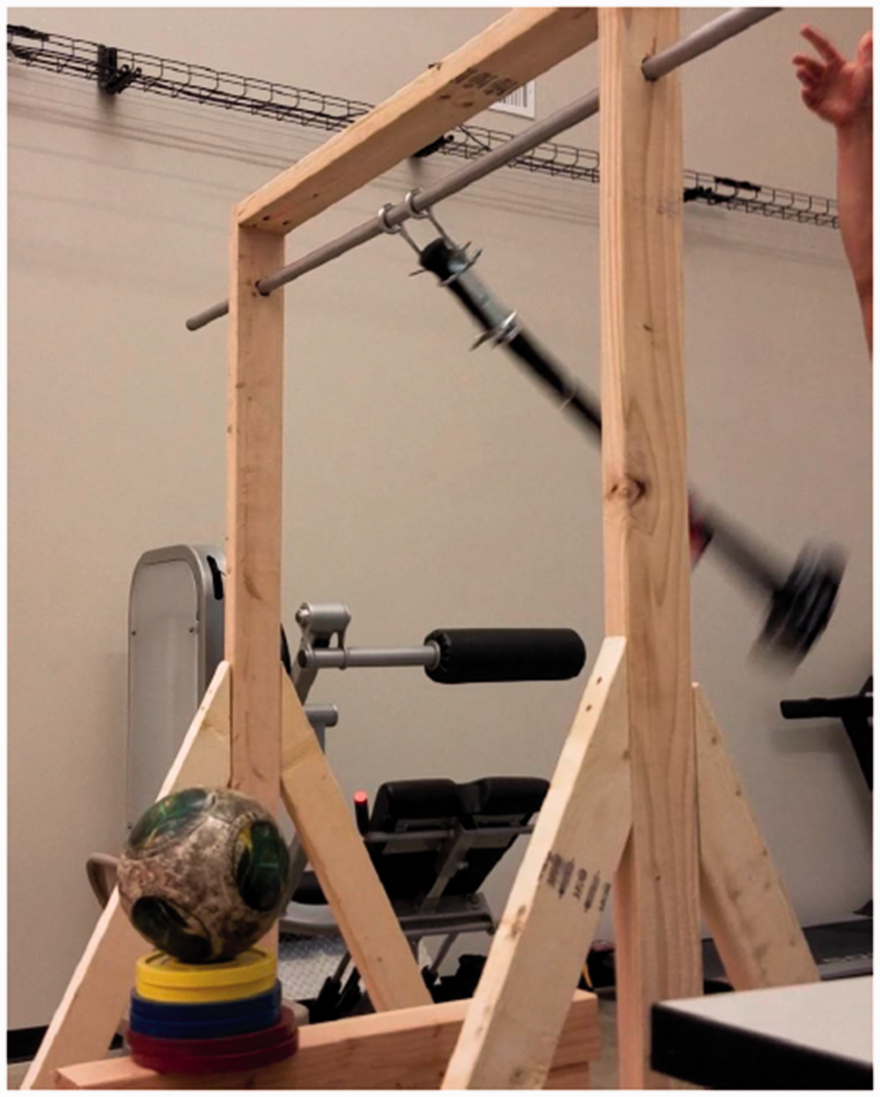
The ball serving pendulum.

The scoring mat (4′×6′) with point zones, 3 is closest to subject. Ball must arrive between the blue tape strips on the bottom of the frame, left gate for left footed subjects (LF), right gate for right footed subjects (RF).
The participant had to execute an inside of the foot trap (See Figure 3) with their dominant foot to control the soccer ball on a 6′×4′ turf mat divided into three scoring zones with 3 points being the closest 2 feet of the mat, 2 points within the middle 2 feet, and 1 point for stopping the ball in the final two foot zone (See Figure 2). Any ball that stopped off of the mat earned zero points. Scoring the highest number of points on each trial was the participant’s goal.
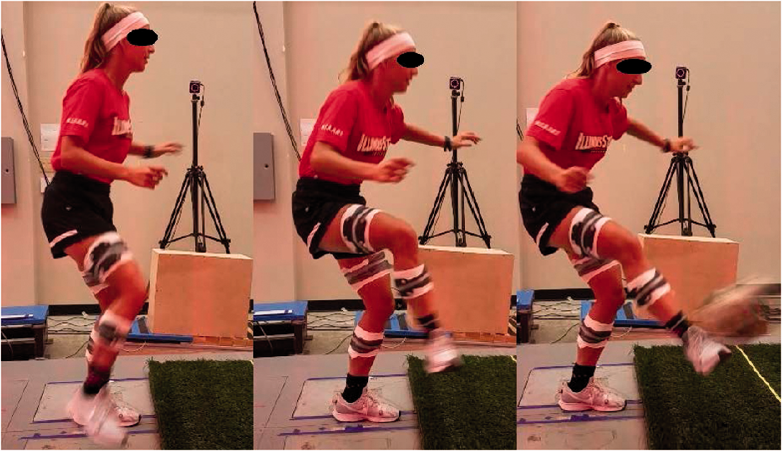
A subject performing a trial during data collection.
There were four conditions: No Cue, the participant was told to simply score the highest amount of points; Internal Cue (IC), the participant was told “to move their foot back when their foot makes contact with the ball;” Holistic Cue (HC), the word “cushion” was told to the participant before each trial; and External Cue (EC), the participant was told to “meet and guide the ball into the scoring zone.” These cues were adapted from an online video teaching the technique of the inside of the foot soccer trap. 22
Data collection
Thirty-three 14 mm retroreflective markers (MKR-6.4, B&L Engineering, Tustin, California, USA) were attached to the participant bilaterally at the anatomical landmarks of 1st metatarsal, 5th metatarsal, heel, medial and lateral malleolus, medial and lateral knee, greater trochanters, iliac crests, and sacrum, (See Figure 4). For the static calibration capture all markers were present, after which markers on the ankles and knees were removed for the dynamic movements. Additionally, two tape retroreflective markers were attached at the equator of the ball, 180 degrees apart, to record the position, velocity, and acceleration of the ball.
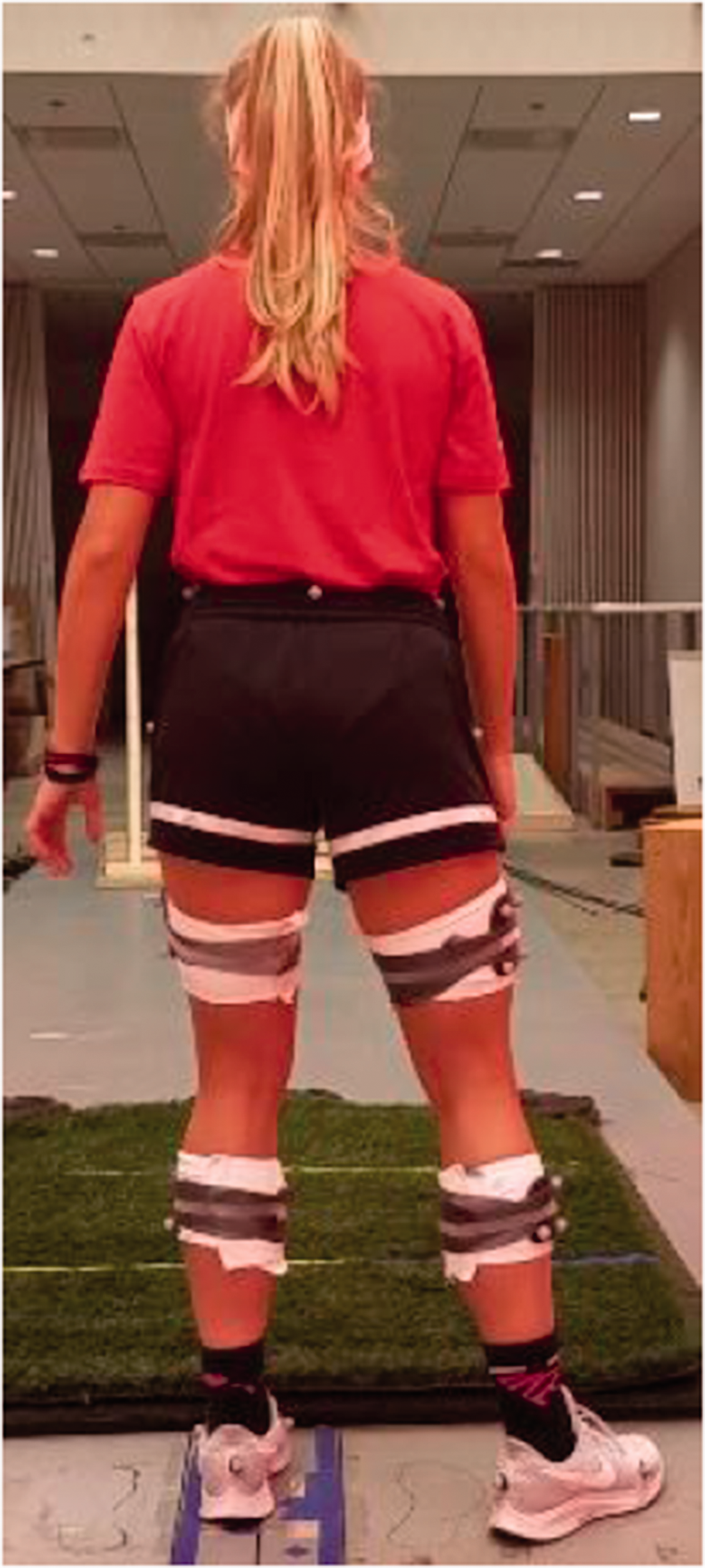
Retroreflective marker placement on the subject.
Participants performed a soccer specific warm up as they would before a game or training session, after which they proceeded to execute 10 familiarization trials of the task. After they were warmed up and familiar with the task, the participants executed five “no cue” recorded trials. After the “no cue” trials, the subsequent cue conditions were randomly assigned at the level of the cue, and each participant executed 5 trials in a row of each cue condition with a 2 minute rest before moving on to the next randomized cue condition.
Score was recorded and told to the participant after each trial.
At the end of the entire protocol, the participants answered a five question survey which served as a manipulation check. The questions were: 1.When the cue mentioned my foot, I focused on my foot while performing the trap; 2. When the cue mentioned the ball, I focused on the ball while performing the trap; 3.When the cue mentioned neither the ball nor my foot, I focused on the whole movement; 4. I simply performed a trap as I normally would regardless of which cue was said to me; and, 5. I focused on the cue from the instructor while I was performing the trap. The survey had a scale of 1–5 with 1 meaning “strong disagreement” with the statement and 5 meaning “strong agreement” with the statement. 23
Measurement of segment motion was obtained using a 16-camera Vicon MX optical motion capture system (Vicon®, Los Angeles, CA, USA) with a sampling frequency of 200 Hz. For each trial, data from the lifting of the participant’s foot until the ball contacted the foot was collected for analysis. Ball contact was inferred through tracking the acceleration of the ball and examining the graph for the point where the horizontal acceleration changes from constant to sharply negative.
Data processing
Marker trajectories were lowpass filtered (6 Hz) and local segment axes were established from global laboratory coordinates using an X-Y-Z Cartesian rotation sequence. Visual 3 D software (C-Motion Research Biomechanics, Germantown, Maryland, USA) was used to calculate relative segment angles. Joint axes were aligned with the z-axis extending vertically, the y-axis extending anterio-posteriorly, and the x-axis extending mediolaterally.
X represented movement in the saggital plane (flexion – negative/extension – positive), Y represented movement in the frontal plane (abduction – positive/adduction – negative at the hip and varus – positive/valgus – negative at the knee), and Z represented movement in the transverse plane (internal rotation – positive/external rotation – negative).
For the ankle complex, X represented dorsiflexion (positive)/plantar flexion (negative), Y represented eversion (positive)/inversion (negative), and Z represented internal (positive)/external (negative) rotation. All data were reported in degrees.
Ball movement data was processed in Vicon Nexus 2.1.1 software (Vicon®, Los Angeles, CA, USA), manually tracking the marker on the ball from 10 frames before ball contact to 10 frames after ball contact. Consequently, the ball displacement data was exported to Visual3D where the second derivative was calculated to obtain an acceleration graph.
The movement was broken down into one phase, from foot up to ball contact (See Figure 4). “Foot up” was determined by when the force plate (Advanced Mechanical Technology, Inc., Watertown, MA, 1000 Hz) on which their foot read zero, and the frame of ball contact was inferred from the acceleration of the ball. More precisely, the ball moved from the launcher towards the participant in the positive direction with a consistent acceleration until there is a sharp negative acceleration in the negative direction that implies a force acting upon the ball. This force is the foot of the participant and the moment where the graph of ball acceleration spiked negative was calculated in Visual3D as the global minimum of the acceleration graph. This phase was normalized from 1–100% and the variables were calculated as the mean of 10 percent intervals of the phase. 24 A MATLAB 9.6 (MathWorks, Natick, MA) script then pulled and aggregated the data from the relevant frames, calculating the means and standard deviations of the 5 trials per condition to arrive at the 90 kinematic variables.
Data analysis
All variables were tested for normality via Mauchly’s test of sphericity with a p-value of .05. A total of 9 omnibus MANOVA test were then used to determine the main effect for Cue, Interval, and Cue-x-Interval for each joint in each plane of movement. One repeated-measures ANOVA was then used at each 10%-time interval to detect differences among each kinematic variable for the four conditions. 24 A p-value of less than .05 was regarded as a statistically significant difference. For the variables that violated sphericity, significance in the proceeding ANOVA was derived from the Greenhouse-Geisser p-value. A post-hoc comparison then established pairwise differences with a Bonferroni correction. Additionally, participant score was averaged over the five trials and then tested with a repeated measures ANOVA by condition. Statistical analyses were conducted with IBM SPSS Statistics v22.0.
The survey data were averaged across participants with standard deviation calculated.
Results
An omnibus MANOVA test revealed a significant main effect of cue type for the ankle joint in the saggital plane (F(3,27) = 4.73, p < .05, ηp2 = .35). A pairwise comparison at the cue level revealed a significant difference between the external cue (75.74 ± 3.15°) and internal cue (75.86 ± 3.3°) conditions (p < .001, 95% CI [−4.223, −2.022]) as well as between the internal cue and the no cue (76.673 ± 3°) condition (p = .03, 95% CI [0.248, 4.133]) (See Figure 5). All other MANOVA tests failed to indicate a significant effect (p>.05) at the hip (See Figures 6 to 8), knee (See Figures 9 and 11) and ankle (See Figures 12 and 13).
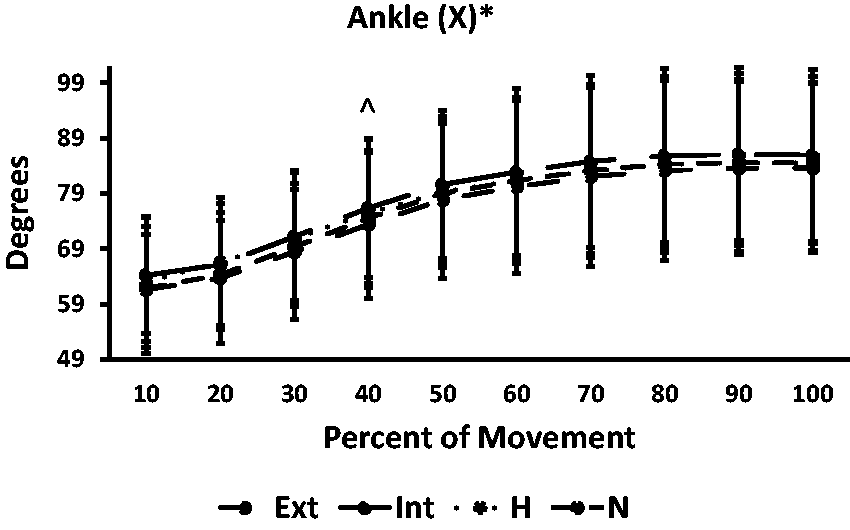
Kinematics of trap over 10% intervals of movement at the ankle joint in the frontal plane. Significant difference detected between external and internal cues. Positive indicates dorsiflexion, negative indicates plantar flexion. * indicates significant difference between external and internal cue, as well as between the internal cue and no cue conditions, ^ indicates significant difference between external and internal cue at time interval.

Kinematics of trap over 10% intervals of movement at the hip joint in the sagittal plane. Positive is hip flexion, negative is hip extension.
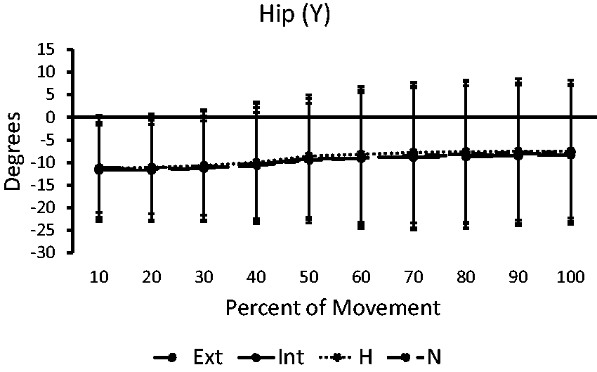
Kinematics of trap over 10% intervals of movement at the hip joint in the frontal plane. Positive is abduction, negative is adduction.
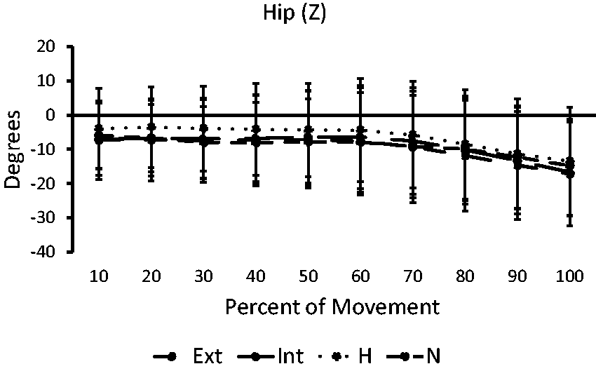
Kinematics of trap over 10% intervals of movement at the hip joint in the transverse plane. Negative is external rotation, postivie is internal rotation.
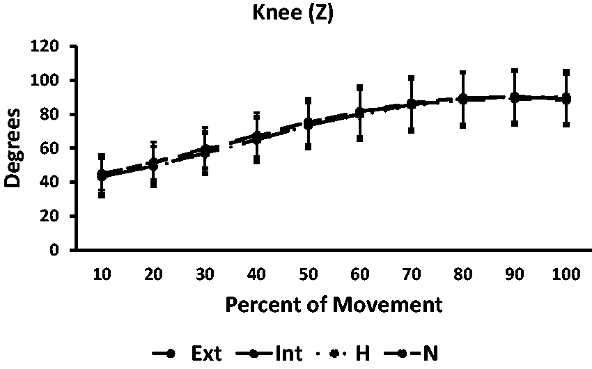
A within subjects ANOVA revealed that there was a significant effect of cue type on the kinematics of the movement for knee joint (See Figure 10) in the sagittal plane for the 41–50% of the movement (F(3,27) = 3.63, p < .05, ηp2 = .29) as well as the ankle joint in the sagittal plane for the 31–40% (F(3,27) = 3.22, p < .05, ηp2 = .26) phase of the movement (See Figure 5).
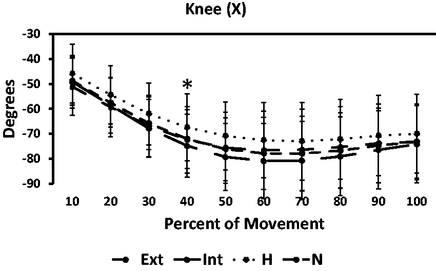
Kinematics of trap over 10% intervals of movement at the knee joint in the sagittal plane. * indicates significant difference between external and internal cue. Negative is knee flexion, positive is knee extension.
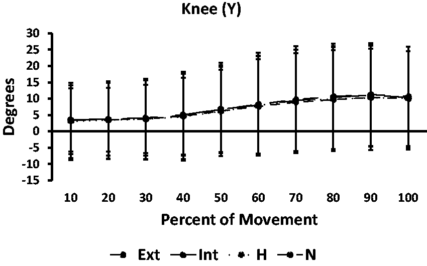
Kinematics of trap over 10% intervals of movement at the knee joint in the frontal plane. Positive is varus, negative is valgus.
Kinematics of trap over 10% intervals of movement at the knee joint in the transverse plane. Negative is external rotation, postivie is internal rotation.
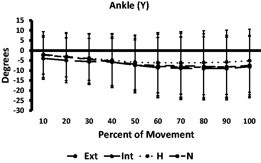
Kinematics of trap over 10% intervals of movement at the ankle joint in the frontal plane. Positive represents eversion, negative represents inversion.
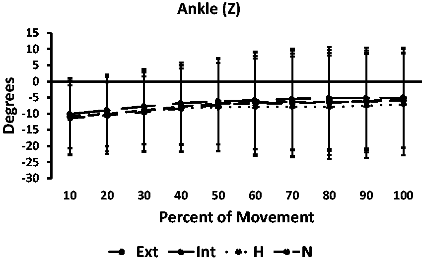
Kinematics of trap over 10% intervals of movement at the ankle joint in the transverse plane. Negative is external rotation, postivie is internal rotation.
Pairwise comparisons revealed that movement during the 40–50% phase in the knee joint sagittal plane, KX, involved less knee flexion when the player received the external cue (−75.57° ± 8.08°) compared to the internal cue (−79.27°±10.24°) (p = .046, 95% CI [0.058, 7.338]). There was, however, no significant difference in movement between the external cue and holistic cue (p = 1), the external cue and no cue (p = 1), the holistic cue and the internal cue (p=.104), and the holistic cue and no cue (p=.532)
Pairwise comparisons revealed that movement during the 30–40% phase in the ankle joint sagittal plane, AX, involved less ankle dorsiflexion when the player received the external cue (73.42°±10.19°) compared to the internal cue (76.39°±11.05°) (p=.01, 95% CI [−5.222, −0.716]). There was, however, no significant difference in the movement between the external cue and holistic cue (p=.66), the external cue and no cue (p = 1), the holistic cue and the internal cue (p = 1) and the holistic cue and no cue (p = 1).
All other repeated-measure ANOVA tests failed to indicate a significant result (p > .05).
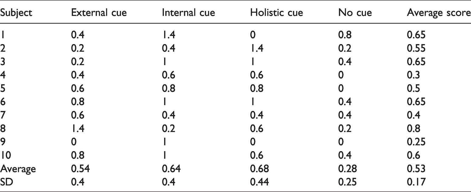
Additionally, the score analysis failed to uncover a significant effect of cueing on performance outcome (p > .05) (See Table 1).
Score data for the participants.
The manipulation check results are listed in Table 2.
Manipulation check results.

Discussion
The purpose of the present research was to employ biomechanical techniques for a quantitative analysis of the effects of attentional focus coaching cues on expert soccer players performing an inside of the foot trapping movement. The hypothesis that there would be kinematic differences in the movement performance between the internal cue and the external cues was confirmed. However, contrary to the original hypothesis, these differences occurred at the knee and ankle joints in the sagittal plane rather than the hypothesized transverse plane of the hip and knee joints. When the participants were instructed with the internal cue, they conducted a greater knee flexion compared to the external cue condition (See Figure 10). Additionally, the participants performed a sharper ankle dorsiflexion after instructed with the internal cue compared to the external cue (See Figure 5). These differences occurred during the middle stages of the movement as opposed to the endpoint when the participant made contact with the ball. As this is the first study to analyze this movement, it is unknown at present what the implications of these kinematic differences are on the effectiveness of the inside of the foot soccer trap.
The implication of the present findings is that coaching cues may affect the coordination and movement of expert soccer players performing common soccer techniques, such as the inside of the foot trap. Although the present study failed to uncover significant performance outcome differences, the kinematic differences suggest that it is therefore important for coaches who are aiding in the mastery of fundamental soccer techniques to be cognizant of the language they instruct and cue players during training and competition. This conclusion echoes the findings of Porter & Sims 25 who asserted that coaches who work with highly skilled athletes “must pay close attention to the verbal instructions they provide” when they uncovered no significant performance differences among expert sprinters who received an external cue versus an internal cue, and ultimately performed better with no cue. Moreover, these findings bolster the understanding in the literature that attentional focus states have consequences on the kinematic performance and coordination patterns of complex motor tasks among expert performers.
With respect to previous kinematic analyses in the domain, the present results are consistent with Zentgraf and Munzert 15 who found kinematic differences in upper body movements between externally and internally focused groups of novice jugglers. Furthermore, the current analysis included a holistic cue condition as theorized by Gucciardi and Dimmock 4 and Becker et al., 5 expanding upon the previously studied conditions of internal and external cueing. Although performance improvements were undetected in contrast the results of the two aforementioned study, the finding of kinematic differences is significant as the underlying movement patterns of experts may be disrupted due to different cueing. This could be the result and demonstration of the underlying coordination pattern change of a skill-oriented attentional focus rather than a “normal” focus which has been associated with decreased performance in expert acrobats. 26 A suggested reason for the lack of a significant different in performance in the present study is the difficulty of the designed task, along with the discomfort of the apparatus which included a raised turf area in front of the participant, rather than the participant being directly on the surface where they performed the movement.
When analyzing an inside of the foot soccer trap, Tahara et al. 18 uncovered an association among ball deceleration and internal and external rotation at the knee and hip joints. For this reason, it was hypothesized that the performance differences among different attentional focus cues would occur in this plane of motion at these joints. However, the observed kinematic differences occurred in the sagittal plane of the knee and ankle joints. This difference could be attributed to the fact that in the Tahara et al. 18 study, the ball was passed on the ground, whereas in the present study, the ball was passed in the air. This suggests that there may be evidence that the inside of the foot soccer trap movement changes depending on if the ball is delivered in the air or on the ground when arriving at the player. The kinematics confirm that there is a triple flexion of the hip, knee, and ankle joints when the ball is arriving in the air. Further research could demonstrate that, although these two skills appear similar, they are in fact controlled differently within the lower extremity.
Between the performer lifting their foot off of the ground to meet the approaching ball and their foot contacting the ball, the hip, knee, and ankle joints conduct a highly specific, coordinated movement. Evident in all trials for all participants in all conditions is a flexion in the hip and knee along with dorsiflexion in the ankle, slight hip abduction, slight knee varus, with slight inversion in the ankle, and external rotation in the hip and knee with slight ankle abduction. The significant difference in attentional focus conditions occurs during the middle of the movement in between lifting their foot and foot/ball contact in the knee and ankle joint in the sagittal plane. This difference in flexion is unobserved at the endpoint of when the ball contacts the participant’s foot. Concurrent with Bernstein’s 13 examination of hammer strikes from expert blacksmiths, differences occur in the movement path but are relatively stable at the end point. For the participants in the present study, there is a greater flexion in the knee in the middle of the movement and then slight extension, leveling out, before contacting the ball during the internal cue, whereas during the external cue, the knee flexion occurs more gradually and is more level to the final ball contact position. Equally at the ankle joint, there is a faster dorsiflexion during the internal focus cue than during the external cue. The suggestion is that the participants are arriving at the same endpoint when their foot contacts the ball but the strategy to move their foot to the required position is changed due to the attentional focus cue.
During the phases of movement where there were differences among the cue types, the holistic and external cues were similar. This suggests that holistic and external cueing might elicit similar attentional focus states. Although the no cue control condition was never significantly different from the holistic and external cues during all phases of the movement, it was never significantly different from the internal cue while only the external cue was different during some phase of the movement significantly different than the internal cue. This could be attributed to the large amount of variation in the movement between all participants and within the individual participant’s trials.
The manipulation check indicates a faithful adherence to the attentional focus cues during the performance of the movement by the participants. They agreed strongly that they were thinking about their foot during the internal focus condition, that they thought about the ball during the external condition, and that they thought about the movement as a whole during the holistic condition. The cueing was consistent with descriptions of attentional focus cues in Wulf, 2 while being adapted from coaching cues from a professional soccer coach. 22
A weakness of the study concerns the task and apparatus whereby the scoring mat onto which the participants were aiming to control the ball was raised and not directly stood on by the participants. Although this was a necessity since they were standing on un-turfed force plates, the positioning was uncomfortable, unfamiliar, and unrealistic to a match scenario where they would be executing the movement. An improvement would be to use a larger turf area that encompasses the force plates so that the participants could stand directly on the surface where they are controlling the ball. This weakness may have led to a decline in the performance and made the movement more consciously controlled than automatic. Regardless of this potential limitation, the study’s apparatus and task were superior to that used in the Tahara et al. 18 study, which had a high variation in ball speed and direction as the ball was kicked anew for each trial and there was no clear target for the participant to aim for, and the Asai et al. 19 study which swung a ball attached to the ceiling via a string to the participant in an unrealistic ball trajectory. Although the ball speed was determined a priori with a consistent parabolic trajectory and relatively uniform participant height, difference in subject height might have influenced participant joint angles.
Other weaknesses include the small sample size as well as that the control condition was conducted first to ascertain a base-level of performance before the randomized cued conditions were conducted. A better design would include more familiarization trials until the participant states that they feel comfortable with the movement, and then randomizing the cueing conditions after a short break.
This study was the first investigation into how attentional focus cues affect the lower extremity kinematic performance of a soccer trap movement among expert soccer players. The results suggest that different attentional focus cues may change the movement strategies of expert soccer players during common techniques. This provided further data for using biomechanical techniques to investigate the effects of attentional focus on complex motor skills. Ultimately, providing expert soccer players with cues which induce a skill-focused attention may disrupt the player’s “natural” attentional focus, resulting in a coordination change. Although the results of these coordination changes must be further investigated, soccer coaches must consider the effects that their cues and instructions have on the coordination patterns and performance of their players during technique execution.
Supplemental Material
sj-pdf-1-spo-10.1177_1747954121994691 - Supplemental material for Effects of attentional focus cues on lower extremity kinematics during inside of the foot soccer trap among expert soccer players
Supplemental material, sj-pdf-1-spo-10.1177_1747954121994691 for Effects of attentional focus cues on lower extremity kinematics during inside of the foot soccer trap among expert soccer players by Ladule Lako LoSarah, Adam E Jagodinsky, Michael Torry and Peter J Smith in International Journal of Sports Science & Coaching
Supplemental Material
sj-pdf-2-spo-10.1177_1747954121994691 - Supplemental material for Effects of attentional focus cues on lower extremity kinematics during inside of the foot soccer trap among expert soccer players
Supplemental material, sj-pdf-2-spo-10.1177_1747954121994691 for Effects of attentional focus cues on lower extremity kinematics during inside of the foot soccer trap among expert soccer players by Ladule Lako LoSarah, Adam E Jagodinsky, Michael Torry and Peter J Smith in International Journal of Sports Science & Coaching
Footnotes
Declaration of conflicting interests
The author(s) declared no potential conflicts of interest with respect to the research, authorship, and/or publication of this article.
Funding
The author(s) received no financial support for the research, authorship, and/or publication of this article.
References
Supplementary Material
Please find the following supplemental material available below.
For Open Access articles published under a Creative Commons License, all supplemental material carries the same license as the article it is associated with.
For non-Open Access articles published, all supplemental material carries a non-exclusive license, and permission requests for re-use of supplemental material or any part of supplemental material shall be sent directly to the copyright owner as specified in the copyright notice associated with the article.