
Abstract
Carbamylation of lysine residues and protein N-termini is an ubiquitous, non-enzymatic post-translational modification. Carbamylation at sites of inflammation is due to cyanate formation during the neutrophil oxidative burst and may target lysine residues within the antimicrobial peptide LL-37. The bactericidal and immunomodulatory properties of LL-37 depend on its secondary structure and cationic nature, which are conferred by arginine and lysine residues. Therefore, carbamylation may affect the biological functions of LL-37. The present study examined the kinetics and pattern of LL-37 carbamylation to investigate how this modification affects the bactericidal, cytotoxic and immunomodulatory function of the peptide. The results indicated that LL-37 undergoes rapid modification in the presence of physiological concentrations of cyanate, yielding a spectrum of diverse carbamylated peptides. Mass spectrometry analyses revealed that the N-terminal amino group of Leu-1 was highly reactive and was modified almost instantly by cyanate to generate the predominant form of the modified peptide, named LL-37C1. This was followed by the sequential carbamylation of Lys-8, Lys-12, and Lys-15 to yield LL-37C8, and Lys-15 to yield LL-37C12,15. Carbamylation had profound and diverse effects on the structure and biological properties of LL-37. In some cases, anti-inflammatory LL-37 was rapidly converted to pro-inflammatory LL-37.
Introduction
Post-translational modifications (PTMs) are pivotal steps in protein maturation. PTMs increase the functional diversity of the proteome and play a key role in multiple cellular processes, including protein–protein interactions, cell signaling, differentiation and regulation of gene expression. 1 Carbamylation is a ubiquitous, non-enzymatic PTM, in which cyanate (OCN−) reacts with primary amino groups (R-NH2) at the N-termini of proteins, and with lysine residues in the polypeptide chain, to generate a protein:homocitrulline complex.2,3 Since urea (a by-product of protein metabolism) and cyanate comprise an equilibrium pair, the level of protein carbamylation is markedly increased in diseases associated with chronic uremia (renal dysfunction). A recent study identified a novel pathway linking inflammation, myeloperoxidase (MPO) and carbamylation. MPO, a heme peroxidase released by activated neutrophils, catalyzes the formation of cyanate from hydrogen peroxide and thiocyanate, ultimately leading to homocitrullination of proteins. 3 The chemical conversion of positively charged Lys residues to neutral homocitrulline residues affects the charge distribution within a polypeptide chain in a manner that often results in impairment or even loss of function. For example, loss of function upon carbamylation has been reported for matrix metalloproteinase-2, inhibitor of metalloproteinase-2 and insulin.4,5
The abundance of MPO at inflammatory foci has sparked significant interest in the role of carbamylation in the context of chronic inflammatory diseases and atherogenesis.3,6,7 Apart from directly affecting protein function and turnover, homocitrulline residues act as neo-epitopes that can trigger primary immune responses, thereby inducing chemotaxis and proliferation of CD4+ T cells and the subsequent production of IFN-γ, IL-10 and IL-17. In addition, Abs against carbamylated residues have clinical value in that they predict a more erosive progression of rheumatoid arthritis.8,9
Cationic antimicrobial peptides (CAMPs) are essential components of human innate immunity and are produced by a variety of cells, including epithelial cells, 10 keratinocytes 11 and neutrophils. 12 In humans, CAMPs are represented by α- and β-defensins, and the cathelicidin-derived LL-37 peptide. The active form of LL-37 is characterized by a high abundance of arginine and lysine residues, which generate a net positive charge of +6 at a neutral pH. Apart from killing a wide spectrum of pathogenic bacteria,13,14 LL-37 neutralizes LPS, functions as a chemoattractant for immune cells (including T cells, monocytes, neutrophils and mast cells),15,16 profoundly affects the course of dendritic cell maturation, 17 stimulates production of cytokines, chemokines and their receptors,18,19 and triggers mast cell degranulation. 16 These effects are mediated, at least in part, by activation of at least four different receptors: formyl peptide receptor-like 1 (FPRL1), epidermal growth factor receptor, P2X7 and CXCR2. Finally, recent studies show that LL-37 has a direct effect on the cells and might be hemolytic or cytotoxic to peripheral blood mononuclear cells (PBMCs) at high concentrations.20,21 At low concentrations (< 5 μM), LL-37 induces rapid secondary necrosis of apoptotic human neutrophils. 22 To prevent collateral tissue damage due to exacerbation of inflammation, the activity of LL-37 is strictly controlled by serum proteins, 23 most likely apolipoprotein-A1 (ApoA-1). 24 Taken together, all available data indicate that the antibacterial activity of LL-37 is secondary to its immunomodulatory functions. In a twist of the paradigm, it is now generally accepted that the major role of CAMPs is not direct killing of invading microbes, but rather acting as signaling molecules for innate and acquired immunity.
Within the inflammatory milieu, the concomitant release of LL-37 and MPO by activated neutrophils may create conditions required for LL-37 carbamylation. Additionally, lysine residues within CAMPs are crucial for peptide structure and activity, and conversion of these positively charged residues to neutral homocitrulline residues would be expected to abrogate the biological activity of LL-37.
Here, we performed mass spectrometry (MS)-based sequence analysis and found that LL-37 undergoes rapid carbamylation in the presence of cyanate in a time- and concentration-dependent manner. Unexpectedly, we found that the free amino group of the N-terminal leucine was most susceptible to carbamylation under conditions that reflected the cyanate concentration in the inflammatory milieu. Prolonged incubation resulted in the generation of a mixture of variably carbamylated LL-37 molecules with impaired antimicrobial activity against both Gram-negative and Gram-positive species.
Taken together, these results suggest that carbamylation of LL-37 within an inflammatory environment might actually exacerbate inflammation and be detrimental to the host.
Materials and methods
In vitro carbamylation of LL-37 and MS analysis
LL-37 (ProImmune, Oxford, UK) was carbamylated by incubation with 10, 50 and 100 mM KCNO (Sigma-Aldrich, Oslo, Norway) in 100 mM HEPES (pH 7.8) for 3 h at 37℃. The reaction was quenched by addition of formic acid to a final concentration of 5% and samples were immediately purified using StageTips (ThermoScientific, Waltham, MA, USA). The samples were lyophilized and either analyzed directly by LC-MS/MS or subjected to proteolytic digestion with Staphylococcus aureus protease V8 (1:25 w/w) at 37℃ for 16 h prior to LC-MS/MS analysis. NanoESI-MS/MS analyses were performed on an EASY-nLC II system (ThermoScientific) connected to a TripleTOF 5600 mass spectrometer (AB Sciex, Oslo, Norway) equipped with a NanoSpray III source (AB Sciex) operated under Analyst TF 1.5.1 control. The samples were suspended in 0.1% formic acid, injected, trapped and desalted on a Biosphere C18 column (5 µm, 2 cm × 100 µm i.d.; Nano Separations, Nieuwkoop, The Netherlands) after which the peptides were eluted from the trap column and separated on a 15-cm analytical column (75 μm i.d.) packed in-house in a pulled emitter with RP ReproSil-Pur C18-AQ 3 μm resin (Dr. Marisch GmbH, Ammerbuch-Entringen, Germany) and connected in-line to the mass spectrometer. The peptides were eluted using a 20-min gradient from either 5–35% phase B or 5–90% phase B (0.1% formic acid and 90% acetonitrile). The collected MS files were converted to Mascot generic format using the AB Sciex MS Data Converter beta 1.1 (AB Sciex). The peptide sequence was identified using in-house Mascot search engine (Matrix Science, London, UK). Search parameters allowed two missed cleaving sites and carbamylation as a variable modification. Peptide tolerance and MS/MS tolerance were set to 10 ppm and 0.1 Da, respectively.
Peptide synthesis
Native and carbamylated LL-37 were synthesized by Proimmune (Oxford, UK) by using Fmoc solid-phase peptide synthesis, diluted in 0.01% v/v acetic acid and stored at −70℃ until use.
Circular dichroism spectroscopy
The secondary structure of the LL-37 analogues was investigated by circular dichroism (CD) spectroscopy. The experiments were performed using a Jasco J-810 spectropolarimeter (Jasco, Essex, UK). Far-UV spectra were acquired at 37℃ in the 195–260 nm range at a scan rate of 50 nm/min and a band width of 1 nm. Three scans were accumulated for each sample and appropriate blanks were subtracted from each spectrum by using the software provided by the instrument manufacturers. The peptides (10 µM) were analyzed upon dilution in a 10 mM sodium phosphate buffer containing 50% v/v trifluorethanol (TFE) or a physiological salt solution resembling plasma (113 mM NaCl, 24 mM NaHCO3, 0.6 mM MgCl2, 1.3 mM CaCl2, 3.9 mM KCl) in 1.0-mm quartz cuvettes (Hellma-Analytics, Oslo, Norway). The mean ellipticity was calculated using the formula [θ] = θ/(10·c·l), where θ is the ellipticity (mdeg), 10 is a scaling factor, c is the protein concentration (M) and l is the path length of the cuvette (cm). The helical content (percentage of helix) was estimated by using the CDNN program from the molar ellipticity θ [deg.cm2 dmol–1].
Broth microdilution assay
Frozen samples of S. aureus LS-1, Escherichia coli ATCC 25922 and Bacillus subtilis ATCC 3366 were cultured on horse blood-agar plates at 37℃ overnight. Few colonies were selected and pre-cultured in 50 ml LB broth in a shaking incubator (220 rpm, 37℃) overnight. The bacteria were diluted 1:100 times in fresh LB broth and cultured to its mid-log-phase. The bacteria were washed four times at 4000 g for 5 min (E. coli and S. aureus) or at 6000 g for 8 min (B. subtilis) at 4℃ and thereafter suspended to 1 × 10⁶ CFU/ml in PBS without calcium and magnesium. Native and carbamylated LL-37 described above were diluted in 0.01% v/v acetic acid containing 0.2% w/v BSA (Sigma-Aldrich). Thereafter, one part of the peptide solutions was mixed with nine parts bacterial solution to get a final peptide concentration of 1 µg/ml (i.e. 0.2 μM). In addition, one positive control sample containing bacterial solution without additives and one negative control without bacteria were prepared. All samples were incubated for 2 h at 37℃. Samples containing B. subtilis were incubated on a shaking plate at 220 rpm. Aliquots (100 μl) of the bacterial mixture were spread on blood agar plates in duplicate or triplicate after being 10-fold diluted in four steps (undiluted, 1:10, 1:100, 1:1000, 1:10,000). The plates were incubated at 37℃ overnight. Colony-forming units were counted and the total number was determined from the dilution factor. The experiments were performed under sterile conditions.
Culture of human monocyte-derived macrophages
Human blood samples were collected from healthy volunteers by using heparin-coated tubes (BD, Franklin Lakes, NJ, USA) and diluted 1:1 in PBS without calcium and magnesium. PBMCs were isolated by density gradient separation on Lymphoprep (Axis-Shield Poc AS, Oslo, Norway) and diluted in macrophage medium (RPMI 1640 containing UltraGlutamin supplemented with 10% v/v autologous human serum and penicillin streptavidin; Sigma-Aldrich). Subsequently, 3 × 10⁶ cells/well were plated in 24-well plates (Sarstedt, Nümbrecht, Germany) and incubated for 24 h at 37℃ in 5% CO2. Non-adherent PBMCs were removed by washing with PBS. Adherent cells were cultured in regularly changed macrophage medium for a minimum of 10 d.
Cytokine production of hMDMs
Following washing (three times) with PBS mature hMDMs were incubated with RPMI medium containing 0.5 µM native or carbamylated LL-37 together with 100 ng/ml E. coli LPS (Sigma-Aldrich) in triplicated wells for 20 h at 37℃ in 5% CO2. RPMI without any additives was used as a negative control and RPMI with LPS was used as a positive control. The supernatants were removed from the wells, centrifuged at 300 g for 5 min and frozen at −70℃ until cytokine analysis. The concentrations of GM-CSF, IFN-γ, IL-1β, IL-2, IL-4, IL-5, IL-6, IL-8, IL-10 and TNF-α were measured using a cytokine human 10-plex panel (Life Technologies, Oslo, Norway).
Hemolysis of erythrocytes
Peripheral blood was collected in heparin-coated tubes (BD) from healthy volunteers and centrifuged at 800 g for 10 min at 10℃. The pellet was gently suspended in PBS, twice the original volume and washed twice by centrifugation. The pellet was once again suspended in PBS to the initial blood volume. Thereafter, 4% v/v erythrocyte suspension was mixed with 1–20 µM native and carbamylated LL-37 diluted in PBS, added to a V-bottomed 96-well plate and incubated for 1–6 h at 37℃. The plate was mixed every 15 min by shaking at 600 rpm. After incubation, the plates were centrifuged at 500 g for 5 min. The supernatant was collected and the released of hemoglobin was measured at 405 nm.
Chemotaxis of neutrophils
The Insall chamber was used to visualize chemotaxis. 25 For each sample, discontinuous Percoll gradient-isolated neutrophils (400 μl in RPMI, final density 1 × 10⁶/ml) were added to acid washed (0.2 M HCl), dried and blocked (7.5%, BSA 400 μl; Sigma) coverslips (22 mm; VWR International, Radnor, PA, USA), which were then incubated at room temperature (approximately 23℃) for 30 min, to allow the cells to adhere. The coverslip was then inverted and placed at the top of the chemotaxis chamber ensuring that the chemoattractant loading bays were exposed. The desired chemoattractant (80 µl), fMLP (10 nM) or LL-37 and its modified forms (used at 20 µM after assessing a range of concentrations) or control (RPMI media) was injected into the chemoattractant channels. The cell movement was analyzed using a Zeiss Primovert microscope (Carl Zeiss Imaging, Thornwood, NY, USA) and images captured every 30 s for up to 40 frames per condition using a Q Imaging Retiga 2000R camera (Qimaging, Surry, Canada).
Binding of LL-37 and ApoA1 by surface plasmon resonance
The surface plasmon resonance experiments were performed using a BIACORE 3000 instrument (GE Healthcare, Little Chalfont, UK). The N-HisTag containing ApoA1 (NH-ApoA1) was immobilized on the surface of a NTA sensor chip (GE Healthcare), according to manufacturer’s protocol. Shortly, for direct capture of NH-ApoA1, the surface of NTA chip was prepared by 1 min injection of 0.5 mM NiCl2, then NH-ApoA1 at concentration of 2.5 µg/ml diluted in running buffer (10 mM HEPES, 150 mM NaCl, and 0.005% surfactant (v/v), pH 7.4) was injected with a flow rate of 5 μl/min to achieve capture level between 800 and 1000 resonance units (RU). The surface-stabilized ApoA1was primed with running buffer before subsequent assay steps. The binding of all LL-37 analogs were each tested in triplicate in a concentration range up to 1000 nM in running buffer. All samples were injected at a flow rate of 5 μl/min. Between experiments, the surfaces were strictly regenerated with two pulses (30 s) of 1 M NaCl at a flow rate of 20 μl/min, followed by an extensive wash procedure with running buffer. All measurements were performed at a constant temperature of 25℃. Sensorgrams corrected for the reference were aligned, and a blank run was subtracted.
Results
LL-37 undergoes rapid carbamylation, which affects the peptide structure
Multiple studies based on circular dichroism,
23
Fourier transform infrared
26
and NMR spectroscopy
27
reveal that LL-37 exists as a linear cationic, amphipathic α-helical structure within vesicles, both within the lipid bilayer and in solutions with an ionic composition resembling that of intracellular fluid or plasma. We used MS to show that LL-37 undergoes rapid modification in the presence of cyanate, with the number of carbamylated residues increasing in a concentration- and time-dependent manner (Figure 1A–D). In the presence of 10 mM KCNO (equivalent to cyanate levels found in the inflammatory milieu), multiple forms of carbamylated LL-37 were detected in the reaction mixture after only 10 min. Closer analysis of the elution pattern of modified peptides from a RP-HPLC column revealed two distinct forms of LL-37 bearing a single homocitrulline, which were the most commonly detected modifications. Although almost 70% of the modifications were represented by a single site substitution, a variety of peptides carrying multiple (two, three or four) carbamylated residues were also detected (Figure 1B). Increasing either the cyanate concentration (to 50 mM and 100 mM) or incubation time shifted the modification pattern. In 50 mM KCNO, previously predominant peptides bearing a single modification were replaced by peptides bearing multiple carbamylations on different Lys residues, resulting in a highly heterogeneous mixture. Peptides of the same molecular mass bearing one or more carbamylated amino groups resolved into several peaks (same color in Figure 1C), which depended on the particular combination of the amino groups modified. This suggests that all six lysine side chains in LL-37 were equally susceptible to carbamylation. This was confirmed by analysis of LL-37 in the presence of 100 mM cyanate, which showed the presence a of a heterogenous population of LL-37 with both five and six carbamylations (several peaks with same color Figure 1C).
The number of carbamylated amino acid residues increases with KCNO concentration. LL-37 was incubated for 3 h with increasing amounts of KCNO. (A) 0 mM, (B) 10 mM and (C) 50 mM. The samples were then analyzed by LC-MS/MS and the m/z values corresponding to LL-37 bearing 0–7 carbamylated amino acid residues were extracted (see legend for color code). KCNO-mediated carbamylation results in a heterogeneous population of carbamylated LL-37 peptides, as evidenced by the appearance of multiple peaks. (D) The N-terminal leucine (L1) and the lysine at position 8 (K8) within LL-37 are highly accessible for carbamylation. LL-37 was incubated with 10 mM or 50 mM KCNO for 1 h at 37℃. The position of the modifications was determined using NanoESI-MS/MS after V8 digestion. Spectral counts were calculated by summing the number of spectra matching the identified peptides.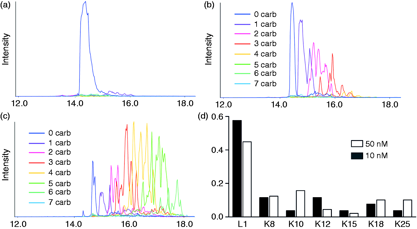
To address the functional significance of LL-37 carbamylation, we focused our attention on modifications identified after incubating the peptide in an environment mimicking that of the inflammatory milieu. To this end, we first identified the most common carbamylated variants of LL-37 under these conditions. KCNO-treated samples were digested with S. aureus protease V8 and analyzed by LC-MS/MS. By calculating the number of spectra matching the identified peptides (spectral count), we showed that LL-37 bearing a carbamylated N-terminus (α-carbamyl-Leu-1 LL-37; LL-37C1) was the predominant peptide form present after 1 h of exposure to 10 mM KCNO. The second most common single-modified peptide was LL-37 bearing homocitrulline residue at position 8 (LL-37C8) (Figure 1D).
LL-37 peptides synthesized and examined in this study.
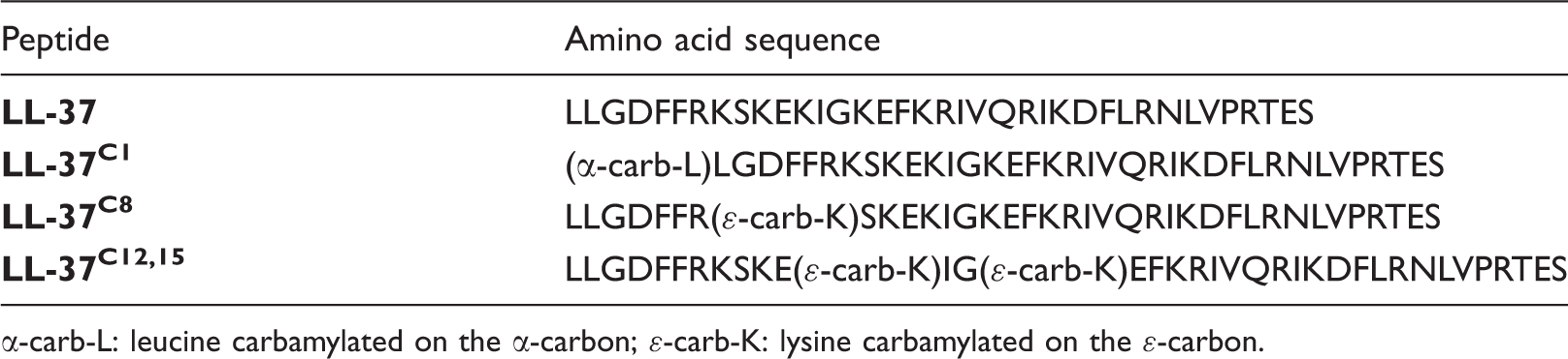
α-carb-L: leucine carbamylated on the α-carbon; ɛ-carb-K: lysine carbamylated on the ɛ-carbon.
In aqueous solution, LL-37 forms a random coil structure but adopts an α-helical conformation under physiological conditions; the α-helical structure is also adopted in TFE or lipid bilayer vesicles.
26
In the latter case, NMR studies show that LL-37 comprises three basic parts: an N-terminal α-helix, a C-terminal α-helix and a C-terminal tail.
27
The hydrophobic surface of LL-37 is bordered by positively charged residues, which enable interaction with negatively charged molecules or structures such as LPS,
28
genetic material, and bacterial cell wall components.13,14 We hypothesized that carbamylation-induced changes in charge and hydrophobicity would have a significant impact on the secondary structure of the peptide and, consequently, its biological activity (which is strictly related to the physiochemical properties of the peptide). We used CD spectroscopy to examine the impact of carbamylation on the capacity of LL-37 to form a helical structure. The far-UV CD spectrum of LL-37 in a physiological salt solution resembling blood plasma showed two minima (at 208 and 222 nm) (Figure 2A). These are characteristic for an α-helical secondary structure. Interestingly, neither single carbamylation of Lys-8 (LL-37C8) nor double carbamylation of Lys-12 and Lys-15 (LL-37C12,15) had any impact on the α-helical structure of LL-37. Conversely, carbamylation of the N-terminal amino group (LL-37C1) led to a significant increase in the propensity of the peptide to adopt an α-helical structure (Figure 2A).
CD spectra of native and carbamylated LL-37. The peptide concentration was 10 μM. The TFE buffer comprised 50% TFE in 10 mM sodium phosphate buffer. The plasma buffer contained 113 mM NaCl, 24 mM NaHCO3, 0.6 mM MgCl2, 1.3 mM CaCl2 and 3.9 mM KCl. (A) Spectra represent the mean of two (50% TFE buffer) or three (plasma buffer) independent experiments. (B) Differences in the predicted α-helical content of the peptide analogues in 50% TFE buffer at 190–260 nm. (C) Difference in the predicted α-helical content of the peptide analogues in plasma buffer at 190–260 nm. (B, C) Data are expressed as the mean ± SD. Statistical significance was evaluated by one-way ANOVA, followed by Tukey’s multiple comparison test. *P < 0.05, **P < 0.01, and ***P < 0.001.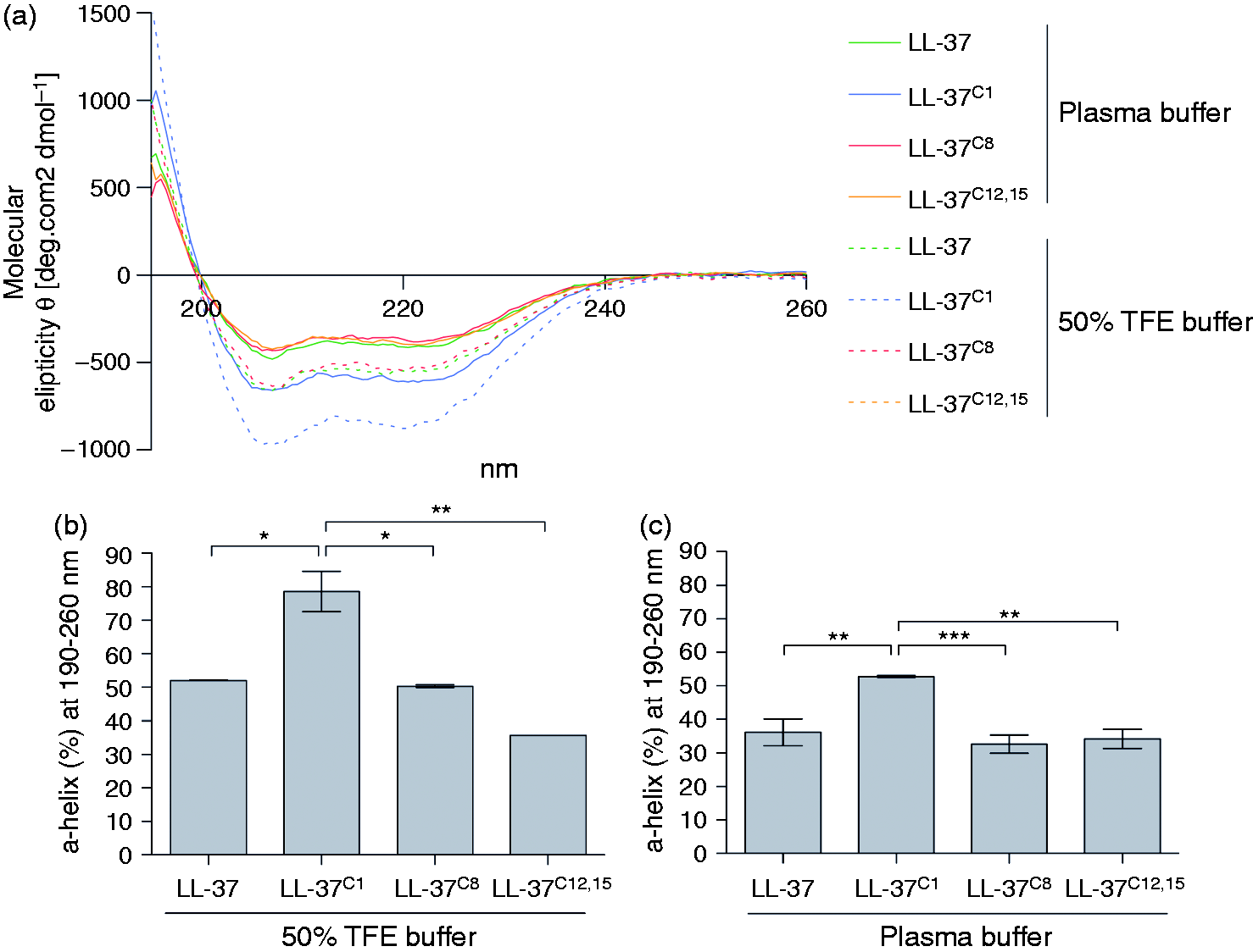
As expected, incubation with TFE increased the ellipticity and the helical content of the peptides beyond that observed in plasma buffer (Figure 2B). Under these conditions, CD analysis of the far-UV spectra predicted an α-helical content of approximately 80% for LL-37C1 but approximately 50% for the native peptide (Figure 2B). However, the difference between the peptides in TFE was similar to that observed in plasma buffer, that is, the α-helical composition of LL-37C1 was about 20-30% greater than that of the other peptides (Figure 2C).
Carbamylation abrogates the bactericidal capacity of LL-37
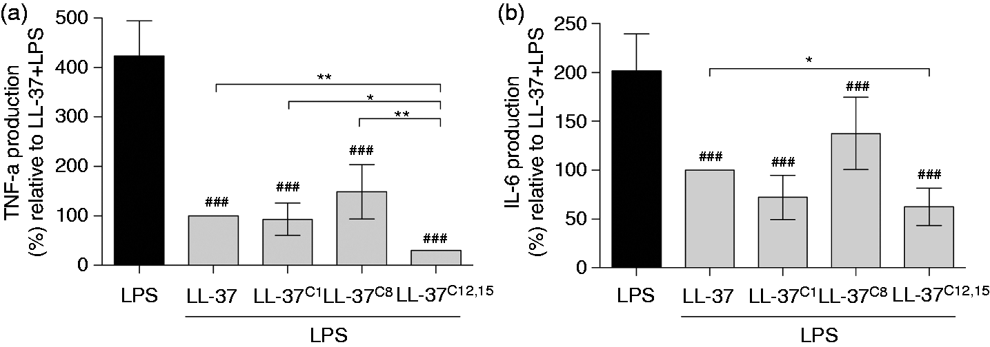
LL-37 interacts with bacteria via electrostatic and hydrophobic interactions resulting in membrane permeabilization and disruption. A broth microdilution assay revealed that the bactericidal activity of LL-37 was profoundly affected by carbamylation. At a peptide concentration of 1 µg/ml (i.e. 0.2 μM), LL-37C8 and LL-37C12,15 demonstrated impaired ability to inhibit bacterial growth of B. subtilis when compared with the native peptide (P < 0.05 and P < 0.001, respectively; Figure 3A). Additionally, the potential of LL-37C12,15 to inhibit the growth of E. coli (P < 0.001; Figure 3B) and S. aureus (P < 0.05; Figure 3C) was significantly decreased compared with native LL-37. Confirming previous observations, that reducing the number of residues on the N-terminus of LL-37 had only minor impact on its bactericidal properties, carbamylation of the N-terminal amino group did not affect the antimicrobial capacity of LL-37C1 compared with that of the native peptide (Figure 3A–C). In addition to its direct microbicidal role, LL-37 is a potent regulator of innate immunity, controlling the response to pathogen-associated molecular patterns, including LPS. Electrostatic interaction of the cationic LL-37 with the strongly anionic lipid A domain of LPS prevents it from binding to toll-like receptors (TLRs) expressed by monocytes and macrophages, a keystone event in inflammatory response. Therefore, we investigated whether carbamylation, and thus conversion of cationic Lys residues into neutral homocitrulline in LL-37 will affect the capacity of LPS to stimulate cells. hMDMs were exposed to LPS (100 ng/ml) in the presence of the either native LL-37 or carbamylated peptides. Thereafter, the cytokine profile of the supernatants was examined by multiplex analysis. LL-37C1 and LL-37C8 attenuated the pro-inflammatory activity of LPS and blocked secretion of TNF-α and IL-6 from the cells as effectively as the native peptide. Interestingly, we observed a statistically significant decrease of TNF-α and IL-6 in supernatants from LPS-stimulated macrophages in the presence of LL-37C12,15 compared with the native peptide (Figure 4A,B). This may indicate that, compared with the native peptide, LL-37C12,15 exhibits higher affinity for endotoxin thereof limiting its binding to the TLR4 receptors.
Carbamylation abrogates the antimicrobial activity of LL-37. (A–C) The number of colony-forming units formed after incubation with (A) B. subtilis, (B) E. coli and (C) S. aureus. All bacterial species were incubated with carbamylated or native LL-37. Bacterial growth in the presence of native LL-37 was set to 100%. Data are expressed as the mean ± SD. Statistical significance was evaluated by one-way ANOVA, followed by Tukey’s multiple comparisons post-test. *P < 0.05, **P < 0.01, and ***P < 0.001; and #P < 0.05, ##P < 0.01 and ###P < 0.001 compared with negative control (broth). LPS-induced cell activation remain supressed in the presence of carbamylated LL-37. Human monocyte-derived macrophages were incubated with 100 ng/ml LPS in the absence or presence of modified peptides. The levels of (A) TNF-α and (B) IL-6 in the supernatants were then measured. LL-37 + LPS was set at 100%. Data are expressed as the mean ± SD. Statistical significance was evaluated by one-way ANOVA, followed by Tukey’s multiple comparisons post-test. *P < 0.05 and **P < 0.01; ###P < 0.001, compared with LPS.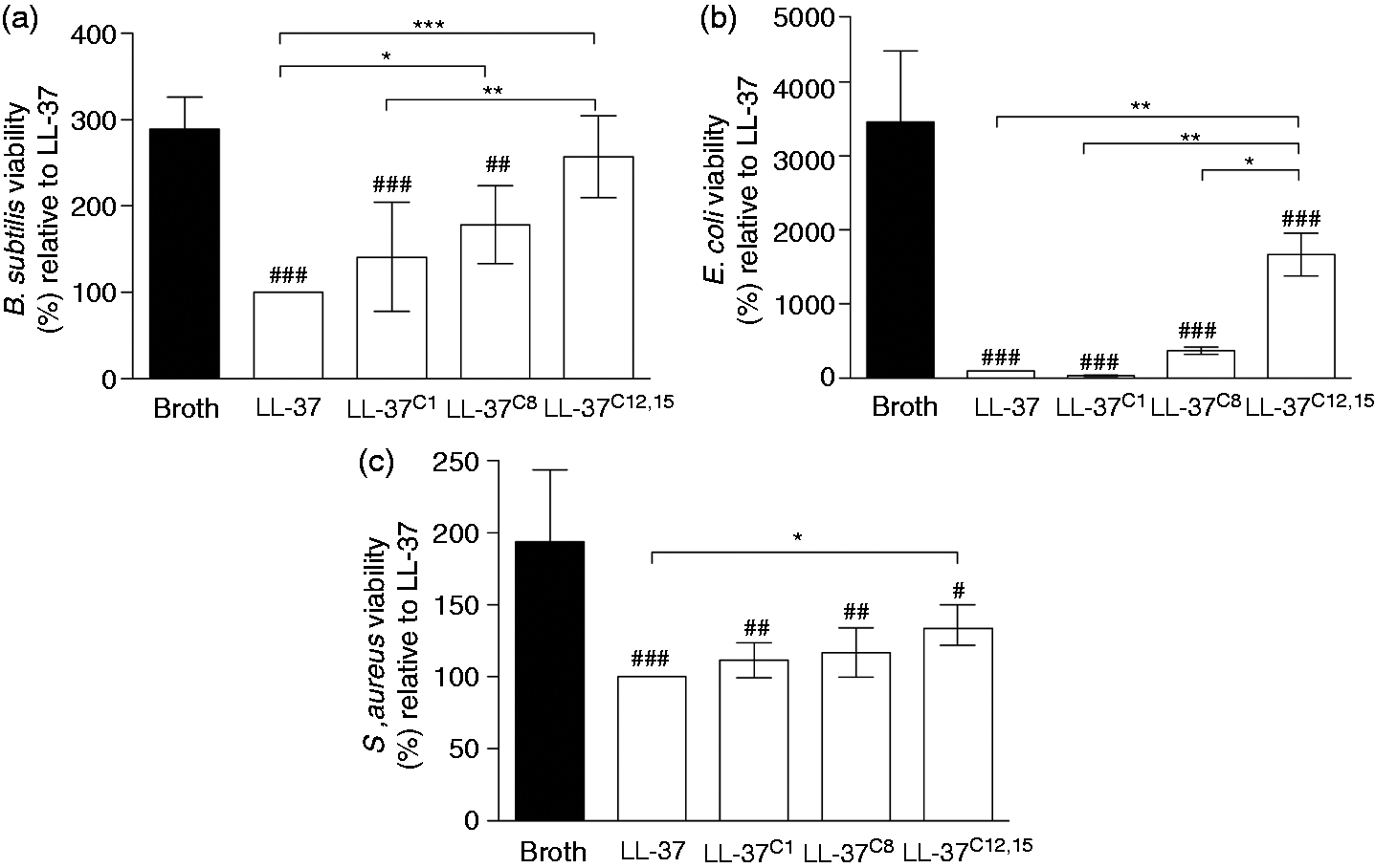
Carbamylation of LL-37 affects its affinity for ApoA1
In vivo, LL-37 circulates in a complex with ApoA1.
24
This carrier protein might function as a scavenger to inhibit the cytotoxic effects of the peptide secondary to its release at inflammatory sites. Therefore, we used surface plasmon resonance to examine whether carbamylation alters the affinity of LL-37 for ApoA1, which would effectively modulate the concentration of the bioavailable peptide within the inflammatory milieu. We found that Lys-12 and Lys-15 are critical for the interaction between the peptide and ApoA1. LL-37C12,15 showed a threefold lower affinity for ApoA1 than the native peptide. By contrast, both peptides carrying a single modification, LL-37C8 and LL-37C1, showed significantly stronger binding (1.5 and three times higher, respectively) than native LL-37. Taken together, these data indicate that any interference with the charged side chains of the amino acids within the LL-37 polypeptide chain has a significant impact on the peptide’s ability to associate with its carrier protein. This may either reduce or amplify the observed in vivo effects by modulating the amount of accessible LL-37 in the environment (Figure 5).
Carbamylation of LL-37 affects its affinity for ApoA1. Surface plasmon resonance curves showing the interaction between ApoA1 and carbamylated and native LL-37 at a concentration of 1 μM.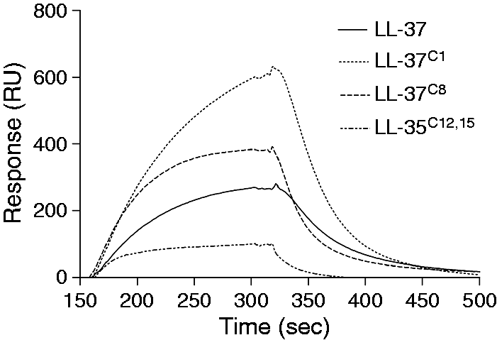
Carbamylation affects the chemotactic capacity of LL-37
The ability of neutrophils to efficiently reach sites of inflammation is pivotal for efficient elimination of pathogens and restriction of potential tissue damage. LL-37 is a strong chemoattractant for neutrophils, monocytes and T cells, via the FPRL1 receptor.
15
To investigate how carbamylation affects the chemotactic activity of LL-37, we used a ‘state-of-the-art’ approach that allowed us to observe cell migration in real time. The Insall chambers used in the study provide a gradient for the cells to migrate against, rather than simply exposing cells to the chemoattractant alone. This, in turn, allows observation of cell migration in more detail as information about speed (average speed of the cell over time in any direction), velocity (average speed of the cell in the direction of the gradient over time), and the directional accuracy of chemotaxis (expressed as the chemotactic index) can be obtained. We found that a peptide concentration of 20 μM was optimal in our assay (tested concentrations, 10–40 μM; data not shown). Both native LL-37 and the carbamylated forms induced neutrophil migration without any apparent toxicity (Figure 6A–C). However, neutrophils were significantly less responsive to the carbamylated versions of LL-37. Both the speed and velocity of migrating neutrophils were significantly lower when LL-37C1, LL-37C8 and LL-37C12,15 were used as chemoattractants. Interestingly, LL-37C1 was not only a significantly weaker chemoattractant than the other carbamylated peptides, but it also showed lower directional accuracy. Taken together, these observations suggest that, although all of the homocitrullinated peptides were able to trigger cell migration, carbamylation of the N-terminal amino group had a strong negative effect on directional accuracy.
Carbamylation reduces the chemotactic potential of LL-37. Analysis of chemotaxis at a peptide concentration of 20 μM. Neutrophils from four healthy volunteers were treated with either native or modified peptide. Values for speed, velocity, chemotactic index, and the resultant vector length were analyzed. The midline of each box represents the median value. Statistical significance was evaluated by one-way ANOVA, followed by Tukey’s multiple comparisons post-test: **P < 0.01, ***P < 0.001 and ****P < 0.0001; compared with the native peptide.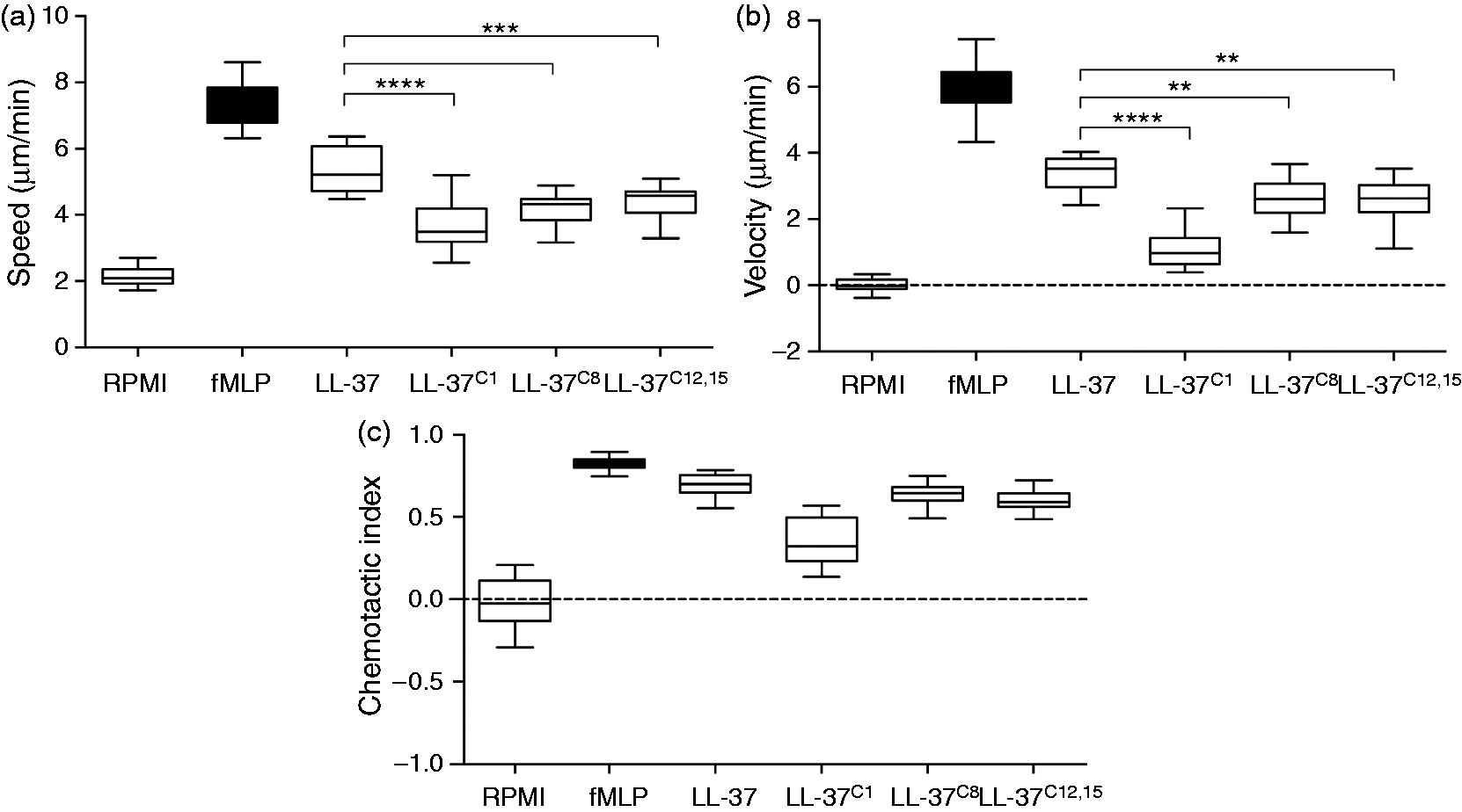
Carbamylation affects the cytotoxicity of LL-37
LL-37 can be cytotoxic to host cells. Therefore, we used a highly sensitive hemolytic assay to examine the impact of carbamylation on the cell-permeabilizing effects of LL-37. Human red blood cells (hRBC) were incubated with the peptide for 2 h at 37℃. The results showed that LL-37C1 exerted a very strong and concentration-dependent lytic effect. The peptide caused a significant increase in hemoglobin release when used at a concentration of 2 μM compared with PBS (P < 0.05; Figure 7). The half-maximal effective concentration for LL-37C1 was 17.9 μM. Conversely, LL-37C8 and LL-37C12,15 had significantly less capacity to induce membrane permeabilization (Figure 7); indeed, hRBC were resistant to permeabilization by LL-37C12,15 at concentrations of up to 20 μM. Given that the concentration of LL-37 can easily reach 20 μM at sites of inflammation (with levels up to 250 μM reported in psoriatic lesions),
29
permeabilization of membranes might facilitate the extracellular release of various potentially deleterious substances from lysed cells, which may then activate neutrophils and eventually lead to further increases in carbamylation.
Carbamylation significantly affects the hemolytic capacity of LL-37. Erythrocytes were incubated for 2 h with each LL-37 analogue at concentrations ranging from 1 to 20 μM. The hemolytic activity of the peptides was evaluated by recording the release of hemoglobin from human erythrocytes at 405 nm. Data are representative of four individual experiments and are expressed as the mean ± SD. Statistical significance was evaluated by one-way ANOVA, followed by Dunnett’s multiple comparisons post-test: **P < 0.01 and ***P < 0.001, compared with LL-37. #P < 0.05 in case of LL-37C1 compared with PBS.
Discussion
Host antimicrobial peptides, in particular LL-37, are potential novel therapeutics, mainly owing to their wound healing, antiseptic and antimicrobial properties.14,30,31 In the era of rapidly increasing drug resistance among pathogens, novel treatments based on LL-37-derived peptides are very much needed. In addition, the immunostimulatory and cytotoxic properties of LL-37 may be useful tools for cancer treatment, 32 or even as a vaginal contraceptive. 33 It is, however, important to keep in mind that the effector functions of LL-37 are most often executed at local sites of inflammation, and mainly in the context of neutrophil activation and subsequent release of MPO and H2O2. 34 Thus, LL-37 is mainly present in microenvironments that foster carbamylation. Therefore, it is important to examine the impact of carbamylation on the immunomodulatory and cytotoxic functions of LL-37 prior to the clinical administration of cathelicidin-derived peptides. Here, we explored the effects of carbamylation on the biological functions of LL-37.
We found that in the presence of cyanate, LL-37 undergoes rapid modification to generate a pool of peptides, each with a diverse carbamylation pattern. MS analyses revealed that the N-terminal amino group of leucine residues is highly reactive and is modified almost instantaneously in the presence of 10 mM KCNO. Thus, LL-37C1 is most likely the predominant form of carbamylated LL-37 in vivo. Even though carbamylation at this site did not affect the bactericidal properties of the peptide, LL-37C1 lost the ability to function as a chemoattractant for neutrophils. Concurrently, LL-37C1 gained significant RBC-lysing capacity; indeed, it was almost threefold more cytotoxic than native LL-37 in a hemolytic assay. Because tissue injury/necrosis results in increased receptor-dependent immune cell migration in response to released intracellular components,35,36 we cannot rule out the possibility that LL-37C1 indirectly triggers cell migration in vivo in response to damage to surrounding tissue. By stark contrast, although the chemoattractive capacity of LL-37C8 and LL-37C12,15 was diminished, neutrophils retained directional accuracy in response to the stimuli. This suggests that, despite the reduced affinity of Lys-8 and Lys-12/Lys-15 carbamylated peptides for FPRL-1, they were still able to bind the receptor. At the same time, LL-37C8 and LL-37C12,15 were significantly less toxic to hBRCs, with the latter exhibiting no hemolytic effects at a concentration of 20 μM. This observation confirms previous studies showing that truncation or blocking of the N-terminus reduces the cytotoxicity of LL-37 while at the same time leaving bactericidal activity unchanged.26,37 Indeed, only LL-37C12,15 showed lower bactericidal activity than the native peptide against B. subtilis, E. coli and S. aureus. In addition, Xhindoli et al. showed that oligomerization of LL-37 results in increased formation of α-helices, leading to an increased capacity to permeabilize erythrocytes and monocytes; this suggests that a parallel arrangement of the peptides favors aggregation via interaction between the N-termini. 26 In agreement with these results, we showed that LL-37C1 was about 50% more α-helical than the native peptide, both in a secondary structure-inducing environment (50% TFE buffer) and in a plasma-mimicking buffer. Since loss of the N-terminal charge due to carbamylation increases the hydrophobicity of this region, 38 it is possible that LL-37C1 is more prone to aggregation due to reduced electrostatic repulsion between the molecules. This would increase its propensity to form α-helices and explain the increased cytotoxicity. Intriguingly, the ability of LL-37C8 and LL-37C12,15 to lyse neutrophils and erythrocytes was different. Neutrophil membranes are zwitterionic and interact with LL-37 independently of the overall charge of the peptide.22,39 By contrast, the erythrocyte membrane contains sialic acid, which results in a negatively charged cell surface; therefore, it is more susceptible to lysis by cationic peptides. Thus, it is not surprising that the reduced cationicity conferred by carbamylation results in impaired hemolysis but does not affect the lysis of neutrophils. Interestingly, this difference was not evident in the case of LL-37C1. However, the uncompromising capacity of LL-37C1 to lyse both erythrocytes and neutrophils (data not shown) emphasizes the involvement of a hydrophobic N-terminus in this process.
The antimicrobial and cytotoxic activities of LL-37 are effectively inhibited in human plasma. This latency is due to the interaction between LL-37 and its carrier protein, apoA-1; 24 this interaction is dependent on the hydrophobicity of the N-terminus of LL-37, 40 as well as on its α-helical content. 41 In line with these results, we found that LL-37C1 showed significantly higher affinity for ApoA1 (300%) than the native peptide, indicating that the cytotoxic effects of LL-37C1 may be limited by an ApoA1-dependent protective mechanism. However, LL-37C1 may be detrimental to the host if generated at inflammatory foci that are poorly infiltrated by blood plasma. By contrast, LL-37C12,15 had lower affinity for ApoA1. Since ApoA1 has a net negative charge, 42 it is likely that the significant loss of electrostatic interactions upon carbamylation of two or more Lys residues impairs LL-37 binding to ApoA1.
Upon carbamylation, the bactericidal activity of LL-37 is compromised. Again, this is most likely related to the loss of two positive charges when Lys residues are converted to homocitrulline. It is likely that the reduced electrostatic attraction between LL-37C12,15 and bacterial membranes results in impaired antimicrobial activity against B. subtilis, E. coli and S. aureus. Interestingly, even though the bactericidal domain of LL-37 has been mapped to the C-terminal region (amino acids 17–29), 43 LL-37C8 showed impaired bactericidal activity against B. subtilis. Thus, the lysine at position 8 is important for efficient killing of this Gram-positive bacterium.
Interestingly, LL-37C12,15 was the most effective LPS-detoxifying agent among all tested forms of LL-37, that is, this carbamylated peptide efficiently attenuated the LPS-induced production of TNF-α, an important mediator of endotoxic shock. 44 In addition to directly binding LPS via electrostatic and hydrophobic interactions, LL-37 also interacts with the LPS receptor, CD14, to block LPS-induced macrophage activation. 45 We hypothesize that carbamylation of Lys-12 and Lys-15 increases the binding of LL-37 to this receptor. From a clinical point of view, it is tempting to speculate that LL-37C12,15 provides therapeutic protection against septic shock without being cytotoxic. Nevertheless, it must be kept in mind that further carbamylation may alter the biological activity of the peptide.
In summary, carbamylation has a profound impact on the bactericidal, cytotoxic and pro-inflammatory activity of LL-37, which may have detrimental consequences for the host. The pattern of carbamylation is dependent on the OCN− concentration and time of exposure, suggesting that the subsequent effects of this modification will be difficult to foresee and therefore control in vivo. Thus, we suggest that caution should be exercised when administering cathelicidin-derived peptides to patients with diseases manifested by inflammation, such as severe infections or sepsis.
Footnotes
Declaration of Conflicting Interests
The author(s) declared no potential conflicts of interest with respect to the research, authorship, and/or publication of this article.
Funding
The author(s) disclosed receipt of the following financial support for the research, authorship, and/or publication of this article: This work was funded by grants from the EC Marie Curie ITN (RAPID no. 290246) and (FP7-HEALTH-F3-2012-306029 ‘TRIGGER’). PM and AH were supported by a grant from the National Science Center (2014/14/E/NZ6/00162, Poland), and PM was supported by the Broegelmann Foundation. JP acknowledges support from the National Science Center (2012/04/A/NZ1/00051), National Institutes of Health, NIDCR (DE 09761 and DE 022597) and Polish Ministry of Science and Higher Education (2975/7.PR/13/2014/2).