
Abstract
Cancers of the kidney may be primary or secondary in origin. The vast majority of primary cancers are renal cell carcinomas, diagnosed incidentally on abdominal imaging. Secondary tumours found in the kidney are evaluated and managed depending on other clinical findings, including extent of the original cancer. The common original sites of spread include breast, lung, skin and lymph. Surgery remains the only curative therapy for renal cancer, and longer-term prognosis remains poor. Cancer referrals and diagnosis are an important aspect to increased successful curative treatment. The aim of this article is to evaluate renal cancer and give an overview of the important aspects from prevalence through diagnosis and staging, to treatment and management.
The GP curriculum and renal cancer
Know the epidemiology of older peoples’ problems presenting in primary care, such as dementia and cancers as well as their risk factors Recognise the common, early, ‘red flag’ symptoms and signs of malignancy (e.g. weight loss, dysphagia, melaena, diaphoresis, etc.), many of which may be non-specific if taken in isolation Know the local rapid access referral pathways and common treatment options, along with their complications and side effects Know that many cancers are more prevalent in the elderly population and may be insidious Understand how co-morbidity will influence the management of existing disease and delay the early recognition of adverse clinical patterns
Background and epidemiology
Renal cancer is the eighth-most-common cancer seen in the UK, with over 10 000 cases diagnosed every year (Cancer Research UK, 2014a). Renal cell carcinoma (RCC) accounts for over 90% of primary tumours involving the kidney (Znaor, Lortet-Tieulent, Laversanne, Jemel, & Bray, 2014) Transitional cell carcinoma is the next most common type, affecting around 7% of those diagnosed with renal cancer (Cancer Research UK, 2014a). There are other rarer forms of renal carcinoma including Wilms’ tumour that present in the young.
A significant number of patients will present with locally advanced or indeed metastatic disease. Boorjian and Uzzo (2009) demonstrated that although the incidence of renal cancer has been increasing, there has been a fall in stage migration, with small renal masses accounting for the largest proportion at diagnosis, most likely due to incidental findings from abdominal imaging. There is a male-to-female incidence ratio of 1.6:1 (Office for National Statistics (ONS), 2011). Evidence collated from all parts of the United Kingdom between 2009 and 2011, found that the incidence of renal cancer is strongly related to age. On average, 35% of renal cancers were diagnosed in those over 75 years old, and 75% of cases diagnosed over the age of 60 (Cancer Research UK, 2014a)
Prevalence refers to the number of people who have previously received a diagnosis of cancer and who are still alive at a given time point. Some patients will have been cured of their disease and others will not (Cancer Research UK, 2014a). In the UK in 2006, there were 26 503 people with a diagnosis of renal cancer still alive 10 years after diagnosis; 16 468 men and 10 035 women (Cancer Research UK, 2014a).
Risk factors
Established risk factors for renal cancer.
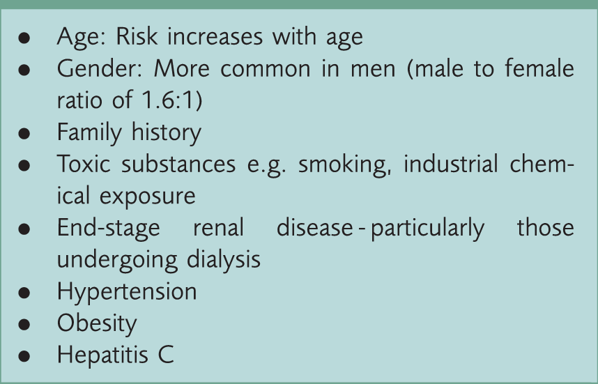
The incidence of RCC increases in older age groups. Regarding family history, the highest risk comes with an affected sibling, but risk is increased if any first-degree relative has a history of renal cancer. There is also an associated increase in the risk of RCC if the patient has family history of other cancers including prostate cancer and leukaemia.
The relative risk of RCC is 1.38 times higher in smokers compared with non-smokers. There also appears to be a strong dose-response relationship that has been recognised i.e. the more and longer someone smokes, the greater the risk.
Exposure to cadmium, asbestos and petroleum by-products all increase the risk of developing RCC. Incidence is particularly increased in smokers exposed to cadmium, occupationally or otherwise.
It is clear that hypertension is an independent risk factor for the development of renal cancer. The underlying pathological process remains largely uncertain. There is growing evidence that the treatment of hypertension with thiazide diuretics contributes to an increase in risk.
Increasing weight and body mass index (BMI) also has a positive correlation with risk of developing renal cancer. There is a step-wise increase in risk starting in those with a BMI of over 25 kg/m2 (overweight).
Pathology
Previously it was thought that kidney lesions of less than 3 cm in diameter signified benign adenomas, however, after further investigation, this assumption has been proven flawed (Schlomer, Figenshau, Yan, Venkatesh, & Bhayani, 2006). As a consequence, the diagnosis of a malignant or benign lesion based on size alone is no longer made and all growths without clear origin or type should be resected or biopsied for a histological diagnosis. The only exception to this rule is if a metastatic deposit can be identified and biopsied.
The aim of RCC classification is to accurately establish the morphology, cell of origin, growth pattern, histochemical and molecular basis of different types of adenocarcinoma. There are two distinct groups of RCC, of which one can then be further divided into subtypes. The World Health Organisation recognises the three major histological subgroups as clear cell, papillary and chromophobe. Figure 1 illustrates the various types of renal cancer and their derivatives.
Renal cancer types and subtypes.
Clear cell carcinomas are by far the most prevalent type of RCC. They are normally found to arise from the proximal tubule and classically have a deletion of chromosome 3p. They are usually solid masses rather than cystic in nature, and can occur in sporadic disease. Poorer prognosis is seen with a high nuclear grade of clear cell carcinoma or the presence of sarcomatoid pattern in early stage disease. A better prognosis is seen with multilocular cystic clear cell cancers compared with other clear cell types; however, this is a rarer form.
Papillary renal cell cancers typically present as small, early stage lesions. They may be bilateral or multifocal. Similar to clear cell cancers, they originate in the proximal tubule but are distinct in their morphology and genetics. Papillary cancers can be further subdivided into types 1 and 2. This is based upon histologic standards and gene expression. Yang et al. (2005) showed a significant difference in prognosis between the two, with type 1 tending to be a low-grade tumour, giving a more favourable outcome.
Chromophobe carcinomas are constructed from sheets of cells. They do not contain the abundance of lipids and glycogen that is usually seen with RCCs of the kidney. They initiate from the intercalated cells of the collecting system. Generally, these carcinomas present at an earlier stage and therefore provide a better prognosis.
Oncocytomas are uncommon and consist of well-differentiated neoplastic cells. They are difficult to histologically differentiate from RCC. Oncocytomas rarely metastasise and behave in a very similar fashion to benign tumours, they are usually well encapsulated even when large. There is also an association with co-existing RCC in up to 30% of patients with oncocytomas. This is important when constructing management plans in this population of patients, who should therefore be closely monitored for any signs of tumour growth.
Collecting duct tumours are rare and seen mainly in younger patients. Initial presentation is typically with gross haematuria and likely advanced disease. There is no current strong evidence for genetic involvement. They resemble transitional cancers more closely than RCC.
Genetic syndromes
There are several genetic syndromes that have been associated with renal cancer. Recognition of these syndromes can be an important step in diagnosis. Family history alongside clinical findings remains an important diagnostic tool for clinicians. Identification of genetic risk factors may lead to a strategy to avoid morbidity.
Von Hippel–Lindau (VHL) disease is specifically associated with clear cell RCC. The VHL gene is found on chromosome 3 and VHL gene alteration can be found in up to 91% of patients with clear cell RCC (Nickerson et al., 2008). Referral to specialist centres should be made for patients suspected of VHL disease for definitive diagnosis and evaluation. Genetic counselling can be undertaken at this stage as VHL is inherited in an autosomal-dominant manner.
Birt–Hogg–Dubé (BHD) syndrome is a result of mutations in folliculin (FCLN) located on chromosome 17. Renal tumour histology in patients affected by BHD varies, however, a mixed pattern of chromophobe and oncocytic is most characteristic. Houweling et al. (2011) suggested that up to 30% of patients with BHD will receive a diagnosis of renal cancer. Typically, renal cancers associated with BHD will be bilateral or multifocal and slow-growing. Tumours are usually observed if sized less than 3 cm diameter, although all tumours should be removed when surgery is advised.
Hereditary papillary renal carcinoma (HPRC) is an autosomal-dominant condition that leads to development of type 1 papillary RCC. The HPRC gene is found on chromosome 7 and manifests most commonly as bilateral multifocal tumours. The tumours are often slow-growing. Nephron-sparing surgeries are preferred in order to maintain renal function.
Clinical presentation and GP referrals
Most renal cancers are asymptomatic and non-palpable until the late stages. The majority are found incidentally on imaging indicated for other clinical reasons. There is a classic triad of symptoms to be wary of in all age groups, consisting of gross haematuria, flank pain and a palpable abdominal mass. However, this is found in the minority of patients presenting with an underlying diagnosis of renal cancer. Some patients present with symptoms of metastatic disease, for example, bone pain. Paraneoplastic syndromes including hypercalcaemia are common and seen in up to one-third of patients. Haematuria is the most common symptom indicative of renal cancer.
Two-week wait referral criteria for potential renal cancer.

In addition, patients with loin pain or a suspected loin mass should be referred for a urinary tract ultrasound scan (USS). This will help to confirm or exclude a renal cancer. If a patient complains of persistent loin pain but the USS suggests a benign cystic pathology, or is normal, routine referral to a general urology clinic for further investigation is appropriate.
Investigation and diagnosis
Clinical examination has a limited role in the diagnosis of renal cancer; however, there are several clinical signs to be wary of when considering renal cancer, which should then lead to radiological imaging. Signs that may be present when considering a diagnosis of renal cancer include:
Palpable abdominal mass Cervical lymphadenopathy Non-reducing varicocele Bilateral pedal oedema (suggesting venous involvement) Bony tenderness
Blood tests to consider when assessing a patient who may have a diagnosis of renal cancer.

Demonstrating the common investigations and the indication.

Renal biopsies of tumours are on the rise (Ljungberg et al., 2010) and are utilised in diagnosis, follow-up surveillance and for consideration of ablative therapies. Biopsy provides high sensitivity as well as specificity with regards to malignancy.
Staging and classification
TNM staging for renal cancer.

Used with the permission of the American Joint Committee on Cancer (AJCC), Chicago, Illinois. The original source for this material is the AJCC Cancer Staging Manual, Seventh Edition (2010) published by Springer Science and Business Media LLC, www.springer.com.
Management
All renal cancers should be discussed in a local multi-disciplinary team (MDT) and the decision regarding treatment regimen communicated to the GP. GPs should be kept informed of the patient’s progress throughout treatment. The ultimate responsibility for a patient’s care during the decision-making process is that of the local MDT.
Localised disease
Localised disease includes T1-T2 disease and it is becoming a more common presentation. The rate of intervention is also increasing. (Chen & Uzzo, 2009). Treatment options have expanded, although surgical intervention remains the treatment of choice and the only therapeutic curative approach for RCC. Open radical nephrectomy (ORN) is arguably no longer the gold standard treatment, as alternatives can now be offered which lessen or avoid the recognised negative consequences including renal failure.
For tumours of less than 7 cm in diameter (T1), nephron-sparing surgery (NSS) should be offered. This encompasses resection of the tumour and uninvolved kidney remains. The oncological outcomes are similar to radical surgery and this method has shown a preservation in renal function over the longer term, leading to stronger overall survival rates (Chen & Uzzo, 2009). NSS is not suitable if the tumour is locally advanced, in an unfavourable location, or the patient has a significant deterioration in their health. In these situations ORN remains the treatment of choice. Absolute indications for NSS include those with one kidney, or bilateral RCC. Relative indications include those with baseline renal insufficiency or coexisting medical co-morbidity, for example, diabetes.
Open partial nephrectomy has been the approach utilised in the majority of centres. Technically, it is a more complex operation than open/laparoscopic radical nephrectomy and has several recognised complications, including fistula formation and ureteral obstruction. In more experienced centres, the complication rate is reduced to that similar to ORN. NSS addresses the risk of overtreatment in lesions that may not be malignant. As well as the previously discussed benefits of preserved renal function, there is evidence to suggest an increase in patient satisfaction and quality of life.
Originally, when ORN was described and established, resection of the ipsilateral adrenal gland and dissection of regional lymph nodes were key elements. Although adrenalectomy and lymphadenectomy may have a role in staging in more advanced disease, these components have no strong advantage over nephrectomy alone in the absence of abnormal imaging or intra-operative findings suggestive of disease. They are therefore reserved for selected cases only.
Laparoscopic radical nephrectomy (LRN) is a minimally invasive technique that was initially described in the early-1990s. Efficacy has been proven similar to that of the open method. It shows better perioperative parameters, including less blood loss and lower morbidity. For almost all organ-confined RCC, LRN can be considered as a possible approach.
Suggested alternatives to surgical therapies have been advanced due to the technical difficulty and morbidity of surgical therapies. Research has developed ideas that can use NSS principles without the morbidity and risk and can be readily performed. Tissue ablative therapies including radiofrequency and cryoablation, which create a hot or cold temperature that is deadly to the target. These methods are similar to those that are used to ablate liver, prostate and lung lesions. A pre-treatment biopsy should be carried out for histological examination. If the tumour can be accessed, usually percutaneously via imaging or abdominal ports laparoscopically, probes are used to deliver the treatment and sedation is all that is needed. The goal is eliminating the RCC in situ, and as field of ablation is small, the majority of the kidney is not affected.
Unfortunately, there are limitations and disadvantages for these procedures. The lesions must be small, usually less than 3 cm, as larger tumours would require multiple probes and increase the risk of inadequate treatment. These approaches do not give a comprehensive staging and must rely on percutaneous biopsy. Long-term outcomes remain unproven and as a result, the current treatment is reserved for the elderly or sick where treatment is deemed necessary, but surgical intervention is not possible.
Advanced and metastatic disease
There are currently no treatments that have been shown to reliably cure metastatic or advanced disease. Once diagnosis has been made, the typical aim of treatment is for supportive palliation to relieve physical symptoms and extend a good quality of life.
Surgical options for people with advanced disease are summarised in Fig. 2. ORN is strongly indicated even for locally advanced disease, where the objective of surgery should still be total excision. In addition, selected patients with limited metastatic disease can be considered for metastasectomy.
Likely treatment routes by stage.
Metastatic renal cancer is generally resistant to chemotherapy, radiotherapy and hormonal therapy. The first-line treatments approved by the National Institute for Health and Care Excellence (NICE) include the tyrosine kinase inhibitors, sunitinib and pazopanib. These drugs prevent the proliferation of tumour cells as well as angiogenesis through inhibition of vascular endothelial growth factor and platelet-derived growth factor, resulting in a significant increase in disease stability (NICE, 2009).
Sunitinib is the most commonly used of these agents. It is taken every day for 4 weeks, with a break in the subsequent 2 weeks, and then the cycle is repeated. Side effects include gastrointestinal events and deep venous thromboembolism.
Prognosis and surveillance
Surveillance after treatment for renal cancer should be based upon the patient, his or her treatment to that point and risk factors for recurrence. Surveillance in patients with a low risk of recurrence does not usually require frequent imaging. Intermediate and high-risk patients typically require regular/routine CT or MRI imaging. Prognosis after a diagnosis of renal cancer will also depend upon the staging and intervention possible. Although there are conflicting published results, overall 5-year survival is thought to be around 55–60% (Cancer Research UK, 2014b).
Key points
Renal cancer is the eighth-most-common cancer in the UK There are many different subtypes of renal cancer, but renal cell carcinoma is the most common Several genetic syndromes are strongly associated with RCC and patients with these syndromes should be referred for renal surveillance Most renal cancers are now picked up incidentally when abdominal imaging is undertaken for other reasons The earlier the disease is diagnosed, the more likely curative treatment can be offered Surgery is the only curative therapy for renal cancer