



Abstract
Trigger finger is a common cause of hand pain. It is caused by stenosis at the level of the first annular (A1) pulley, which interrupts the smooth gliding of the underlying flexor tendons at the level of the metacarpophalangeal joint. Patients present with locking of the finger in flexion and clicking or snapping. It is important that GPs have an understanding of this condition, in order to help their patients deal with its sequelae. In this article the pathophysiology, clinical assessment and management of patients with suspected trigger finger are discussed.
The GP curriculum and trigger finger
Diagnose common, regional soft-tissue problems that can be managed in primary care (e.g. tennis elbow, trigger finger) Assess the importance and meaning of the following presenting features:
■ Pain: Nature, location, severity, history of trauma ■ Variation of symptoms over time ■ Symptoms which help distinguish inflammatory from non-inflammatory conditions ■ Loss of function: Weakness, restricted movement, deformity and disability, ability to perform usual work or occupation Understand that reducing pain and disability rather than achieving a complete cure could be the goal of treatment
Relevant anatomy
In order to understand trigger finger, it is important to know the anatomy of the flexor tendons and the pulley system of the fingers. Flexor digitorum superficialis (FDS) and flexor digitorum profundus (FDP) are responsible for flexion of the proximal interphalangeal and distal interphalangeal joints in the finger, respectively. They glide within a fibro-osseous tunnel, the flexor sheath. The floor of this tunnel consists of the phalanges of the fingers. The roof comprises a series of soft-tissue fibrous pulleys. There are five annular pulleys and three cruciform pulleys, which facilitate smooth gliding of the FDP and FDS tendons allowing finger flexion without bowstringing of the tendons. The pulley system is shown in Fig. 1. The A1 pulley arises from the palmar plate and proximal portion of the proximal phalanx, and it overlies the membranous sheath at the level of the MCP joint. It is the A1 pulley that is surgically released during treatment of trigger finger.
Anatomy of the flexor pulley system in the finger.
Epidemiology
Trigger finger is a common cause of hand pain in adults; it is more common among women and men in the fifth and sixth decades of life (Moore, 2000). The condition can also be seen in children, where it commonly affects the thumb (Saldana, 2001).It affects approximately 2% of the population. Trigger finger can affect multiple digits, and can be bilateral. Trigger finger is more common among patients with other conditions, such as diabetes and rheumatoid arthritis.
The aetiology in the majority of patients with trigger finger is idiopathic (McAuliffe, 2010). There have been some reports that associate trigger finger with repetitive activities, however, the exact causal link has not been identified (Moore, 2000). Patients usually report developing symptoms spontaneously without any prior history of trauma. Histological analysis of patients suffering from trigger finger shows fibrocartilaginous metaplasia of the ligamentous layer of the A1 pulley (Sbernardori & Bandiera, 2007). This results in a reduction in the cross-sectional area underneath the A1 pulley, thus causing mechanical impingement that leads to entrapment of the underlying tendon and results in functional impairment in finger flexion and extension.
Presentation and examination findings
Patients with trigger finger initially report an atraumatic catching, locking of the affected digit in flexion and painless snapping. This can often progress to an inability to actively extend the affected digit, and this may require passive extension in severe cases, which can be painful. Patients describe a pain that is localised over the palmar aspect of the metacarpophalangeal (MCP) joint and radiates into the palm or the distal finger (Blazar & Aggarwal, 2015). Patients can wake with a finger locked in a partially flexed position that gradually can be extended as the day progresses. It is important to manage patients with trigger finger early in their presentation, as the reluctance to fully flex and extend the digit, due to pain, can lead to the development of a secondary contracture of the proximal interphalangeal (PIP) joint. Patients with trigger finger can present with multiple affected digits.
Diagnosing trigger finger is based on a history of locking, clicking or snapping during movement of the affected digit. The patient’s hands should be placed on the examination table with the palm facing up, then ask the patient to fully flex and extend their fingers and observe for any evidence of active triggering. Triggering of the affected digit may not occur with every repetition. In addition, palpate over the PIP joint, MCP joint and in the palm while the patient is actively flexing and extending their finger, noting the presence or loss of smooth tendon motion or a clicking sensation. The patient may also complain of pain and tenderness at the level of the MCP joint, and a tender nodule indicating thickening of the tendon may be palpable in the palm.
Performing X-rays of the affected digit is seldom used and not necessary in diagnosing patients with suspected trigger finger, however, it can be useful in diagnosing underlying causes of pain in the hand, such as rheumatoid arthritis and osteoarthritis (Katzman et al., 1999).
Differential diagnosis
Differential diagnosis of trigger finger.
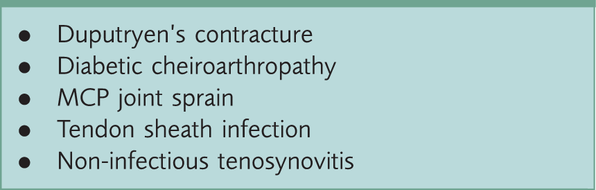
Dupuytren’s contracture is a painless condition, in which there is a loss of full extension of the affected digit at the level of the MCP joint. In Dupytren’s contracture, nodular lesions are palpable in the palmar fascia, and these can progress to form a fibrous cord that extends from the palm to the digits. Dupytren’s contracture can be distinguished from trigger finger, as the loss of extension is fixed and chronic, whereas in trigger finger it is dynamic and episodic.
Diabetic cheiroarthropathy is typically painless; it results in a limited ability to fully extend the MCP joints and/or interphalangeal joints of all fingers. Diabetic cheiroarthropathy may progress to fixed flexion contractures of the finger joints and may present as the ‘Steeple’ or Prayer sign, in which patients are unable to completely close gaps between opposed palms and fingers when pressing their hands together. A tight, waxy skin surface over the dorsum of the hand is usually observed in diabetic cheiroarthropathy; this is not observed in trigger finger. Patients with diabetic cheiroarthropathy are more likely to develop trigger finger (Kameyama, Meguro, Funae, Atsumi, & Ikegami, 2009).
MCP joint sprain presents with tenderness on either side of the MCP joint; it is associated with loss of full flexion of the digit in the absence of triggering with a history of recent trauma.
Flexor sheath infections present with severe pain and tenderness along the course of the flexor sheath in the hand. There is usually a history of preceding puncture or bite wound involving the finger or hand. Flexor sheath infections can be distinguished from trigger finger, as it often leads to more severe pain, tenderness, erythema and swelling along the volar aspect of the affected digit.
Non-infectious tenosynovitis presents with pain, tenderness and swelling along the flexor tendon in the hand. This may be associated with an underlying inflammatory arthritis, such as rheumatoid arthritis or reactive arthritis. Tenosynovitis improves with treatment of the underlying arthritis with non-steroidal anti-inflammatory drugs (NSAIDs), disease-modifying anti-rheumatic drugs, and systemic glucocorticoids.
Management of trigger finger
The aim of managing patients with trigger finger is not only to relieve pain, but also permit smoother movement of the finger, thus improving hand function. Initially patients should be managed with a conservative approach by activity modification, splinting, using NSAIDs and glucocorticoid injections. Surgical treatment is reserved for patients who have persistent symptoms despite conservative management.
Conservative treatment
Patients presenting with acute symptoms should be counselled on activity modification and use splints. Activity modification involves continuing with their normal activities while avoiding movements such as pinching or grasping with the affected finger that can aggravate their symptoms. Observational studies have demonstrated that immobilisation of the MCP joint with a splint can relieve pain and reduce triggering (Makkouk, Oetgen, Swigart, & Dodds, 2008). A single-group pre-post study with 28 participants performed by Colbourn, Heath, Manary, and Pacifico (2008) demonstrated an improvement in triggering function in 93% of patients after wearing a splint for 6 to 10 weeks, with complete symptom resolution being observed in 54% of patients. The splint should be worn with the MCP joint in slight flexion and should be used based on the pattern of triggering and patient preferences. The total duration of splinting is commonly between 3 and 6 weeks (Rodgers, McCarthy, & Tiedeman, 1998). The concurrent use of NSAIDs can also aid in pain relief.
Patients with persistent symptoms after 4 to 6 weeks despite the use of splinting and NSAIDs can be given glucocorticoid injections (Peters-Veluthamaningal, Winters, Groenier, & Jong, 2008). Intermediate-acting glucocorticoid injections, such as triamcinolone can be used (Anderson & Kaye, 1991).
Glucocorticoid injections can be offered sooner in patients presenting with severe locking and incomplete finger flexion or extension (Peters-Veluthamaningal, van der Windt, Winters, & Meyboom-de Jong, 2009). The majority of patients experience significant improvement in symptoms after a single injection: with symptom relief, which continues beyond 12 months, occurring in approximately half of patients in the study performed by Rozental, Zurakowski, and Blazar (2008) and lasting up to 10 years in the study carried out by Wojahn, Foeger, Gelberman, and Calfee (2014). The total number of injection treatments does not tend to exceed three in clinical practice. Patients should be counselled on the side effects of glucocorticoid injections prior to treatment, including short-term pain and tenderness that is self-limiting, infection, bleeding, fat atrophy and hypopigmentation. Patients should avoid strenuous use of the affected digit for several days after the injection, to reduce the risk of tendon rupture.
Surgical management
Surgery is recommended for patients when symptoms persist despite using conservative measures and glucocorticoid injections (Ryzewicz & Wolf, 2006; Wang, Zhao, & Liang, 2013). Patients with persistent symptoms should be referred to a tertiary hand specialty centre for consideration of surgery. Surgery involves the release of the A1 pulley and this can be carried out using an ultrasound-guided percutaneous or open surgical release (Bain, Wallwork, & Percutaneous, 1999; Turowski, Zdankiewicz, & Thomson, 1997). The recurrence rate of symptoms is approximately 3%, with no observed comparable difference in the failure rate and complications between the percutaneous and open surgical technique (Guler, Kose, Ercan, Turan, & Canbora, 2013). Complications of surgery include infection, digital nerve injury, flexor tendon bowstringing and tendon scarring. A1 pulley release should be avoided in patients with rheumatoid arthritis, as it will exacerbate ulnar drift of their digits. The pathology in patients with rheumatoid arthritis and triggering relates to synovitis, and therefore, synovectomy and control of inflammation is more appropriate.
Key points
Trigger finger is a common cause of hand pain in adults, it presents with an initial painless snapping, catching or locking of the affected digits in flexion Trigger finger is caused by the flexor tendon catching when it attempts to glide through a stenotic flexor sheath under the A1 pulley at the level of the MCP joints The diagnosis of trigger finger is made on the history of locking or clicking and the examination findings of observed locking when the patient opens and closes their hand Differential diagnoses of trigger finger include Duputryen’s contracture, diabetic cheiroarthropathy, MCP joint sprain, flexor sheath infection, calcific periarthritis or non-infectious tenosynovitis Conservative treatment should be used initially in patients with trigger finger, these include activity modification, splinting, NSAIDs and the use of glucocorticoid injections Surgical release of the A1 pulley should be considered in patients with persistent pain and locking despite the use of conservative therapy