



Abstract
Epiglottitis is the inflammation of the epiglottis and adjacent supraglottic structures. Early recognition of specific symptoms, as well as the systemic signs of sepsis, can help prevent serious complications. A good understanding of the steps in the management of the condition aids early intervention and treatment. The priority of treatment is to secure the airway. Serious complications can occur in addition to the severity of the disease. Symptoms can be more prolonged depending on the aetiology. If the epiglottis is still swollen after 72 hours of treatment, then further investigation should be carried out.
Clinical case scenario
Harry, a 5-year-old boy, presents with his parents to an urgent appointment with increasing difficulty breathing. On reviewing his record, he has no medical conditions and vaccination records are up to date.
His parents are very worried about him. He is sitting forward and is drooling saliva. On arrival to the surgery, the nurse recorded a systolic blood pressure of 110 mmHg, a heart rate of 120 beats per minute, oxygen saturation of 92%, respiratory rate of 30 breaths per minute and a temperature of 38.1℃.
Anatomy
The epiglottis is an elastic fibro-cartilaginous structure that lies behind the tongue and the body of the hyoid bone. It has several attachments, inferiorly to the thyroid cartilage below the thyroid notch, in the midline to the thyroepiglottic ligament and the hyoid bone, anteriorly to the hyoepiglottic ligament (Gleeson and Scott-Brown, 2008). The vallecular space is formed by the back wall of the epiglottis and the base of the tongue. The epiglottic cartilage is covered anteriorly by a stratified squamous epithelial layer and posteriorly on the superior third by respiratory epithelium. On the lingual surface, the epithelium and lamina propria are loosely attached, thus creating a potential space for oedema and fluid to collect (Woods, 2019) (Fig. 1).
Anatomy of the larynx.
Pathogenesis
Epiglottitis is inflammation of the squamous layer of the epiglottis, and is a cellulitis. It can spread to involve the aryepiglottic folds, and other adjacent tissues, the lingual tonsils and the false vocal cords, thus called supraglottitis (Gleeson and Scott-Brown, 2008). It is usually caused by a bacteraemia or direct access of pathogenic organisms to the epithelial layer (Woods, 2019). An upper respiratory tract infection is usually the primary source of pathogens, however, microscopic trauma to the epithelial surface, whether due to a previous or ongoing viral infection or from deglutition, may be one of the predisposing factors. The epiglottic infection causes inflammation and swelling of the potential space in the lingual surface of the epiglottis.
Among the upper respiratory tract infections, Haemophilus Influenzae Type b (Hib) is the most prevalent cause of epiglottitis (Sack and Brock, 2002). Multiple pathogens have been identified as the cause and these include: Group A Streptococci, Streptococcus pneumoniae, Staphylococcus aureus, and Klebsiella pneumoniae. Neisseria meningitidis can cause a life-threatening supraglottitis (Gleeson and Scott-Brown, 2008; Sykes, 2018; Woods, 2019). However, other less common pathogens can still cause an infection, which is usually seen in immunocompromised patients (Gleeson and Scott-Brown, 2008). Non-infectious causes of epiglottitis have been described: these include thermal injury, foreign body ingestion and caustic ingestion (Harjacek et al., 1992; Lai et al., 2000; Kavanagh and Batti, 2008).
Epidemiology
After the introduction of the Hib conjugate vaccine, the epidemiology of epiglottitis has changed with a dramatically decreased incidence of epiglottitis in children. A study reported that following the Hib vaccination the incidence of epiglottitis decreased from 10.9 per 10 000 admissions before 1990 to 1.8 per 10 000 admissions from 1990 through 1992 (Gorelick and Baker, 1994). In the United States with adult and paediatric population estimates of 217 400 000 and 82 000 000, respectively, in 2006, there were about 1.6 cases of epiglottitis per 100 000 adults and 0.5 cases per 100 000 children (US Census, 2007).
Clinical features
As epiglottitis is a medical emergency, it is important to be able to recognise and know how to initially manage these patients, with the goal of minimising morbidity and mortality. It is essential to have a full set of observations to get a better idea of the status of the patient. Some patients present with a septic picture (fever, hypotension, desaturation, and tachycardia).
On presentation, it is imperative to establish a definitive diagnosis of epiglottitis before complications occur, such as airway obstruction. Signs and symptoms of epiglottitis differ with age, severity and aetiology. For children, the classic presentation is in a ‘tripod’ (trunk leaning forward, neck hyperextended, and chin thrust forward) or ‘sniffing’ position, which are both positions of respiratory distress (Woods, 2019). This position is meant to increase the diameter of the obstructed airway. One of the most common presentations is the drooling of saliva with the absence of a cough. Some patients present with a severe sore throat, but normal oropharyngeal examination. In children the progression of epiglottitis happens rapidly, within hours and abruptly, the common symptoms are dysphagia, drooling and distress (the three Ds). Patients can also present with high-grade fever, sore throat and odynophagia. In adults, presentation is usually more insidious, with the most common presenting features being sore throat, high-grade fever, muffled voice, facial drooling, stridor and respiratory distress (with signs including laboured and rapid breathing, nasal flaring, intercostal recession and diaphoresis).
Examination
In children with suspected epiglottitis, examination should be conducted in a calm setting to avoid distressing the child, as this can precipitate serious airway compromise. Some features associated with a serious condition should be identified as soon as possible and will require emergency hospital transfer. These features include: Stridor, altered level of consciousness, significant respiratory effort, elevated respiratory rate, oxygen saturation below 92%, cyanosis, tachycardia, hypotension and peak expiratory flow rate below 50% of predicted level (National Institute for Health and Clinical Excellence (NICE), 2015).
Palpation
Examination includes thorough inspection of the neck with palpation of the supraclavicular, cervical, submandibular, submental, occipital and parotid nodes. The presence of any lumps or swelling should be noted. Findings may help identify the cause of the infection or the presence of other underlying pathology.
Oropharynx
It is important to note the dentition of the patient when examining the oropharynx, as this can be a source of infection. It is also important to look for Stensen’s duct (parotid gland duct), Wharton’s duct (submandibular gland duct), the tonsils and the mucosa of the oropharynx. In a patient with epiglottitis, the only positive finding is usually erythema and inflammation of the oropharyngeal mucosa. When examining the oropharynx, especially in children with suspected croup or epiglottitis, the use of a tongue depressor is hazardous and should be avoided, as it can exacerbate symptoms by either the manual compression of the airway or by increasing patient anxiety.
Flexible naso-endoscopy
A definitive diagnosis can be made through direct visualisation using flexible naso-endoscopy (FNE) to assess the epiglottis and adjacent structures (the vallecula, the pyriform fossa, the arytenoids, the vocal cords and the patency of the airway). On FNE examination, pooled secretions may be noted around the pyriform fossa as well as an inflamed, oedematous epiglottis extending to the supraglottic structures, the aryepiglottic folds and the arytenoid cartilages. Where FNE is not possible, for example in children, a lateral neck X-ray can be helpful in diagnosis (Gleeson and Scott-Brown , 2008; Woods, 2019).
Investigations
If the patient presents with respiratory distress, invasive, diagnostic procedures should be delayed and airway management started (Woods, 2019). The features of epiglottitis visible on an X-ray scan include thickening of the epiglottis (the thumb sign) and absence of clear, defined vallecula (the vallecula sign) (Fig. 2) (Sack and Brock, 2002: 81-86).
Neck X-ray showing swollen epiglottis (E) and obliteration of the vallecular.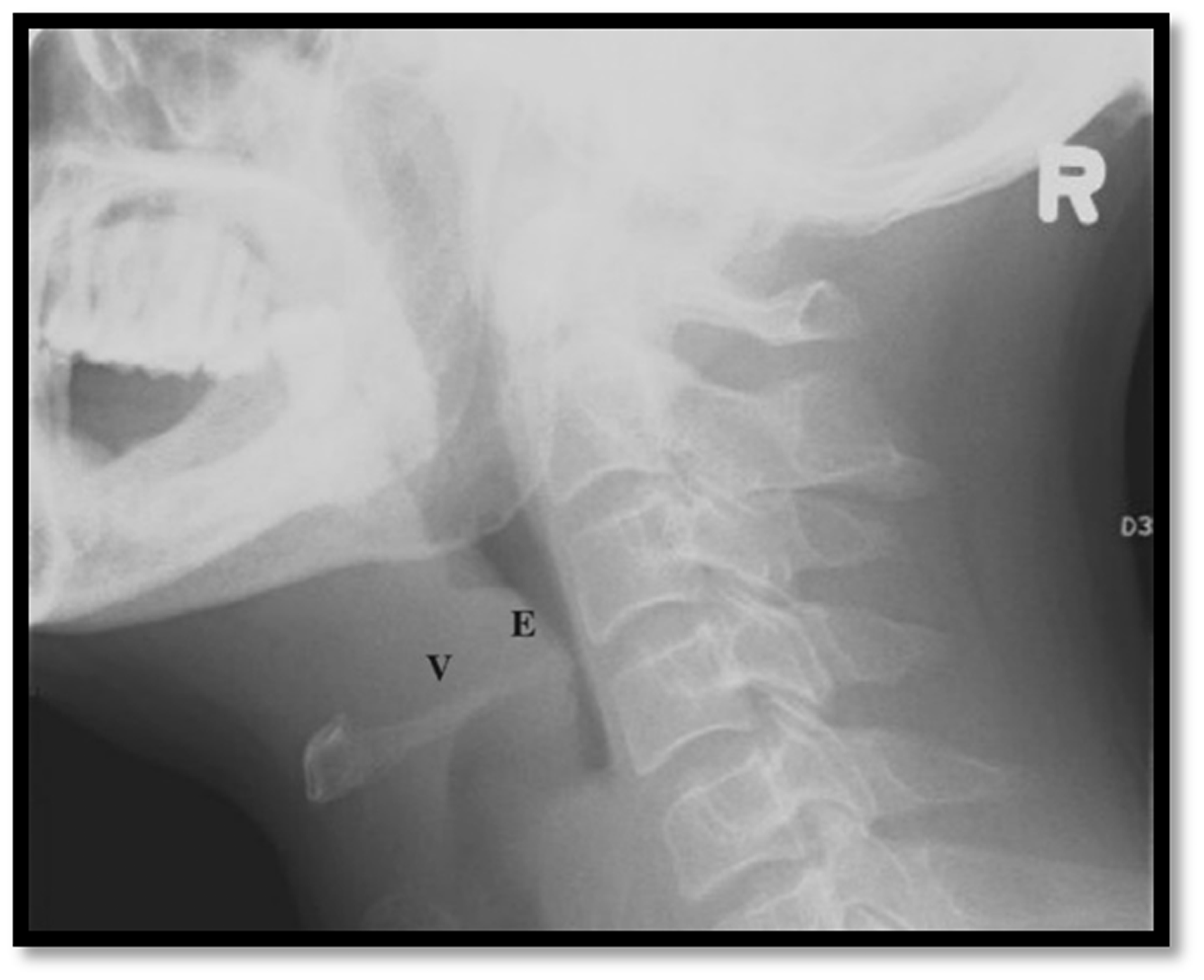
Other investigations that could be requested include blood cultures and throat swab, however, they are often negative (Gleeson and Scott-Brown, 2008). After stabilisation of the patient the following blood tests are essential: Full blood count, urea, creatinine, electrolytes, and C-reactive protein. Lactate should also be added to measure the severity of the hypoxia. If no definitive diagnosis has been made after blood tests, the FNE and X-Rays then a computerised tomography scan of the neck may be be appropriate.
Differential diagnosis
The differential diagnosis varies according to symptoms. When a patient presents with acute upper airway obstruction other pathologies should be considered (Woods, 2019):
Laryngotracheitis (more commonly known as croup or spasmodic croup) Uvulitis Bacterial tracheitis Peritonsillar or retropharyngeal abscesses Foreign body lodged in the larynx or vallecula Angioedema whether anaphylactic or hereditary or drug-induced Congenital anomalies of the upper airway Diphtheria Trauma whether thermal or chemical or mechanical
Differential diagnosis of acute upper airway obstruction.

Reprinted from Adil EA, Adil A, Shah RK. Epiglottitis. Clinical Pediatric Emergency Medicine, 16(3), 149-153, Copyright 2015, with permission from Elsevier.
Management
Primary care
In primary care, if epiglottitis is suspected, arrange a blue-light ambulance transfer to the ear, nose and throat (ENT) or paediatrics team via the accident and emergency department (A&E). The potential severity of this condition requires the A&E department to be contacted and informed about the patient’s arrival. While awaiting hospital transfer, an ABCDE approach should be followed. For the management of the airway, two manoeuvres can be used to facilitate air entry. The head tilt and chin lift manoeuvre and the jaw thrust manoeuvre can help to maintain the airway. Supplemental oxygen can be given to prevent hypoxia. Children may not tolerate a face mask. If the child or adult deteriorate, oral dexamethasone (0.15 mg/kg) may be used. However, if the child is too unwell for oral medication, then inhaled budesonide (2 mg nebulised as a single dose) or intramuscular dexamethasone (0.6 mg/kg as a single dose) can be given to decrease upper airway inflammation.
Secondary care
Epiglottitis is an ENT emergency. If early intervention is not initiated serious complications and airway compromise can ensue. Management of epiglottitis is approached according to airway patency (Woods, 2019):
Management of patients who cannot maintain their airway
Maintaining the patency of the airway is the clinical priority when managing epiglottitis. This requires intervention of specialists in ENT and anaesthetics, and may be achieved by invasive and non-invasive means (Woods, 2019):
Bag-valve-mask ventilation Endotracheal/nasotracheal intubation Tracheostomy
It is important to note that patients with an upper respiratory tract obstruction should not have any supraglottic airway device inserted, for example, a laryngeal mask, as these devices can worsen the patient’s condition by increasing obstruction or distorting the airway anatomy (Woods, 2019).
Management of patients who can maintain their airway
If the patient can maintain their airway, then supplemental humidified oxygen should be given, and the appropriate specialist team contacted. After the airway is established, blood cultures and epiglottic swabs for both aerobic and anaerobic organisms should be obtained. The patient should be managed in a closely monitored specialist setting (High Dependency Unit, ENT) for 24 to 72 hours. Daily examination of the oropharynx and larynx is necessary to check the response to therapy and can help identify complications such as epiglottic abscess or delayed airway obstruction.
In the initial treatment of epiglottitis, racemic epinephrine can be administered to reduce oedema. However, in children the use of nebulised medication can have an unwanted effect causing anxiety exacerbating hyperventilation and respiratory distress (Woods, 2019).
Parenteral glucocorticoids may be added to reduce inflammation. However, studies showing benefit are limited (Baxter and Dunn, 1988).
These patients will also need broad-spectrum intravenous antibiotics to cover the most likely organisms: Hib, S.pneumoniae Group A streptococcus and S.aureus (including methicillin-resistant strains). Usually, third-generation cephalosporins such as ceftriaxone or cefotaxime are recommended in addition to an anti-staphyloccocal agent such as Vancomycin or as per local microbiology guidelines (Woods, 2019).
Complications
The complications of acute epiglottitis include:
Airway obstruction Epiglottic abscess Secondary infection Necrotising epiglottitis
Airway obstruction can lead to hypercapnia, hypoxaemia and severe respiratory distress, life-threatening complications associated with acute epiglottitis.
An epiglottic abscess can result from either underlying epiglottic infection or may be secondary to an epiglottic mucocele. The presence of an abscess complicates up to 30% of cases (Berger et al., 2003). A study published in 2003 showed a rise in acute epiglottitis and epiglottic abscesses (Berger et al., 2003). Airway compromise was a delayed result of such complication with the abscess developing from a few hours to days after the onset of epiglottitis. In a few cases, epiglottitis can spread and lead to secondary infections. Pneumonia and cervical lymphadenitis were the most common complications followed by exudative tonsillitis and otitis media (Molteni, 1976).
Outcomes
If treated promptly, before hypoxic injury occurs, patients with acute epiglottitis recover without residual airway or other problems. The most effective prevention of complications is achieved with a secured airway and prompt antimicrobial therapy.
Prevention
The best way to prevent epiglottitis is with immunisation against Hib.
KEY POINTS
Hib vaccine has decreased the incidence of epiglottitis, especially in children Detection of specific clinical features aids early diagnosis, essential for correct and timely management Patients with acute epiglottitis can deteriorate very rapidly, and inadvisable or incorrect examination can compromise the airway and cause rapid deterioration in the patient’s condition Early, prompt secondary care referral prevents serious complications If treated promptly, patients with acute epiglottitis recover without any sequelae Complications of epiglottitis can occur hours or days after onset of the disease
