



Abstract
Female genital prolapse is a common presentation to general practice. Although frequently asymptomatic, genital prolapse causes some women significant, distressing and debilitating symptoms. Discomfort and feeling a ‘lump or bulge’ in the vagina or ‘something coming down’ is the most commonly reported symptom. Urinary and bowel dysfunction, including frequency and incontinence can occur and dyspareunia, change in body image and psychological symptoms are common. All of these can have detrimental effects on a woman’s wellbeing, quality of life and relationships. Multiple treatment strategies exist for prolapse, including reassurance, pelvic floor muscle training, pessary use and surgery. These can be discussed to facilitate a shared decision about the right management strategy with the patient. This article aims to give an overview of female genital prolapse and its management, and to increase knowledge and confidence for management of the condition in primary care.
Clinical case scenario
A 38-year-old woman presents with feeling a ‘lump’ in the vagina. Further questioning reveals stress urinary incontinence and dyspareunia. Previously, she has had two vaginal deliveries, one requiring forceps.
Examination reveals a utero-vaginal prolapse, the most distal part of the prolapse visible <1 cm from the hymen. You diagnose a stage 2 uterovaginal prolapse.
You discuss treatment options with her. The prolapse is affecting her quality of life and relationship significantly, so she wants to try something, but at this stage wants to avoid surgery. She is young and sexually active, and as such you both agree a pessary would not be suitable.
You discuss conservative management, including keeping a healthy weight and preventing constipation. She is referred to the physiotherapist for supervised pelvic floor muscle training.
She returns after 12 months. There has been some symptomatic improvement, particularly the incontinence after the supervised pelvic floor exercises. However, she is still struggling with feeling a ‘lump’ and this is affecting her body image, confidence and relationship. At this stage, you agree on referral for consideration of surgical repair.
Definition
The pelvic organs are supported by the levator ani muscles and the endopelvic fascia. Female genital prolapse is the downward descent or displacement of the pelvic organs into or through the vagina (Barber, 2016). This may include descent of the uterus, bladder, vaginal vault and/or the rectum (Barber, 2016). This is usually accompanied by local urinary, bowel and/or sexual symptoms (Knott and Tidy, 2019).
Types of prolapse
Types of female genital prolapse.

Epidemiology
Prolapse commonly affects 40–60% of parous women (Maher et al., 2016), and 30–76% of women attending for routine gynaecological care (Barber, 2016). The true incidence is difficult to determine, but is likely higher, as many women do not report prolapse symptoms or seek medical advice. Studies suggest the prevalence of female genital prolapse increases by 40% with each decade of life (Swift et al., 2005). The most common type of prolapse is a cystourethrocoele, followed by uterine prolapse and rectocoele (Knott and Tidy, 2019).
Causes and risk factors
Developing female genital prolapse is a multifactorial process, often involving weakening of the muscular support structure through ageing, trauma, neuropathic injury or stretching (Knott and Tidy, 2019).
The most consistently identified risk factors within published studies are increasing age, menopause, vaginal childbirth, increasing body mass index and previous hysterectomy (Barber, 2016). There is weaker evidence supporting other potential risk factors including forceps delivery, high infant birth weight, prolonged second stage of labour, family history, occupations involving heavy lifting, shape of the pelvis, constipation and connective tissue disorders (Barber, 2016).
Principles of assessment
Female genital prolapse can have a significant effect on the quality of life of affected women. It may cause physical symptoms and associated psychological, sexual, emotional and relationship problems. The aim of an assessment is to understand the symptoms a woman is experiencing and the impact they have on her daily life, to sensitively diagnose prolapse and to establish the nature or ‘type’ of prolapse. Clinicians should provide information about the next steps, signpost and support women leading to a shared management decision.
History and symptoms
A thorough history should be taken sensitively for all women with genital prolapse. Questions may include exploration of the physical symptoms. These may include a dragging sensation, feeling of a vaginal bulge, reports of a palpable or visible bulge or pelvic/lower back pain.
The most commonly reported symptom of genital prolapse is a feeling of ‘something coming down’ the vagina or seeing or feeling a bulge in the vagina. A feeling of pelvic or vaginal pressure or heaviness is also frequently reported. Some women experience pelvic or lower back pain (Barber, 2016) or report difficulty retaining tampons (Knott and Tidy, 2019).
Ask directly about urinary, bowel and sexual symptoms when taking the history. Urinary frequency, urgency, stress incontinence, voiding difficulties and repeated urinary infections are commonly associated with genital prolapse. Manual reduction of the prolapse or position change in order to void the bladder may be required (Barber, 2016).
Bowel symptoms are common with posterior compartment prolapse or rectocoele; women may report incontinence of flatus or stool, incomplete emptying, straining or needing to apply pressure to the perineum or reduce the prolapse manually to defecate. Women with pelvic floor deficiencies may present with rectal prolapse. This may cause symptoms of obstructed defecation, mucous, blood and constipation.
Dyspareunia, reduced vaginal sensation, vaginal flatus and reduced arousal may all result from prolapse. Psychological symptoms and altered body image are common. These can have a significant effect on relationships and quality of life. Ask about vaginal dryness, discharge or inter-menstrual or post coital bleeding; this may be from ulceration of cervix in cases of a severe degree of prolapse.
Past surgical and gynaecological history should be considered. Obstetric history is particularly important and includes attention to gravity and parity status, modes of delivery, obstetric anal sphincter tears and difficult prolonged labour with forceps. Occupation, diet, weight and exercise habits are important to consider. Those with occupations, lifestyles or hobbies involving heavy lifting are more likely to develop a prolapse. There is some limited evidence of an association between anorexia nervosa and pelvic floor dysfunction. A family history of collagen disorders, including Ehlers–Danlos, may contribute to pelvic floor dysfunction (Carley and Schaffer, 2000).
A review of other systems and symptoms should rule out potentially serious underlying causes, such as intra-pelvic malignancy. Consider asking about bloating, early satiety and unexplained abdominal pain, for example, which may point to a sinister ovarian cause. Unexplained weight loss, a palpable pelvic mass or unexplained post-menopausal bleeding all require 2-week wait investigation.
Genital prolapse can have a significant impact on quality of life. Asking how the prolapse impacts the woman’s daily life and routine can yield useful biopsychosocial information and build a holistic picture.
Examination and signs
If a genital prolapse is suspected from the history, examination should be offered to the woman. General Medical Council (GMC) guidance on intimate examination and chaperones should be followed (GMC, 2013). A chaperone should be offered, the examination clearly explained and consent gained.
Abdominal examination should be performed, to assess for scars, masses or evidence of ascites. A pelvic examination should then be offered. The aim of examination is to establish the extent of the prolapse and whether the anterior, posterior or apical segment of the vagina is affected.
POP-Q classification system for female genital prolapse.

Use of a speculum
A Sims’ speculum should ideally be used to assess the anterior and posterior vaginal walls (Barber, 2016). Using a bivalve speculum enables visualisation of the cervix, but is less useful for identifying anterior or posterior compartment prolapse. Inserting the Sims’ speculum along the posterior vaginal wall and asking the woman to strain allows assessment of the anterior wall and vaginal vault. A tissue bulge visible on straining in the anterior wall suggests an anterior compartment prolapse, urethrocoele, cystocoele or urethrocystocoeles. Insertion along the anterior wall and asking the woman to strain allows posterior wall assessment; a tissue bulge here identifies a posterior compartment prolapse such as rectocele or enterocoele (Knott and Tidy, 2019).
During the speculum examination, the general health of the vagina and cervix should be observed. Vaginal discharge or dryness should be noted and enquired about.
Additional investigation
In most cases the history and examination should yield a diagnosis and further investigation is not usually required. In those with lower urinary tract symptoms, consider urinalysis, pre/post void scanning and urodynamic studies. Rarely, kinking of ureters in cases of procidentia can affect renal function. With significant rectal symptoms or faecal incontinence associated with a prolapse, a per rectum examination may be indicated. If there is significant vaginal discharge then swabs for chlamydia and gonorrhea, or candida can be offered. If vaginal dryness and atrophy is noted, then menopausal symptoms and local treatment with vaginal oestrogen should be discussed further.
Classification and grading of prolapse
The pelvic organ prolapse quantification (POP-Q) system is a graded objective measure for the assessment of female pelvic organ prolapse (Knott and Tidy, 2019; Madhu et al., 2018) (Table 2). The system enables a uniform recording method for clinicians (Knott and Tidy, 2019; Madhu et al., 2018).
Treatment options
A crucial first step is explaining the diagnosis of prolapse to the women. Many women are not troubled by their prolapse, especially once the diagnosis has been explained and the benign and often non-progressive nature discussed (Dietz, 2015). Treatment is not always necessary if symptoms are mild, regardless of prolapse stage. Some women with either a stage 3 or 4 prolapse may report few or no symptoms. If this is the case, then monitoring is appropriate (Barber, 2016).
Conservative management
If a woman develops symptoms attributable to the prolapse, conservative management should be discussed. The goal of this conservative management is to improve symptoms, reduce progression and minimise or delay the need for surgery (Kuncharapu et al., 2010).
Although avoidance of heavy lifting is commonly advised, the evidence supporting heavy lifting as a risk-factor is weak and there is no evidence for this measure improving symptoms (Barber, 2016). There is no evidence that weight loss either treats or leads to a regression in female genital prolapse, but it may reduce symptoms and should be recommended preoperatively (Greer et al., 2008). Conservative management and advice will often be dependent on the symptoms experienced. If the prolapse is small, and relatively asymptomatic, reassurance may be all that is required. Lifestyle advice can be helpful, and may include advice about maintaining a healthy weight with dietary advice. Management of bowel and bladder symptoms may be helpful for women with symptomatic prolapse, including appropriate advice about avoiding constipation, dietary change, laxatives, adequate fluid intake and avoidance of potential bladder irritants such as caffeine. For women with significant bowel symptoms, using a stool to lift the knees above hip height when defecating opens the ano-rectal angle and is a useful conservative measure to discuss.
Pelvic floor muscle training
Pelvic floor exercises or Kegel exercises refer to a series of exercises aimed at strengthening the pelvic floor musculature including levator ani and coccygeus muscles (Huang and Chang, 2021). The exercises involve muscular contractions and relaxations that stop or slow urination.
Pelvic floor muscle training (PFMT) refers to a supervised programme of exercises where a woman is monitored and supported to perform pelvic floor exercises consistently and correctly by a specially trained physiotherapist. This may involve measuring the strength of contractions and biofeedback to ensure that the exercises are being completed correctly (Huang and Chang, 2021). PFMT may increase pelvic floor muscle bulk and resting tone of the levator ani muscles, which may reduce symptoms of prolapse (Dietz, 2015).
There is evidence supporting the use of supervised PFMT for treatment of female genital prolapse compared with observation alone (Hagen et al., 2014; Li et al., 2016). A systematic review and meta-analysis of 13 randomised control trials, found that women receiving PFMT gained greater improvements in prolapse symptom score and greater improvement in POP-Q stage. Those receiving PFMT were more likely to report their prolapse was getting better; symptoms from the vagina, bladder and rectum were less frequent compared with controls (Li et al., 2016). The results from trials are based on supervised PFMT, evidence supporting non-supervised pelvic muscle exercises is lacking (Barber, 2016).
Referral to specialist pelvic floor physiotherapy to undertake PFMT should be considered, even in a patient with mild or moderate symptoms from a genital prolapse (Dietz, 2015). This may be particularly beneficial for women wishing to improve their symptoms but to avoid surgery. If referral to specialist physiotherapy is not available, or has a long waiting list, advice to start pelvic floor exercises without supervision or training from a physiotherapist is unlikely to be harmful (Barber, 2016). The ‘Squeezy’ app resource has been designed by specialist physiotherapists to help support women in completing pelvic floor exercises frequently and correctly (www.squeezyapp.com).
Vaginal oestrogens
If vaginal atrophy is noted, particularly around the peri or post-menopausal stage, topical vaginal oestrogens are indicated. These include oestriol 0.1% and 0.01% creams, gels, pessaries and estradiol pessaries 10 mcg dose (licensed for indefinite use) and an oestrogen-releasing ring (each ring lasts for 3 months, licensed use is for 2 years). They are usually applied nightly for the first 2 weeks and twice weekly thereafter. These will help to treat the vaginal atrophy and urinary symptoms, which may be exacerbated by prolapse (Weber et al., 2015). Primary care clinicians should be aware that previous breast cancer is a contra-indication to prescribing these. Some patients with breast cancer on tamoxifen can use local estrogens, after discussion with their oncologist or menopause specialist.
It has been suggested that topical vaginal oestrogens may assist in managing female genital prolapse, alongside treating vaginal atrophy, for example, by improving the strength of weakened supporting structures within the vagina (Ismail et al., 2010). Few studies have assessed the effects of topical oestrogens on symptoms and progression of prolapse (Ismail et al., 2010; Weber et al., 2015) and further research is needed to establish their effectiveness for objective and subjective improvement of prolapse.
Pessaries
A pessary is a mechanical device that is inserted into the vagina to reduce the prolapse, support pelvic structures and relieve pressure on the bladder/bowel (Barber, 2016; Bugge et al., 2020; Knott and Tidy, 2019). Pessaries are available in several sizes, shapes and varieties, including ring-shaped, gelhorn and doughnut (Fig. 1). There are very few contra-indications to pessary use, meaning they are suitable for most women. Pessaries should not be placed if there is evidence of pelvic infection, severe ulceration or patients who are unable or unlikely to attend for follow-up (Jones and Harmanli, 2010).
Commonly used pessaries: ring (top right), doughnut (top left), supported ring (bottom right), Gelhorn (bottom left).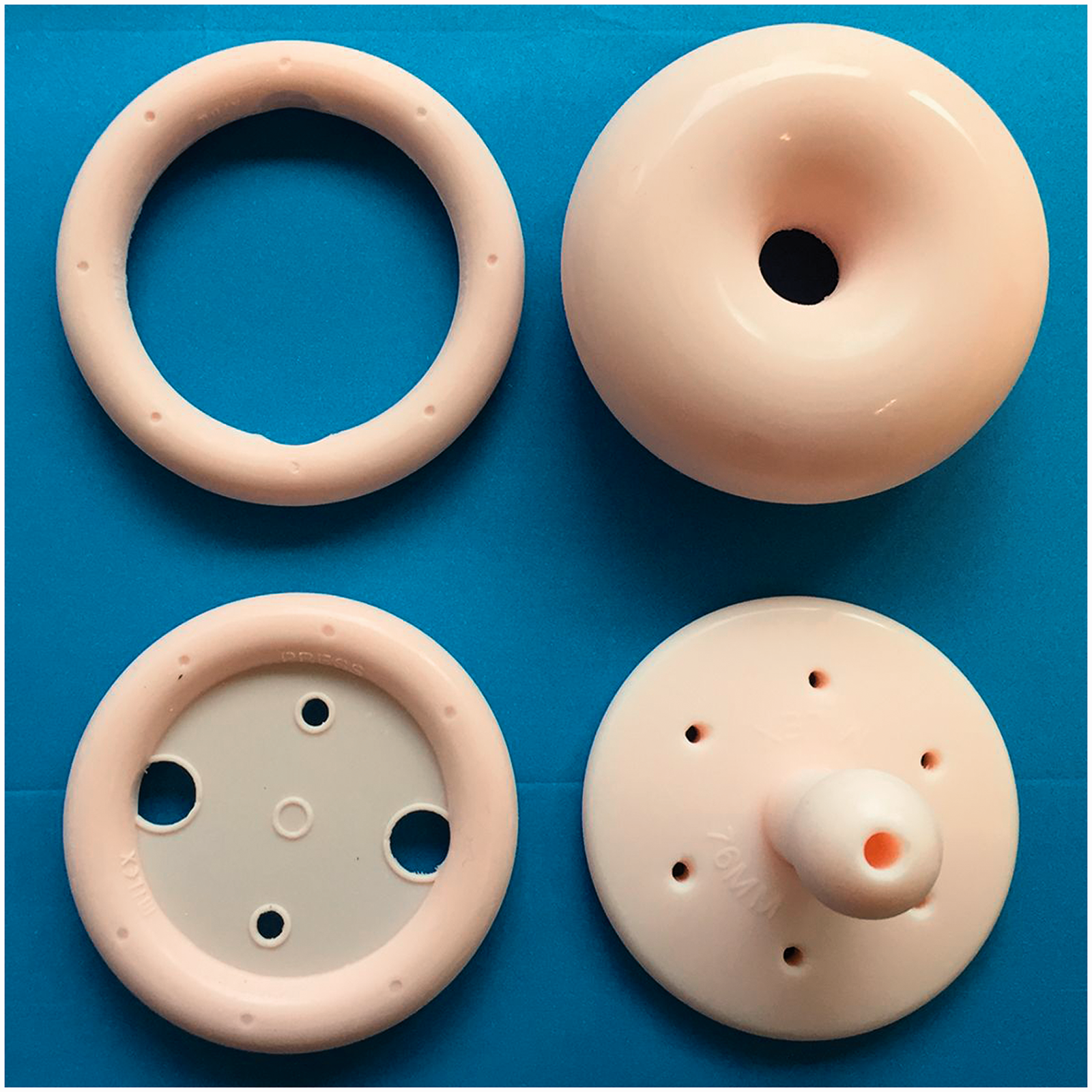
Pessaries are indicated depending on patient choice, as treatment prior to surgery, if a woman is unfit for surgery or wishes to delay surgery (Bugge et al., 2020). They are also indicated in situations where childbearing is not complete, during pregnancy and can be used as a therapeutic test to aid pelvic floor assessment.
In primary care, the ring pessary is most commonly used. PVC and silicone ring pessaries are available. Some silicone pessaries (such as Milex® pessaries) can be reused if not damaged or discoloured. These pessaries are powdered and need to be washed with gentle soap and water before insertion. Ring pessaries with support can be used for patients with first degree and second degree prolapse with cystocele; ring pessary without support is highly recommended for first degree and second degree prolapse (Jones and Harmanli, 2010).
Ring pessaries are relatively easy to change and insert and are unlikely to cause complications. Self-management, where the woman removes and reinserts the pessary herself, is sometimes possible (Dietz, 2015). Sexual activity is possible with this pessary in situ. If fitting a pessary in primary care, a ‘rule of thumb’ for choosing the size is to measure the distance between the urethral meatus and the anus on Valslva in cm, and select a pessary sized just below this value (Dietz, 2015). Sizing can also be established with an internal examination and measuring the distance between posterior fornix and pubic arch. Pessary diameter can vary from 50–95 mm; the sizes come in 5 mm increments. Explain to the woman that more than one attempt may be required to select the correct pessary size and fit the pessary.
To fit a ring pessary, initially it should be compressed by twisting it into a figure of eight shape to reduce its size and improve the ease and comfort of fitting it. Running it under warm water for a few minutes prior to fitting can make the pessary more pliable and make the fitting more comfortable. Once in the compressed position, the pessary can be introduced into the vagina. Once more than half of the pessary is in the vagina, it can be gradually released as it is inserted further. The correct position for the pessary is with its posterior edge within the posterior vaginal fornix, with the anterior aspect behind the pubic symphysis.
After fitting, ask the woman to stand, cough and void the bladder, to ensure the pessary is retained within the vagina without discomfort (Knott and Tidy, 2019). To remove the pessary, the index finger should be hooked around its anterior aspect. The pessary should then be gently brought down to the introitus, compressed and then eased out of the vagina.
The patient can be followed-up in 48 hours to check the pessary is comfortable, and thereafter, followed-up at 4 weeks. At follow-up visits, check for comfort, position, effectiveness and bleeding. Speculum examination to rule out ulceration or erosion of the vagina should be performed. A new PVC pessary should be inserted every 3–6 months.
Changing the pessary at least once every 3–6 months reduces the risk of complications such as erosions, infection or fistulae if left in place without follow-up. If the woman is menopausal, offering treatment with a topical vaginal oestrogen prior to the fit may make fitting/using the pessary more comfortable.
Self-management can be encouraged with some pessaries. For example, some silicone ring pessaries can be washed and reused for up to 5 years, unless damaged or discoloured. Women can remove the pessary weekly, wash it and reinsert weekly after leaving it out overnight.
Gelhorn, doughnut, shelf, cube and other types of pessaries may be indicated in some women, for example, with stage III or IV prolapse or those who are not sexually active (Jones and Harmanli, 2010). Typically, these are fitted in gynaecology clinics. Women are not usually able to remove or reinsert these on their own (Barber, 2016). An article by Jones and Harmanli (2010) gives a useful overview of the use of pessaries, the different types available and their indications.
Surgical management
Indications for surgery.

The type of surgery offered depends on the prolapse’s extent and nature, whether the woman is sexually active, if her family is complete, her preferences and general physical health. Surgery may be transabdominal (Barber, 2016), but 80–90% of procedures are carried out transvaginally (Knott and Tidy, 2019).
Before referring a woman for prolapse surgery, primary care clinicians should discuss the options. The National Institute for Health and Care Excellence (NICE) has created a decision aid for women about genital prolapse surgery, to support shared decision making (NICE, 2019). Prior to referral, women should be informed about the potential benefits of surgery, alongside possible complications, uncertainty about long-term effectiveness and differences in terms of type of anaesthetic, likely length of hospital stay and alternatives to surgery.
Anterior compartment prolapse: cystocoele, urethrocoele and cystourethrocoele
Options for surgical management of anterior prolapses include anterior colporrhaphy and colposuspension. Anterior repair (colporrhaphy) is a transvaginal technique involving central plication (folding) of the fibromuscular layer to strengthen and tighten the anterior vaginal wall. This may be with or without mesh reinforcement (Knott and Tidy, 2019). Colposuspension is performed for urethral sphincter incompetence and involves approximation of the paravaginal fascia to the pelvic side wall, with sutures through the iliopectineal ligament (Knott and Tidy, 2019).
Uterine prolapse
Several procedures exist for correcting uterine prolapse. Four of these are detailed in the NICE patient decision aid (NICE, 2019). Evidence for the effectiveness of one surgical procedure over another is limited, with few studies comparing the individual types of surgery to each other (NICE, 2019). Evidence is also limited in terms of risk of recurrence (NICE, 2019).
Hysterectomy is an option for treatment of uterine prolapse, usually in older women, and in those whose family is complete. A vaginal approach can be used. Sacrospinous fixation is a further transvaginal option, which involves fixation of the uterus to the sacrospinous ligament with sutures (NICE, 2019). This procedure can also be used for vault prolapse. If the woman wishes to retain her uterus, sacrohysteropexy can be performed. This may be an open abdominal or laparoscopic procedure. This procedure involves attaching the uterus to the anterior longitudinal ligament over the sacrum and being held in place using a mesh.
Posterior compartment prolapse: Rectocoele/enterocoele
Posterior repair (colporrhaphy) is recommended for repair of rectocoele/enterocoele. This involves repair of fascial defects or plication of the levator ani musculature, to strengthen the posterior vaginal wall, with or without a mesh for support.
A further option is colpocleisis. This procedure is highly effective in reducing prolapse and has reduced peri-operative morbidity, but involves completely and permanently closing off the vaginal canal, meaning vaginal intercourse is no longer possible. Pre-operative counselling is recommended if a woman prefers this procedure (Knott and Tidy, 2019).
Possible complications of prolapse surgery
Complications of prolapse surgery include wound complications, infection and pain. Dyspareunia is a further complication, and damage to the sciatic or pudendal nerve is possible during sacrospinous fixation (NICE, 2019). Stress urinary incontinence, urinary frequency and faecal incontinence are further potential complications. These are discussed in further detail in the NICE decision aid (NICE, 2019).
Mesh surgery
Recently, there has been controversy surrounding the use of meshes in prolapse surgery, with reports of serious complications resulting from the use of synthetic meshes and a paucity of long-term safety and efficacy data. Reported complications include mesh exposure or extrusion several years after the surgery, causing significant physical and psychological morbidity. Further complications include pain, dyspareunia and sexual dysfunction; neurological deficits, bowel and bladder dysfunction have also been reported. The rates of mesh-related complications are estimated to be between 1 and 10% of surgeries by NICE (NICE, 2019). Rates of mesh-related complications reported in retrospective and cross-sectional studies are lower (Baines et al., 2019; Izett-Kay et al., 2020).
Patients may raise concerns about mesh surgery. A GP’s role is to explain to the patient that they will have a detailed discussion with their consultant surgeon about the type of mesh used and the risks and benefits of proceeding with this type of surgery. The patient decision aid provided by NICE may help structure discussion (NICE, 2019).
Conclusions
Female genital prolapse is an important problem in primary care. The severity of symptoms experienced by women varies, and management strategies should be guided by the woman’s preferences. Primary care clinicians have an important role in reassurance, giving lifestyle advice and supporting and advising around conservative management. When indicated, referral for surgical management is important, and detailed, patient-centred counselling is crucial to help a woman reach the right decision for her. Several resources, such as the NICE patient decision aid, are available to promote shared decision making. It is important clinicians are aware of these to aid appropriate signposting.
KEY POINTS
Female genital prolapse is common, with vaginal delivery being the most important risk factor Some women are asymptomatic and can be reassured The most reported symptom is a feeling of a lump or bulge in the vagina, but urinary, bowel, sexual and psychological symptoms are also common Treatment is very much dependent on the symptoms experienced and dictated by patient preference Conservative management can be helpful and is supported by evidence Surgical management is appropriate in some cases, but there is limited evidence on which surgical options are superior in terms of outcome, complications and recurrence
