



Abstract
Bioengineering aims to develop biomaterials that closely mimic the native extracellular matrix (ECM) to support tissue regeneration. This study presents a detailed protocol for producing hydrogels derived from decellularized bovine placental cotyledons. Bovine placentas at 4–5 months of gestation (n = 10) were subjected to vascular perfusion with increasing concentrations of sodium dodecyl sulfate (0.01–1%) and Triton X-100 (1%), which effectively removed cellular components. Decellularization efficacy was confirmed by histological (hematoxylin and eosin and 4′,6-diamidino-2-phenylindole [DAPI] staining), molecular, and structural analyses, including residual genomic DNA quantification averaging 9.1 ng/mg of dry tissue. The ECM scaffolds were enzymatically digested using 0.1% (w/v) pepsin in 0.01 M HCl and reconstituted with sodium alginate at concentrations of 5%, 8%, 10%, and 12% (w/v). Crosslinking was achieved with 1% calcium chloride. Among the tested formulations, hydrogels containing 12% alginate demonstrated greater mechanical stability and preserved three-dimensional architecture, including interconnected porosity, as evidenced by scanning electron microscopy. Cytocompatibility was evaluated by culturing canine adipose-derived mesenchymal stem cells on both decellularized biomaterials and hydrogels. DAPI staining revealed nuclei after 7 and 25 days of culture, indicating cell presence and distribution throughout the constructs. These results indicate that bovine cotyledon-derived ECM hydrogels maintain structural and biochemical features favorable for cell interaction and may serve as adaptable platforms for tissue engineering, dermal repair, and three-dimensional cell culture.
Impact Statement
This workflow generates sterile, bioactive, and structurally stable scaffolds and hydrogel.
Introduction
Tissue bioengineering has advanced significantly in the development of biomaterials capable of providing structural and biochemical support for tissue regeneration. 1 Among the available platforms, scaffolds derived from the extracellular matrix (ECM) have gained increasing attention due to their intrinsic bioactivity and ability to mimic the native microenvironment of target tissues.2,3 Decellularization techniques enable the efficient removal of cellular components from biological tissues while preserving the biochemical composition and structure of the ECM.1,4
The bovine placental cotyledon has emerged as a promising source of ECM for tissue engineering applications due to its wide availability, low cost, and rich biochemical composition,5–7 including collagen, elastin, laminin, fibronectin, and glycosaminoglycans (GAGs), key components involved in maintaining bioactivity and cellular support.8,9 Anatomically, each bovine placental cotyledon consists of a fetal villous tree that interdigitates with maternal caruncles, forming a synepitheliochorial placentome. This structure provides a large surface area for nutrient exchange and is particularly rich in ECM proteins, making it an ideal candidate for biomaterial development. When subjected to optimized decellularization protocols, placental tissue retains its native three-dimensional architecture and displays mechanical properties compatible with soft tissues, making it an attractive candidate for the formulation of biomimetic hydrogels. These hydrogels combine biocompatibility, in situ remodeling capacity, tunable physicochemical properties, and potential for controlled drug delivery, supporting their use in soft tissue regeneration.10,11
The methodological standardization (Fig. 1) of such processes is essential to ensure reproducibility, safety, and consistent performance of bovine placental cotyledon-derived scaffolds and hydrogels in both experimental and, potentially, clinical settings.5,7 This study presents a reproducible method for producing hydrogels from decellularized bovine placental cotyledons. The protocol encompasses all steps from tissue harvesting and decellularization to sterilization, enzymatic digestion, and final formulation, aiming to establish a versatile ECM-based platform for bioengineering applications. Previous work from our group has demonstrated the potential of decellularized placental tissue as a biomaterial (Barreto et al., 2018), supporting the relevance and feasibility of using bovine placental cotyledon in tissue regeneration strategies.
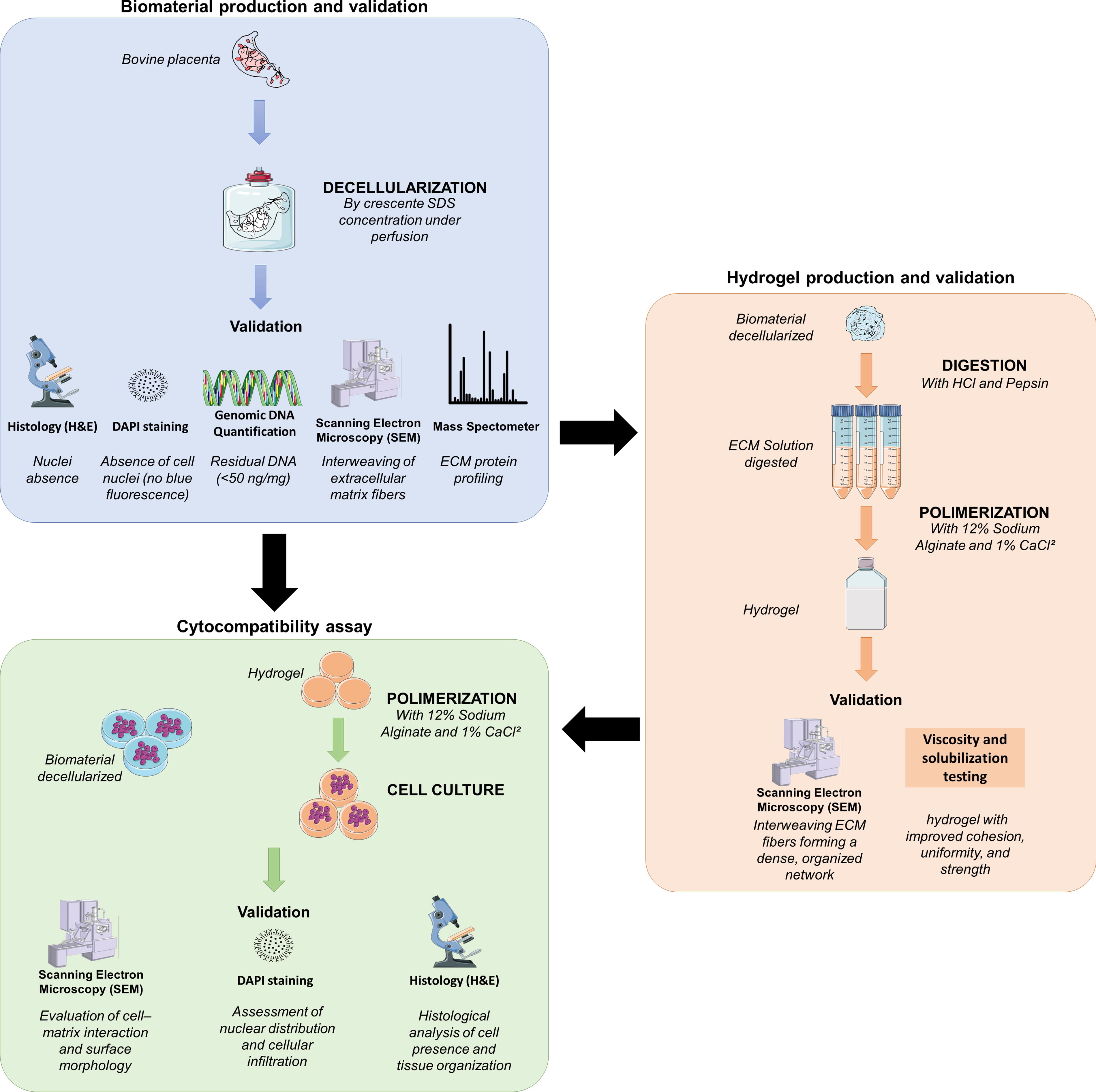
Schematic steps involved in the establishment of biomaterial and hydrogel from decellularized bovine cotyledons.
Materials and Methods
Tissue collection
This study was approved by the Animal Ethics Committee of São Paulo State University (UNESP-FCAV) under protocol 3236/2024. Bovine placentas (n = 10) at 4–5 months of gestation, determined by crown-rump length (Evans and Sack, 1973), were obtained from local slaughterhouses and transported on ice to the Laboratory of Regenerative and Reproductive Biotechnology (UNESP-FCAV). Decellularization and hydrogel production were repeated in triplicates using tissue from three independent animals (biological replicates), totaling, at least, three batches of biomaterials.
Placental decellularization
Umbilical arteries were cannulated with 14G catheters and perfused using a peristaltic pump. Tissues were first washed with phosphate-buffered saline (PBS; 136.9 mM NaCl, 26.8 mM KCl, 14.7 mM KH2PO4, and 8.1 mM Na2HPO4·7H2O; pH 7.2) at 0.5 mL/min for 24 h, followed by sequential perfusion with sodium dodecyl sulfate (SDS) in increasing concentrations: 0.01% (24 h), 0.1% (48 h), 0.25% (48 h), 0.5% (72 h), and 1% (24 h). Residual lipids were removed by perfusion with 1% Triton X-100 for 3 h. The decellularized tissues were washed in sterile PBS for 24 h to eliminate detergent residues. 5 Cotyledons were then dissected from the allantochorionic membrane and stored at −20°C for up to 3 months in sterile, sealed polypropylene tubes until further use.
Validation of the decellularization process
Histological analysis
Representative samples of native and decellularized cotyledons were fixed in 4% buffered paraformaldehyde for 48 h, dehydrated through a graded ethanol series (70%, 80%, 90%, and 100%), cleared in xylene, and embedded in paraffin. Sections (5 µm) were cut using a semiautomatic microtome (Leica RM2265), mounted on glass slides, and stained with hematoxylin and eosin (H&E) to assess tissue architecture and the presence of residual cell nuclei.
DAPI staining
To detect residual nuclear material, sections were stained with 4′,6-diamidino-2-phenylindole (DAPI). Cotyledon fragments were cryopreserved in liquid nitrogen, sectioned in a cryostat (–25°C), and incubated in DAPI solution (1 µg/mL) for 10 min at room temperature, protected from light. Slides were washed, mounted, and examined under an epifluorescence microscope (Nikon Eclipse 80i). The absence of nuclear fluorescence indicated successful decellularization. Cell nuclei were quantified in five random fields per sample (400× magnification), and the average number of nuclei per field was calculated. A significant reduction was observed in decellularized tissues (native: 147.3 ± 15.2 nuclei/field; decellularized: 4.7 ± 2.1 nuclei/field; p < 0.01).
Scanning electron microscopy
Scanning electron microscopy (SEM) was used to evaluate the preservation of ECM structure. Samples were fixed in 4% buffered paraformaldehyde (pH 7.4) at 4°C for 24 h, dehydrated through a graded ethanol series (70–100%), and subjected to critical point drying (Leica EM CPD300). Samples were mounted on stubs using double-sided carbon tape, sputter-coated with gold (Emitech K550), and imaged with a scanning electron microscope (JEOL JSM-6390LV) to assess fibrous architecture and structural integrity.
Reminiscent genomic DNA analysis
Genomic DNA (gDNA) extraction was performed using a saline precipitation protocol adapted from Olerup and Zetterquist 12 Tissues were digested with proteinase K (20 µg/mL), 20% SDS, ethylenediaminetetraacetic acid tetrasodium, and 0.375 M NaCl, followed by centrifugation, ethanol precipitation, and elution in ultrapure water. Quantification was performed using a Nanodrop 2000 spectrophotometer (ThermoFisher Scientific, Waltham, USA). Triplicate samples were analyzed for each batch, and the mean residual DNA content was 35.2 ± 7.9 ng/mg of lyophilized tissue. To obtain values <50 ng of gDNA per milligram of lyophilized biomaterial. 13
ECM-reminiscent protein by mass spectrometry
According to Hedrick et al. 14 and Barreto et al., 15 native and decellularized cotyledons were sampled, lysed, urea reduced, acetone precipitated, dithiothreitol reduced, iodoacetamide alkylated, trypsin digested, and C-18 column purified. Finally, 3 µg of protein were loaded in the OrbitrapFusionLumos spectrometer (ThermoScientific). Spectra were exported to MaxQuant software (v1.6.10.43) for protein list production of each sample, and LFQ intensities were statistically (p > 0.05) analyzed by Inferno software (v.1.1.6970). After, proteins related to ECM and cellular junction ontologies were filtered and manually annotated using DAVID Bioinformatics Resources 6.8.
Hydrogel preparation
Biomaterials were subjected to three cycles of 5-min ultrasonic baths, followed by incubation in sterile PBS supplemented with 2% penicillin/streptomycin (10% v/v). Dehydration was performed by critical point drying (Leica EM CPD300, Wetzlar, Germany), following protocols adapted from Nakayama et al.16,17 Sterilized samples were stored at 4° C for up to 1 week or at −20°C for no longer than 3 months. 18
A total of 500 mg of dried biomaterial was added to 50 mL of 0.01 M HCl containing 50 mg of pepsin (1 mg/mL, Sigma-Aldrich, St. Louis, USA) and minced with sterile scissors to a paste-like consistency. Digestion occurred at 37°C under agitation for 24 h. The pH was then neutralized to 7.4 using 0.1 N NaOH.
To induce polymerization, sodium alginate (C6H7NaO6, #1003669272, Sigma–Aldrich, St. Louis, USA) was added at 5%, 8%, 10%, and 12% (w/v). Cross-linking was achieved by adding 200 µL/mL of 1% calcium chloride (CaCl2, PM 110.98, #13-10490-05, Nova Biotecnologia, Campinas, Brazil). Aliquots of 1 mL were dispensed into 12-well plates and incubated at 37°C in 5% CO2 for 24 h. 19
After polymerization, hydrogels were fixed in 4% paraformaldehyde vapor, freeze-dried in a lyophilizer (OPERON FDB5503), sputter-coated with gold (Emitech K550), and examined using an SEM (Leo 435 VP) to evaluate porosity and three-dimensional network organization.
Cytocompatibility assay
Previously to cell culture, biomaterial and hydrogels were incubated for 7 days in Dulbecco’s modified Eagle’s medium low glucose supplemented with 10% of fetal bovine serum (FBS #12657-029, Gibco, Grand Island, USA) and 0.5% penicillin/streptomycin (10,000 U/mL and 10 mg/mL, respectively; #15070-063, Gibco, USA) at 37°C in a 5% CO2 atmosphere. Changes in turbidity, color, and transparency of the medium were monitored daily as indicators of contamination.
For biomaterials testing, fragments of approximately 1 cm3 were cultured in the complete medium (DEMEM Low Glucose + 10% FBS + 0.5% penicillin/streptomycin) (#10-013-CV, Corning, NY, USA) with 2.6 × 10⁵ canine adipose-derived mesenchymal stem cells (MSCs) per fragment. The MSCs used were isolated from healthy adult donor dogs under approved ethical protocol. Cells were used between passages 4 and 6, and were previously characterized for surface markers and trilineage differentiation potential. Culture was maintained at 37°C and 5% CO2, with medium changes every 2–3 days for 25 days. On days 15 and 25, fragments were collected for analysis of cell viability, distribution, and scaffold integration.
For hydrogel formulation, 1 mL was dispensed into 12-well plates and seeded with 5 × 10⁵ MSCs suspended in 1 mL of complete medium. Cultures were maintained at 37°C and 5% CO2 for 7 days, a period chosen based on preliminary optimization assays indicating this as the optimal timeframe for initial cell adhesion, viability assessment, and early matrix remodeling without compromising hydrogel integrity or inducing significant degradation.
Results
Biomaterial production and validation
Throughout the decellularization process, a progressive reduction in tissue volume and a distinct whitening of the bovine cotyledons were observed (Fig. 2A and 2B). These macroscopic alterations are indicative of effective removal of cellular components. Notably, the tissue maintained the gross anatomical integrity and elasticity, suggesting preservation of major ECM constituents. The whitening effect may be attributed to the removal of blood and chromophore cellular components, further evidencing the efficacy of the protocol in preparing a suitable acellular scaffold (Fig. 2A).
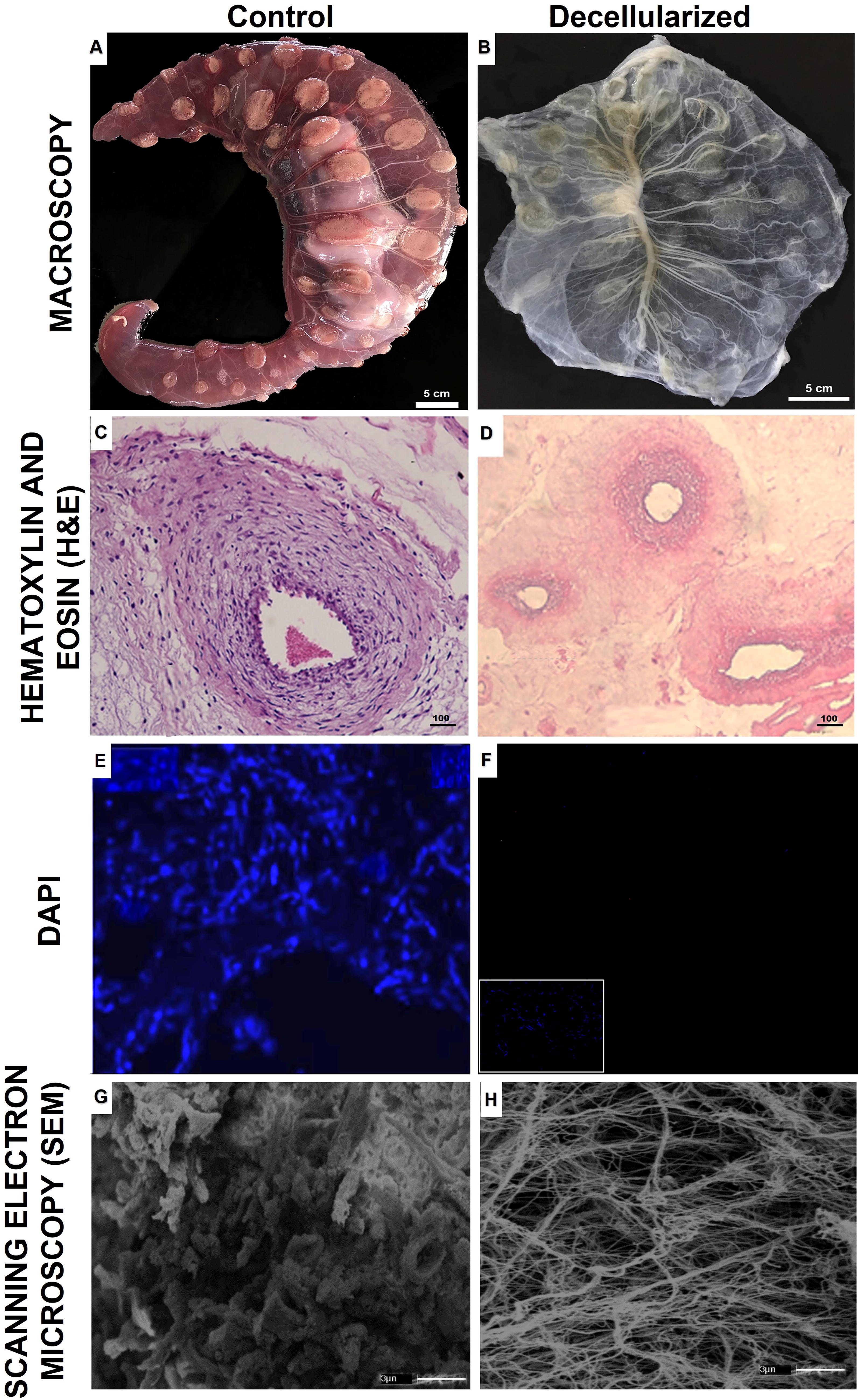
Decellularization of bovine cotyledons. Bovine allantochorionic sac at 4 months gestation before
Histological analysis using H&E staining revealed a complete absence of nuclear basophilia, with tissue architecture maintained and devoid of cellular remnants (Fig. 1C and 1D). DAPI staining confirmed the lack of nuclear DNA, supporting the H&E observations (Fig. 2E and 2F). These findings collectively validate the efficiency of the decellularization process in eliminating nuclear material, while preserving key ECM structures, including the vascular framework.
Quantification of residual DNA revealed an average concentration of 9.1 ng/mg of dry tissue. This value is significantly below the widely accepted immunogenicity threshold of 50 ng of gDNA per mg of dry tissue, reinforcing the potential of the scaffold for biomedical applications. The low DNA content further corroborates the histological and structural analyses and indicates a minimal risk of host immune activation upon implantation (Fig. 3).
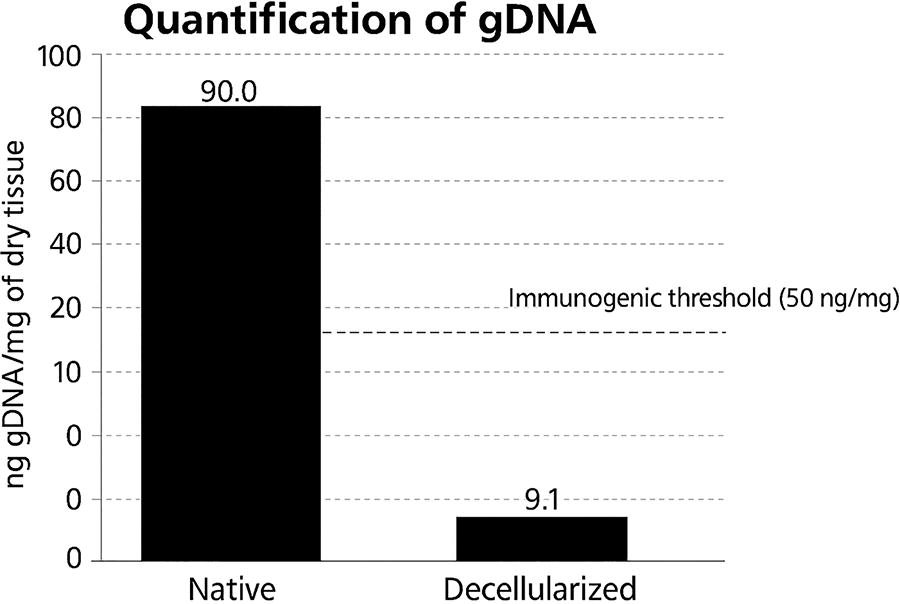
Quantification of genomic DNA. Concentration of genomic DNA (gDNA) in native and decellularized bovine cotyledon. Decellularization led to a significant reduction in the residual gDNA, resulting in an average of 9.1 ng gDNA/mg of dry material, which is lower than the established immunogenic limit (<50 ng gDNA/mg of dry material). The dashed line represents the reference value considered safe for tissue bioengineering applications.
SEM demonstrated a fibrillar ECM with well-preserved collagen bundles and an absence of cellular debris (Fig. 2G and 2H). The structural integrity of the decellularized tissue suggests that the protocol preserved the native architecture essential for cell adhesion and migration. The open and organized fiber arrangement supports the potential for subsequent recellularization and nutrient diffusion.
By mass spectrometry, 144 unique proteins (related to ECM or cell adhesion ontologies) were identified in the biomaterials in comparison with native bovine cotyledon. From those proteins, majorly several adhesive glycoproteins (fibronectins, laminins, and vitronectin), cell adhesion proteins (integrins, dystroglycan, epithelial cell adhesion molecule, and thigh junction proteins), collagen types (I, III, IV, V, VI, VIII, XII, and XVIII), cytoskeletal proteins (actins and myosin), growth factors (tumoral growth factor beta, von Willebrand factor A, and von Willebrand factor), and GAGs (biglycan, decorin, heparan sulfate, nidogens, and tenascin) were detected. Highlighting that even after the decellularization process, not only were the ECM structures maintained, but several pivotal ECM components for cell adhesion, maintenance, and proliferation.
Hydrogel production and validation
Following enzymatic digestion of the decellularized ECM, a viscous, collagen-rich solution was obtained. This material was then blended with sodium alginate at concentrations of 5%, 8%, 10%, and 12% (w/v) and crosslinked with calcium chloride to form hydrogels. All formulations polymerized; however, distinct macroscopic differences were observed (Fig. 4). The 12% alginate formulation yielded a hydrogel with enhanced cohesiveness, greater structural uniformity, and superior mechanical integrity, whereas lower concentrations resulted in less stable and more fragile constructs (Fig. 5A and 5B).
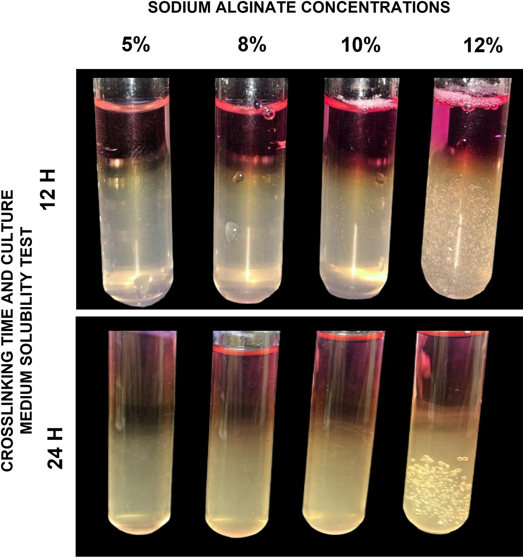
Polymerization test of hydrogel from bovine decellularized cotyledon. Concentration test (5%, 8%, 10%, and 12%) of sodium alginate in digested bovine cotyledon extracellular matrix solution. Representation of the concentrations, respectively. It is possible to observe the difference in viscosity between the formulations, evidenced by the variation in the color and firmness of the gels. Also noticeable is the formation of bubbles due to the cross-linking process with calcium chloride (CaCl2).
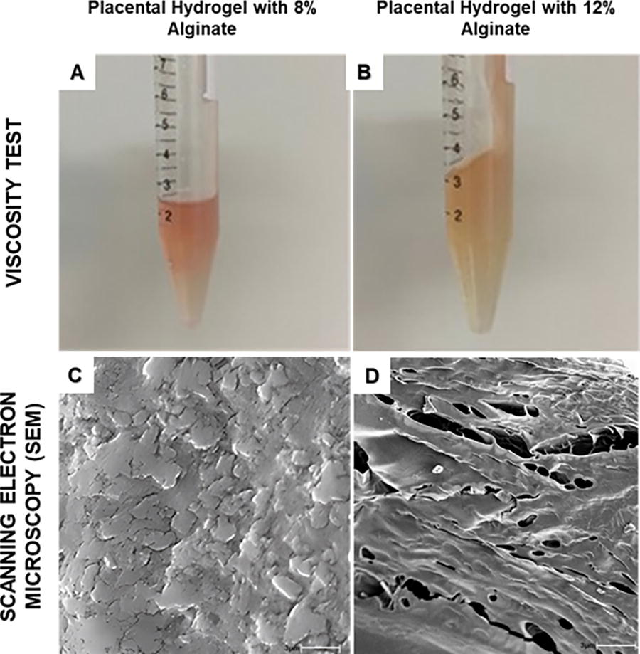
Hydrogel structural validation.
SEM imaging of the hydrogel surfaces revealed clear differences in microarchitecture across formulations. The 12% alginate hydrogel displayed a highly porous and interconnected network, characterized by uniformly distributed pores conducive to cell infiltration and nutrient diffusion (Fig. 5C). In contrast, the 8% formulation exhibited reduced porosity and irregular pore distribution, suggesting limited spatial organization and compromised structural functionality for tissue engineering purposes (Fig. 5D).
Biomaterial and hydrogel cytocompatibility assay
The combination of antibiotic immersion, ultrasonic cleaning, and critical point drying sterilization procedure does not compromise the gross integrity of the biomaterials and hydrogels. After 7 days, under standard cell culture incubation, the culture medium from the biomaterial or hydrogel do not presented turbidity or color change, and no evidence of contamination was observed under microscopy.
The cytocompatibility assay showed cells all over the biomaterial surface, such as cells that were able to migrate into the central areas (Fig. 6A and 6B). Visually, by days 15 and 25 of the culture, the number increased, indicating that cells were capable of adhering, maintaining, and proliferating themselves. In the hydrogel, the cells also migrate through central areas and are able to adhere and proliferate (Fig. 6C and 6D).

Cytocompatibility assay on biomaterial and hydrogel.
Discussion
The decellularization protocol applied to bovine placental cotyledons resulted in scaffolds with well-preserved macro- and microstructural features, consistent with the essential characteristics required for tissue engineering applications. The progressive whitening and reduction in volume observed during the process are consistent with the removal of cellular components while retaining ECM elements such as collagen and GAGs, as reported in similar studies involving fetal membranes and placental tissues.1,5,20 These macroscopic changes served as initial visual indicators of successful decellularization, subsequently confirmed by molecular and histological analyses.6,7
Histological staining (H&E and DAPI) revealed the absence of cellular nuclei and nuclear remnants, which is an established benchmark for decellularization quality.1,4 Furthermore, quantification of residual genomic DNA demonstrated values substantially below the accepted immunogenicity threshold of 50 ng/mg dry ECM proposed by Crapo et al. 2011, 1 with an average of 9.1 ng/mg. These findings reinforce the reliability of the protocol adopted here and underscore the potential of the material for biomedical use.1,5–7,19
The preserved ECM architecture, including the native vascular framework, enhances the biomaterial’s potential for future recellularization strategies and regenerative applications. 3 SEM confirmed a well-organized, fibrillar network devoid of debris, which supports favorable cell–ECM interactions mediated by integrins, particularly through fibronectin and laminin-binding sites.3,4,20 These matrix proteins, along with other structural and signaling molecules identified by mass spectrometry, contribute to the maintenance of essential cellular functions such as adhesion, migration, and proliferation. 15 The biomechanical and topographical integrity of the ECM is also known to influence mechanotransduction, with implications for guiding cell fate and functional behavior.3,21
To generate a more versatile and injectable formulation, the decellularized ECM was enzymatically digested and combined with sodium alginate in varying concentrations to produce hydrogel scaffolds. Herein, the 12% alginate concentration yielded the most structurally stable and morphologically uniform hydrogels for general usage. These hydrogels exhibited enhanced handling properties and architectural consistency, aligning with previous reports that associate sodium alginate concentration with hydrogel performance.5,6,21,22 However, for future and specific approach, the sodium alginate concentration could be altered to reach the natural ECM shiftiness, varying from 0.2 kPa in brain and 106 kPa in bone, to regulate cell behavior of a specific cell lineage or purpose. 23 In this late scenario, all mechanical tests (e.g., rheology, compressive modulus, or tensile strength) need to be performed to support the similarities of the natural ECM and the manufactured hydrogel. SEM analysis of the 12% formulation revealed a highly porous and interconnected microstructure favorable for cell infiltration, diffusion, and vascular ingrowth, key parameters for supporting tissue regeneration.5,6 By contrast, lower concentrations produced less defined networks, which may be suboptimal in terms of mechanical support and biological integration.
The decontamination protocol, which combined antibiotic perfusion, ultrasonic bathing, and critical point drying, proved effective in ensuring material integrity while minimizing potential damage to ECM components. This method provides a gentler alternative to conventional sterilization approaches, which are known to induce protein denaturation and compromise biomaterial quality.6,24,25 Even no evidence of microbial contamination was observed under cell culture conditions, for clinical application, it is necessary to check mycoplasma contamination and specific culture in bacterial medium (i.e., tryptic soy broth and thioglycollate medium).26,27 This step is critical to reduce the risk of infection in in vivo applications and to maintain regulatory compliance for clinical translation.
The biological performance of the scaffolds and hydrogels was further evaluated through in vitro assays, where MSCs demonstrated interaction with the constructs, including adherence, survival, and migration toward internal regions of the matrix. These outcomes highlight the favorable cell-material interactions promoted by the preserved ECM and hydrogel architectures and reinforce the potential application of these materials in tissue repair and regenerative strategies.28–30
Conclusion
Taken together, these results suggest the feasibility of producing bovine cotyledon-derived hydrogels with structural preservation and favorable in vitro performance. Their preserved ECM structure, cell-supportive features, and mechanical versatility support their potential as a scaffold for soft tissue regeneration, dermal repair, and possibly organotypic modeling. Future work should aim to evaluate the behavior of these hydrogels in vivo, particularly regarding immune response modulation, vascularization potential, and integration with MSCs and other therapeutic cell types. The incorporation of bioactive compounds, such as antimicrobial or anti-inflammatory agents, may further enhance their therapeutic value. 21 Therefore, their use as a tridimensional microenvironment for cell culture, organoid production, organ-on-a-chip development, and bioprinting is also an approach that may be effective given the structural and biochemical characteristics retained after processing. Altogether, these findings highlight the translational potential of the developed scaffold as a promising platform for regenerative medicine and in vitro bioengineering, pending further validation of sterility, biocompatibility, and functional reproducibility.
Footnotes
Acknowledgment
The authors thank Rose E.G. Rici from Advanced Center of Image Diagnosis of University of Sao Paulo for microscopy analysis supporting. The authors thank Edmar Delegá da Silva for all technical supporting in the experiments. ChatGPT (by OpenAI in San Francisco, CA, USA) assistance was used as language editing in some sentences.
Authors’ Contributions
R.A.Q.C. executed the experiments, optimizations and data analysis, and wrote the article. I.S.C. executed the experiments, optimizations and data analysis. M.A.M. funding acquisition and article revision. R.S.N.B. supervision, project administration, conceptualization, funding acquisition and article revision.
Data Availability
The article includes all datasets generated or analyzed during this experiment.
Disclosure Statement
No competing financial interests exist.
Funding Information
The authors gratefully acknowledge financial support from CNPq (grant number 409630/2023-3 and 141249/2023-4) and FAPESP (grant number 2021/05445-7).