
Abstract
The development of effective biomaterials for bone defect repair remains challenging due to limitations in mechanical properties, bioactivity, and degradation characteristics. We summarize recent progress in synthetic bone materials, including metals, ceramics, and polymer composites, critically analyzing their clinical strengths and weaknesses. This review presents the fabrication of a new generation of mineralized collagen materials through biomimetic mineralization, demonstrating that their composites exhibit promising clinical application potential. Inspired by the hierarchical architecture of natural bone, a multiscale cascade regulation strategy is further proposed to achieve multidimensional mimicry in composition, structure, mechanical properties, and biological functionality. Special attention is given to multidimensional biomimetic strategies integrating nano-scale molecular self-assembly, electrospinning, and macroscale pressure-driven fusion to construct artificial lamellar bone and artificial cortical bone. In summary, this article provides valuable insights into understanding artificial bone repair materials and their development trends, offering significant guidance for the development of new degradable biomimetic artificial compact bone materials.
Impact Statement
This paper reviews the clinical needs for bone repair materials and the limitations of current options, emphasizing the potential of biomimetic approaches inspired by natural cortical bone’s hierarchical structure. It introduces the mineralization process and mechanical properties of compact bone, evaluates biomimetic mineralized collagen fibers, and summarizes preparation methods. A multidimensional strategy is proposed for constructing high-strength, biodegradable, and biocompatible artificial compact bone, offering insights into future development and clinical application.
Keywords
Introduction
Bone defects caused by trauma, infection, congenital deformity, and other factors are a common occurrence and often require the implantation of bone repair grafts.1–6 Autologous bone has long been considered the gold standard for clinical bone repair, yet due to its limited availability, inability to match defect shapes, and high risk of secondary surgeries, it fails to meet the needs for various types of bone defect repairs.7–11 The development of artificial bone repair materials and orthopedic implants is currently receiving significant attention and has substantial market demand. In recent years, the market size in fields such as orthopedics, neurosurgery, and oral surgery has shown a significant growth trend.12–14 According to incomplete statistics, the domestic market size for bone repair materials and medical devices has exceeded 30 billion yuan. 15
Due to the wide range of sources of materials, ease of processing, and ability to load a variety of bioactive molecules, artificial bone materials are used more than autogenous bone and allogeneic bone in clinical applications and have occupied more than 60% of the market share so far.16–18 There are many types of artificial bone repair materials commonly available. These include bioceramics, polyetheretherketone (PEEK), 19 polymethylmethacrylate (PMMA), 20 porous polyethylene, 21 titanium (Ti) and its alloys, 22 and bioactive glass. 23 Although these materials exhibit good biocompatibility, they typically function merely as simple fillings or provide mechanical support, yet they lack osteoinductivity, osteogenic activity, and adequate biomechanical properties.24–26 Hence, creating artificial bone materials that are biodegradable, possess osteogenic inducibility, and have superior mechanical characteristics is essential to addressing this difficulty. Ideal bone repair materials should gradually degrade during the osteoinduction process, meeting the dynamic requirements for “regenerative repair” and “growth and development” at the site of the bone defect.27–29
Biomimetic bone materials have become the trend in bone implant materials due to their natural bone-like components, structures, mechanism, functionality, and their superior degradability.30–32 Among them, biomimetic mineralized collagen (MC) artificial bone repair materials feature a composition and micronano hierarchical structure of collagen/nano-hydroxyapatite that closely resembles natural bone.33–35 Clinical applications have demonstrated that these materials can induce new bone formation and accelerate bone healing when implanted at bone defect sites, making them a promising biomaterial. However, MC fibers are typically in the form of micronano scale powder, which makes it challenging to meet the rigid requirements for macroscopic mechanical support; therefore, they are commonly combined with synthetic polyester materials for application, which diminishes the advantages of mineralized collagen materials.36–38 Consequently, the biomimetic fabrication of multidimensional high-performance bone structural materials and the three-dimensional manufacturing of a new generation of functional biomimetic MC-based bone defect regeneration repair materials have significant scientific and clinical value.
Synthetic Bone Repair Materials for Clinical Applications
Currently, with an aging population, the problem of bone damage is particularly serious. Although autogenous bone grafting is the gold standard of bone repair, the prolonged hospitalization caused by donor bone procuring is still a problem.39–41 Therefore, artificial bone graft materials have gradually become the best choice for bone defect repair. Currently, according to the material composition, synthetic bone repair materials are mainly divided into metal-based, bioceramic-based (calcium phosphate/calcium sulfate bone cement), and composite structural materials (bioglass, synthetic polymers).42–45
Metal-based bone repair materials
Due to properties of strong corrosion resistance, high mechanical strength, and fatigue resistance, metals are usually selected for internal fixation or large bone defect repair materials. 46 Currently, metal-based bone repair materials are mainly divided into two categories: nondegradable materials (Ti and tantalum [Ta]) and degradable materials (magnesium [Mg] alloy, zinc [Zn]-Mg alloy, etc.). These materials are usually precisely machined into mesh plates with periodic patterns or fabricated into controllable mesh-cage structures using 3D printing technology. 47 Ti has advantages in biocompatibility, osteointegration, low density, high mechanical strength, and a series of excellent biological properties.47–49 It is generally used in permanent bone implants to repair critical bone defects and has been seen to achieve good repair results.50–52 Ta metal implants have been used in clinical practice as early as the mid-20th century. Its excellent inertness and mechanical properties are considered to be a potential biomaterial for bone defects.53–56 Although Ti and Ta alloys are widely used in bone repair due to their excellent mechanical strength and favorable biocompatibility, their inherent bioinertness limits effective osseointegration. Moreover, their elastic modulus, which is significantly higher than that of native bone, can induce stress shielding, leading to localized bone resorption at the implantation site.57–59 In addition, their nondegradable nature poses potential risks for long-term implantation. The high density of Ta alloys may also result in postoperative discomfort, particularly in load-bearing regions. 60 Notably, the use of Ti mesh for cranioplasty is not recommended in children under the age of 11.61–63
Many research focus on finding a metal structural material that has good supporting function, biocompatibility, and biodegradable properties. Therefore, the research and development of Mg and Zn alloys as bone repair scaffolds has gradually deepened.64–66 With the development of technology, additive manufacturing has enabled the expanded application of porous metal bone repair materials, achieving complex bone defect structures while ensuring mechanical strength.67–69 It has been reported in the literature that the preparation of porous metal structures has a relatively low elastic modulus, so it can alleviate the stress shielding and bone resorption caused by modulus mismatch. 70 Yu et al. 71 prepared MgO-doped Zn scaffolds by 3D printing technology, which took advantage of the fact that Cl− in the body fluids is more likely to react with the MgO nanoparticles to release the Mg2+ and form in-situ pores, thus improving the bioactivity and osteogenic capacity of the composite and accelerating the degradation etching rate. In addition, the design of porous Zn scaffolds faces similar challenges to those of Mg scaffolds mentioned above, with equilibrium mechanics, degradation, and surface activity still not clearly standardized. 64 Mg alloys degrade at an excessively rapid rate, often resulting in substantial hydrogen gas evolution, which can lead to the formation of gas cavities and local pH fluctuations, thereby eliciting inflammatory responses. Although Zn alloys exhibit a more moderate degradation behavior, pure zinc possesses inherently low mechanical strength. Even after alloying, further improvements are required to enhance both mechanical performance and biosafety. Additionally, elevated concentrations of zinc ions have demonstrated cytotoxic effects, and thus Zn-based alloys remain primarily in the stages of large-animal studies and early clinical investigation. 72
Ceramics bone repair materials
There are many types of ceramic-based bone repair materials, which usually simulate the inorganic components in the natural bone matrix, including calcium phosphate and silicate. 73 The types of bioceramics formed are roughly divided into hydroxyapatite (HA), Ca3PO4, CaSO4, and bioglass.74–77 The advantage of ceramic bone repair materials lies in the wide range of material sources, and the porous structure formed by sintering, allowing for suitable support strength and bone conductivity. However, these materials also have disadvantages, being their brittleness and difficulty in degradation.78–80 Other forms of ceramic repair material include cement bone repair products prepared from powdered ceramic materials, which are injectable. The clinical application scenarios of inorganic bone cements prepared from ceramic powder and hydrogel are also gradually increasing. Due to their advantages, such as injectability, curability, and plasticity, they are used in neurosurgery, maxillofacial surgery, orthopedics, and other fields. Many studies have focused on the problem that ceramic structures intrinsically have, which is their difficulty in degrading. 81 Thus, a series of low-crystalline ceramic structures such as ACP and β-TCP have been developed to adapt to applications in more scenarios; however, the difficulty in applying to load-bearing bone defects remains a problem. 82
Composite bone repair materials
Composite structural materials are broadly defined, usually referring to the combination of two or more materials to form a composite structure that combines the excellent physical and chemical properties of both materials.83–85
In the field of tissue engineering, composite materials are commonly created through a combination of inorganic materials and polymer materials to form structural materials with excellent biological properties.
86
Bio-friendly polymer materials mainly include processed natural polymers such as collagen, hyaluronic acid, and chitosan, as well as synthetic polymers such as polycaprolactone (PCL),
Biomimetic Mineralized Collagen Materials for Bone Defect Repair
Biomimetic preparation has emerged as a significant approach for addressing the myriad challenges confronting materials utilized in bone defect repair. 94 Initially, a biomimetic MC fiber structure was successfully devised, employing the fundamental principles of mimicking natural bone at the micronano scale, and garnered favorable clinical evaluations. A comprehensive comprehension of the interplay between collagen and ion nucleation and growth during the mineralization process is imperative. Zhang et al. 95 investigated the nucleation sites of calcium phosphate crystals during the initial phases of collagen mineralization in vitro. They discovered that, in addition to carboxyl groups, carbonyl sites on the surface of collagen fiber molecules also undergo chemical interactions during the calcium phosphate nucleation process. Notably, chelation of calcium ions with carbonyl groups at nucleation sites induces a red shift in the amide I peak, reflecting weakened C=O bond strength during early-stage mineralization. 96 The aforementioned research plays a pivotal role in comprehending the organized crystal growth within MC fibers and the biomimetic synthesis procedure. In subsequent investigations, Cui et al. 97 capitalized on collagen templates’ capacity to absorb calcium ions in an acidic milieu, introduced phosphate groups to modulate pH, and ultimately achieved “self-assembly” to obtain biomimetic MC. TEM observations revealed periodic contrast lines of approximately 67 nm within the collagen interior, while selected area electron diffraction (SAED) exhibited an oriented crystal structure, indicating a (002) preferred orientation. Consequently, the synthesized MC exhibits a composition and crystal structure akin to authentic bone structure at the micronano scale. 98
Biomimetic MC formulations typically exist in powder form and are commonly amalgamated with synthetic polymer materials to address substantial bone defects.99–102 Drawing from the anatomical and physiological attributes of bone and inspired by the extracellular matrix, composite materials are engineered to possess strength akin to natural bone. Wang et al. 37 pioneered the fabrication of cortical MC scaffolds (compact mineralized collagen, cMC) and porous MC scaffolds (porous mineralized collagen, pMC). The pMC was prepared by mixing MC powder with PCL/1,4-dioxane solution and followed by freeze-drying. And cMC was prepared by mixing MC powder with melted PCL. MC powder and PCL are at a weight ratio of 1:1. Of course, an increase in MC content will also lead to an improvement in the strength of the scaffolds. 36 Mechanical evaluations in this investigation demonstrated that both cMC and pMC scaffolds exhibited compressive strength and elastic modulus levels comparable with those of natural cortical and cancellous bone, respectively. In vitro experiments revealed favorable biocompatibility, along with heightened osteogenic and osteoinductive activities, and a degree of biodegradability for both scaffold variants. In vivo assessments demonstrated the potential of biomimetic bone materials in regenerating and repairing large-sized skull defects in developing sheep. A sheep skull defect model, established at one month of age, served as a platform for evaluating the biological attributes, osteogenic inducibility, and biodegradation kinetics of the two scaffolds. Over a 6-month period of in vivo observation, both materials exhibited promising capabilities for bone repair. Although the acidic environment resulting from polymer degradation may potentially affect bone defect repair, the incorporation of MC fibrils may help buffer local acidity. As a result, preliminary in vivo evaluations revealed no significant cytotoxicity or pro-inflammatory responses. Notably, clinical follow-up of 105 patients who underwent cranioplasty using pMC, cMC, and biphasic MC (bMC) demonstrated relatively favorable outcomes, with evident new bone regeneration at the defect sites. 103 The results indicated that pMC is more suitable for the repair of small-scale defects, cMC is appropriate for large-area defect reconstruction, and bMC is particularly well-suited for repairing cranial defects in younger pediatric patients.
Hierarchical Structure of Natural Bone Inspires the Biomimetic Construction of Next-Generation Artificial Bone
The cortical bone is mainly composed of three main components: mineral component (∼65 wt%), organic component (∼25 wt%), and water (∼10 wt%). 104 The mineral component in bones is mainly composed of ion-substituted carbonated apatite (ic-HA) with slight impurities, such as carbonate (4–6%), sodium (0.9%), and Mg (0.5%) ions. 33 Type I collagen (97%) is the most abundant collagen and serves as a substrate for the formation of structures during bone mineralization. 105 Weiner et al. 106 defined bone structure into seven levels through a multilevel structural characteristic from nanoscale to micron scale to macroscale. Simply described as: Level 1, collagen fibers (50–70 nm in diameter) and ic-HA nanocrystals (2–3 nm thick); Level 2, MC fibrils structures; Level 3, the fibril bundles (micron-scale); Level 4, bundles were organized as different array patterns (3- to 7-µm thick); Level 5, Haverson system (200–300 µm diameter); Level 6, cancellous or cortical bone structural units; and Level 7, whole bone tissues. 35 Most intriguingly, beyond the multilayered structural order characteristic of the architectural hierarchy, HA crystals exhibit a specific crystallographic orientation in relation to collagen. SAED patterns revealed a pronounced (002) preferential orientation, growth along their crystallographic c-axis. 107 Despite its relatively simple components, achieving a truly bionic bone structure remains a challenge, with limited reports in the literature documenting successful attempts thus far. In this context, we propose multidimensional criteria for bionic bone repair materials, encompassing component structure bionics, performance bionics, and multifunctional bionics. 108 MC microfibril, which mimics the micronano structure and composition of natural bone, particularly collagen/HA, has garnered attention as a potential biomaterial for bone repair. 109 Nonetheless, pure micronanoscale structures struggle to effectively convey the macrostructural mechanical characteristics of materials. Thus, there is a proposal for higher-dimensional bionic bone structural strategies. Currently, research on select natural materials such as bones, nacre, and lobster cuticle offers boundless inspiration for the development of novel bionic structural materials.110–112 However, the integration of mechanical properties with functional attributes through the precise control of hierarchical biomimetic nanocomposite design remains a formidable challenge. Hence, the primary focus lies in advancing the development of multilayered bionic bone structural materials. Concurrently, the creation of “natural bone structures” possessing multidimensional bionic significance holds promise in addressing the intricate challenges encountered in the realm of bone repair materials.
The lamellar structure stands out as the predominant fiber arrangement in bone, prevalent in both cortical and cancellous bone. It comprises approximately five consecutive and parallel layers of MC Microfibrils, each layer rotating by approximately 30°, creating a plywood-like configuration. This anisotropic and densely packed layered arrangement is crucial for the outstanding mechanical properties observed in natural bone. To replicate the intricate plywood-like structure of natural bone, a meticulously controlled approach termed the “multiscale cascade regulation strategy” has been devised. 113 This strategy integrates nanoscale molecular self-assembly, micronanoscale electrospinning techniques, and macroscale pressure fusion methods to precisely guide the assembly process of collagen molecules and nano-hydroxyapatite crystals at room temperature (Fig. 1). Specifically, MC fibrils were synthesized via cotitration of a collagen/phosphate solution and a saturated Ca(OH)2 solution at a molar ratio of Ca:P = 5:3. For the synthesis of element-doped MC microfibrils, designated cations and anions were introduced into the respective solutions, and mineralization was carried out under mildly alkaline buffered conditions. The electrospinning precursor solution was prepared by homogenously dispersing a defined amount of wet-state MC microfibrils into a 7 wt% collagen/hexafluoroisopropanol (HFIP) solution. Electrospinning was conducted under an applied voltage of 19 kV to achieve aligned fibrous sublayers, which were subsequently stacked with a 30° rotational offset. 115 And the MC microfibrils align in a parallel orientation within the electrospun fiber bundles due to shear-induced forces at the nozzle tip. The multilayered structure was then consolidated under ambient pressure at room temperature to form a bulk material exhibiting a characteristic Bouligand structure. 116
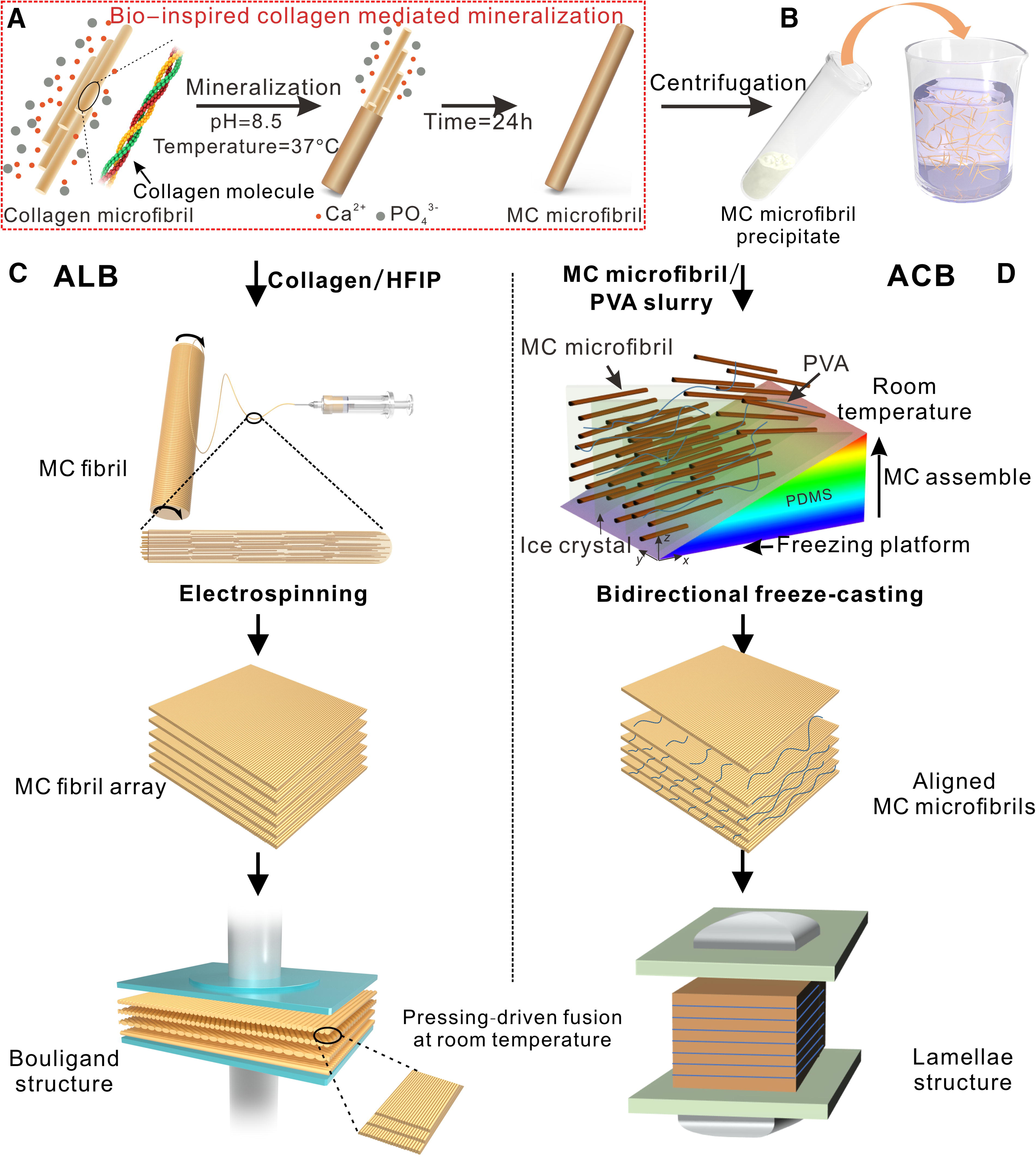
Fabrication process of ALB and ACB.
Figure 2 illustrates the chemical composition and hierarchical assembly structure of the resulting centimeter-scale large artificial lamellar bone (ALB). 113 The synthetic ALB closely mirrors the organizational pattern of natural lamellar bone, faithfully reproducing the hierarchical arrangement of MC microfibrils from the nano- to the macroscale. At the nanoscale, evidence from HRTEM morphology and fast Fourier transform (FFT) patterns verifies the in situ coassembly process of nano-hydroxyapatite and collagen microfibrils. The SAED pattern displays characteristic diffraction rings of nano-HA (nHA). Moreover, the electrospun collagen microfibrils exhibit a uniform diameter distribution of around 100 nm, resembling natural self-assembled mineralized fiber bundles. These microfibrils are aligned parallelly during the electrospinning process, resulting in a preferred orientation of the crystallographic plane (002) of HA approximately parallel to the longitudinal axis of the collagen fiber. At the micrometer scale, the electrospun collagen microfibrils are assembled into layers with ordered orientation, akin to the fiber array layer structure found in natural bone tissue. These oriented microfibril layers are then stacked incrementally at 30° orientations to simulate lamellar bone sublayer units, ultimately forming compact biomimetic bone structures at the centimeter scale after pressure-driven fusion. Importantly, the resulting sublayers maintain their original fiber orientation and structural arrangement. In subsequent work, 114 the research team dispersed MC microfibrils in a 5 wt% PVA solution and subjected the slurry to gradient freezing. Following freeze-drying, a bulk material with parallel lamellar porosity was obtained, wherein the intralamellar MC fibrils exhibited aligned orientation. Upon compression at room temperature, an artificial cortical bone (ACB) structure with a mineral content of 60–70% was achieved. Compared with the ALB, ACB demonstrated a significantly higher mineral content; however, this enhancement came at the expense of interlamellar misalignment. The increased mineral content also resulted in reduced swelling capacity and degradation rate of the ACB structure. From a mechanical perspective, both ACB and ALB exhibited hierarchical architectures that conferred bone-matching strength and toughness, with bending strength exceeding 90 MPa and fracture toughness surpassing 2 MPa·m1/2.
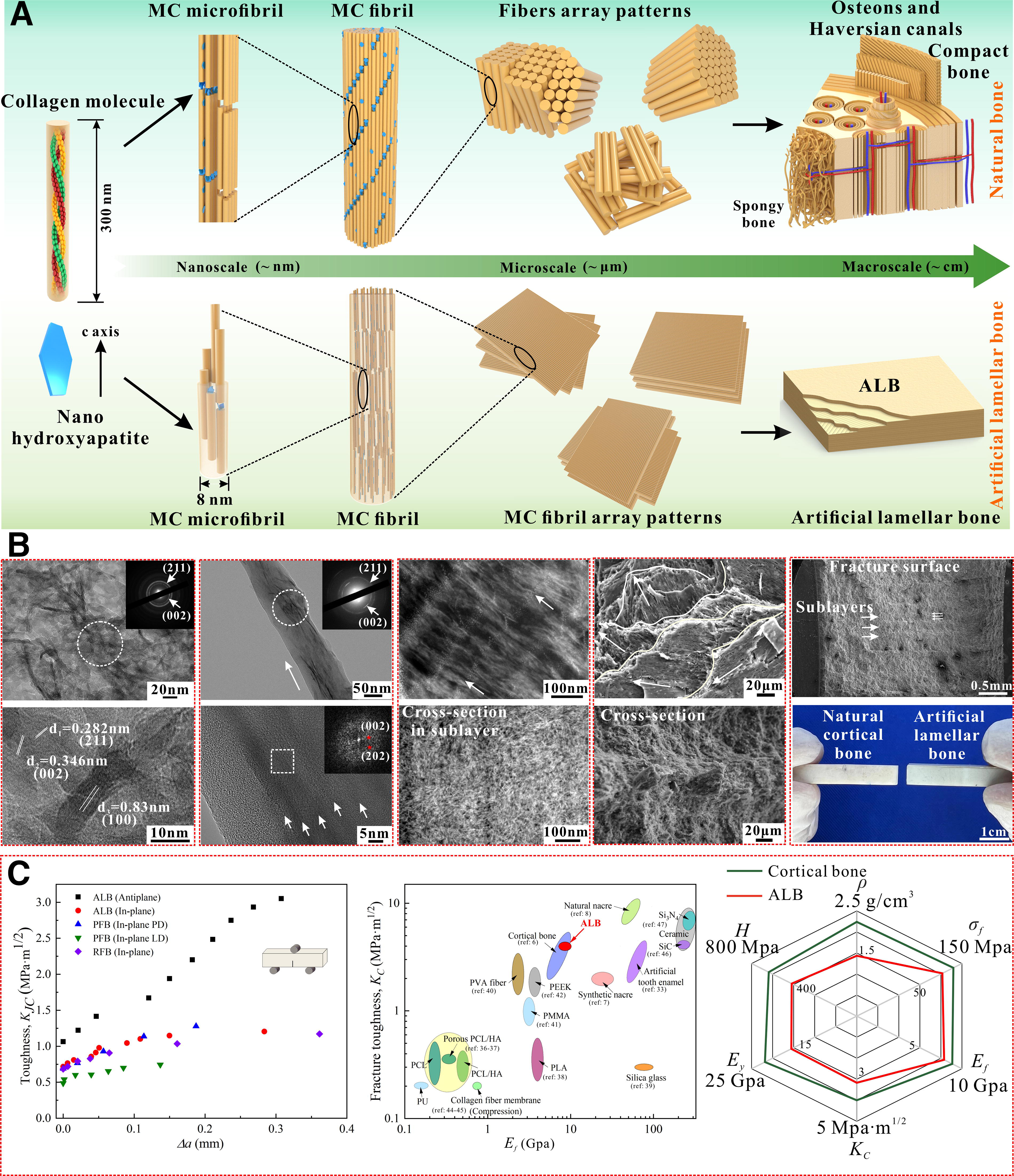
Hierarchical morphologies and mechanical properties of ALB at multiple scales.
Discussion
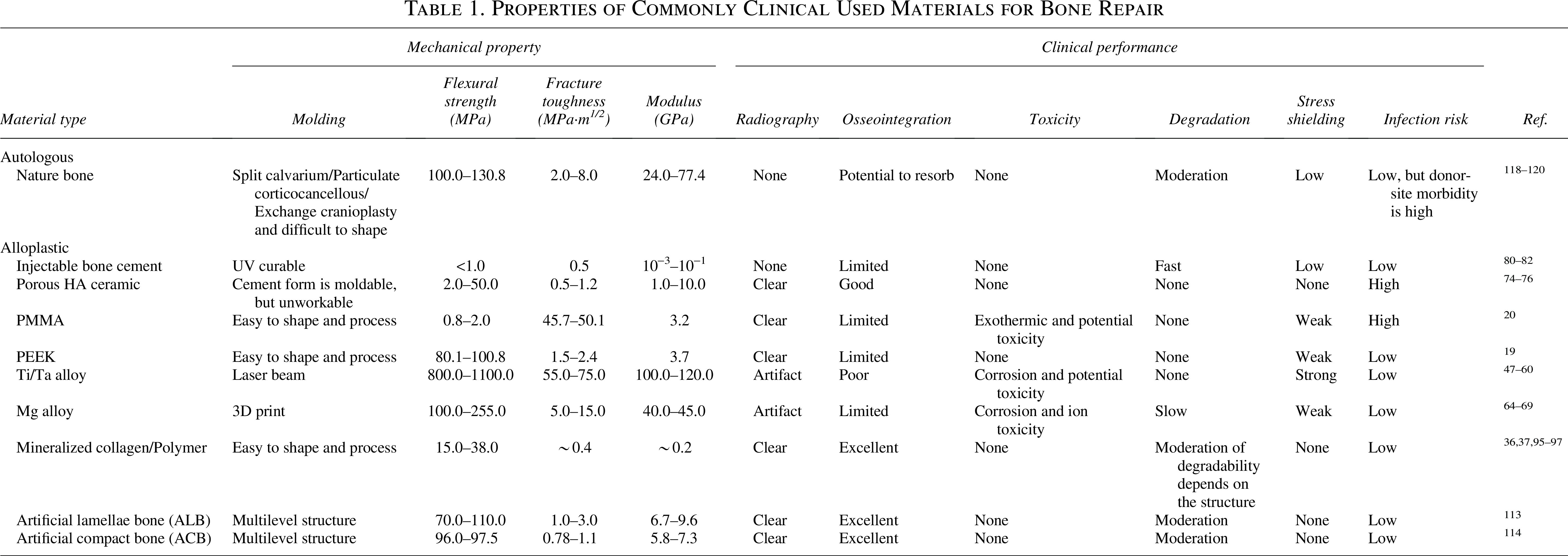
Although these bone repair materials encompass various categories, including metals, ceramics, and composites, they all exhibit essential biological properties such as soft tissue protection and osseointegration. 117 Table 1 compares the overall performance of several clinically used bone repair materials. Among them, the incorporation of MC fibers has significantly enhanced the clinical performance and application potential of polymer-based systems.121–123 Although ALB and ACB have not yet been applied clinically, their favorable composition, architecture, mechanical properties, and degradability collectively demonstrate strong potential for clinical translation. The comprehensive performance is shown in Figure 3. Of particular interest, MC fibers substituted with bioactive elements can exhibit additional functional properties, bringing them closer to the biomimetic characteristics of ic-HA and native bone.
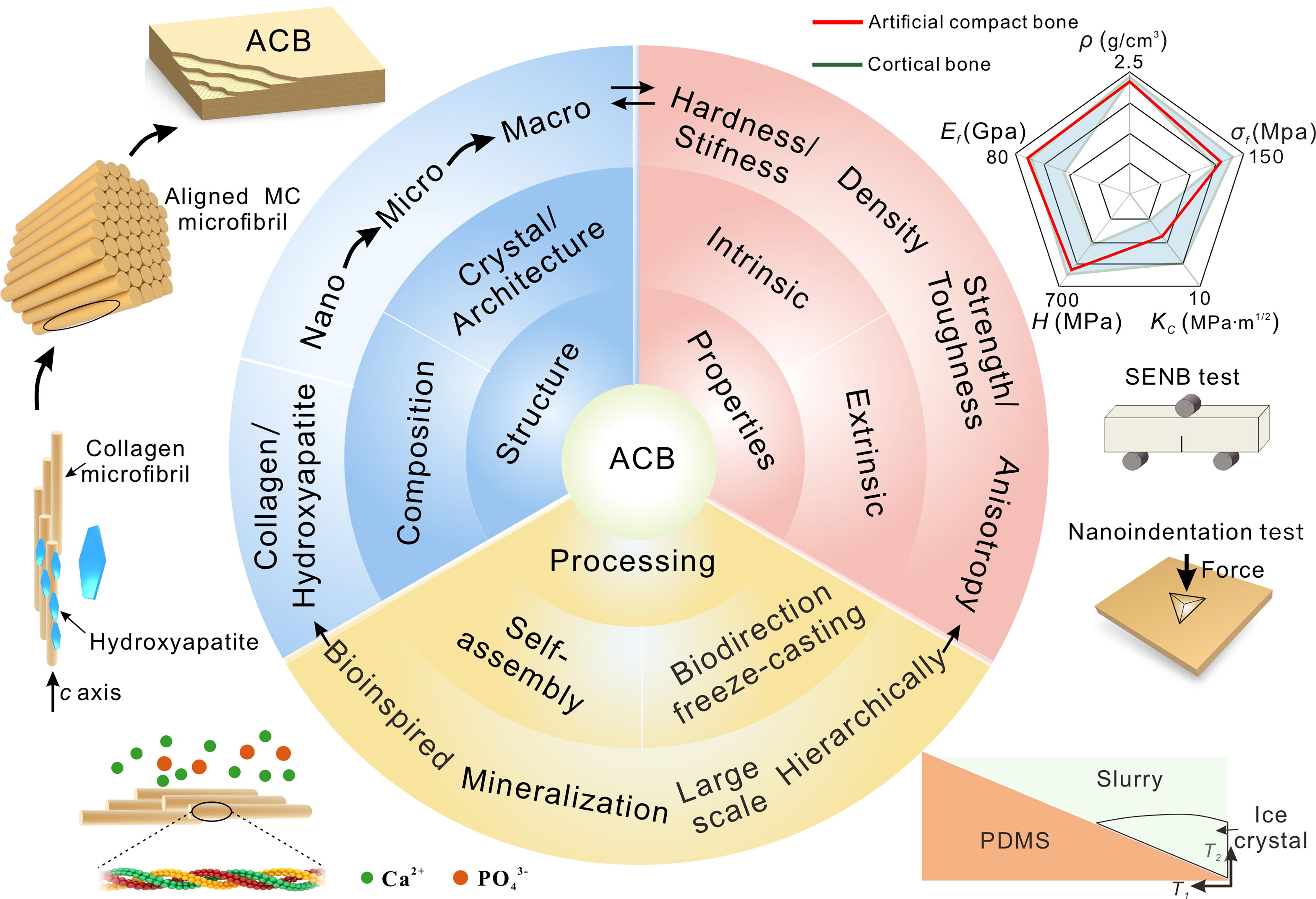
Multidimensional bionic bone structure synthesis strategy. The multidimensional biomimetic construction of bone structures achieves the preparation of biomimetic bone structures in terms of component structure biomimetic, mechanical biomimetic, and biological function biomimetic by integrating biomimetic mineralization and assembly processes.
Properties of Commonly Clinical Used Materials for Bone Repair
Hard materials found in nature have excellent damage resistance and achieve an optimal combination of hardness, strength, and toughness to cope with changes in the natural environment, such as seashells, teeth, and lobster claws.118–120 One factor behind this ability is the multilevel structure of most biological and natural materials, which have unique structural features at multiple scales from molecules to near-macroscopic dimensions. 124 The biomimetic bone structure within the realm of bone tissue engineering diverges from material systems such as shells and teeth, given the more intricate multilevel composition of natural bone. The specificity of structure originates from the organized assembly of inorganic minerals by organic tissues acting as templates during the biological evolution process. These structures are closely related to the mechanical properties of multiscale reinforced materials. The difficulty in understanding the fracture mechanisms of these materials lies in determining the relative importance of these microstructural levels on crack initiation, crack propagation, and unstable fracture, as well as the impact of different levels of structure on critical fracture behavior.125–127 Many studies have shown that these materials rely on both intrinsic and extrinsic toughening effects.128–130 Human cortical bone provides a good example and can provide a feasible solution for the development of bionic materials. 131 The origin of the intrinsic toughening mechanism is often at the smaller submicron length scale, while the extrinsic toughening and fracture processes occur at larger scales, usually up to micron level. Robert et al. 132 conducted a systematic study on the anisotropic mechanical properties and fracture mechanism of bone structure. The process of fracturing in bone mainly originates from the external toughening mechanism, shown in crack bridging and crack deflection. This is caused by the deflection of the growing crack that encounters the mineralized interface of the bone structure. Since the size and spacing of bone structural units range from tens to hundreds of microns, the characteristic scale of this toughening method can approach millimeter dimensions.133–135 The intrinsic toughening method of bone originates from the fiber sliding mechanism on the scale of tens to hundreds of nanometers, being the scale range related to MC fibers. 136
Building upon the understanding of the superior strengthening and toughening design advantage in natural bone fractures, it is essential in the design of biomimetic artificial bone materials to possess a certain level of strength and toughness to meet the service requirements during implantation in the body. By emulating the layered structure inherent in natural bone, the ALB and ACB present an opportunity for synergistic effects from both intrinsic and extrinsic toughening mechanisms.137–139 Fracture analysis reveals that cracks initiate from notches ahead of the tip and progress along irregular paths. Various toughening mechanisms, such as crack deflection, twisting, microcracking, MC microfiber bridging, and crack branching, are evident, contributing significantly to extrinsic toughening. Intrinsic toughening primarily manifests in the behavior of MC microfibrils, involving sliding, bending, and pull-out during the yielding phase. This fundamental mechanism stems from sliding occurrences at the interface of HA and collagen, effectively dissipating energy and enhancing fracture resistance. Thus, the combined action of intrinsic and extrinsic toughening mechanisms can effectively redistribute and mitigate high stresses across multiple scales. The simultaneous presence of multiscale, multiphase, and microfiber conditions ultimately enhances the toughness of artificial bulk bone structures.41,140,141
Although the assembly strategy is not difficult to understand, the advantages of natural materials remain challenging to surpass, especially in functions such as self-repair, recycling, and external adaptation. Moreover, the advancement of biomimetic materials is impeded by the absence of scalable manufacturing techniques and dependable design methodologies capable of mimicking diverse levels of ordered structures. Despite recent research endeavors in this domain, there exists an urgent imperative to innovate scalable manufacturing processes to propel the development of high-performance synthetic materials. 3D printing technology exhibits significant advantages in controlling the macroscopic structural precision and porosity of complex bone scaffolds.76,142–144 However, it still faces substantial challenges in achieving nanoscale mineralization and precise regulation of the organic–inorganic interface. In contrast, biomimetic mineralization strategies offer effective simulation of the micro and nanoscale features of native bone tissue, particularly excelling in the orientation control of MC microfibrils and interfacial optimization. Nevertheless, biomimetic strategies alone are limited in their ability to precisely tailor the macroscopic architecture required for patient-specific bone scaffolds. Recent studies have demonstrated that integrating 3D printing with multiscale biomimetic mineralization enables the synergistic advantages of both technologies, combining the macroscopic precision of additive manufacturing with the micro/nanoscale structural and functional mimicry afforded by biomineralization. This integrated approach allows for coordinated biomimicry and performance enhancement across composition, structure, mechanical properties, and biological functionality and thus represents a promising direction for the development of next-generation high-performance biomimetic bone repair materials.
Conclusion
With the growing clinical demand for bone repair, current artificial bone scaffold materials still face limitations in terms of mechanical properties, bioactivity, and degradability. This review systematically evaluates the advantages and disadvantages of various classes of bone repair materials, including metals, ceramics, polymers, and their composites. Particular emphasis is placed on biomimetic MC materials, which exhibit promising clinical potential due to their excellent biocompatibility, osteoinductive capacity, and controllable degradability. However, conventional single-scale biomineralization strategies remain insufficient to fully replicate the complex hierarchical architecture of native bone tissue. To address this challenge, we propose a multiscale cascade regulation strategy that integrates nanoscale molecular self-assembly, electrospinning-based alignment, gradient freezing, and macroscale compression. This approach enables the fabrication of next-generation ACB materials with hierarchical structures closely resembling those of natural bone, achieving significant improvements in the balance between mechanical strength and toughness. Furthermore, the combination of this multiscale biomimetic strategy with advanced manufacturing techniques such as 3D printing is expected to overcome the limitations of individual approaches, offering synergistic enhancement in both macroscopic structural precision and microscale functional mimicry. This integrated technological framework provides a promising direction for the clinical translation of high-performance bone repair materials.
Authors’ Contributions
Y.Z.: Data curation, formal analysis, funding acquisition, investigation, methodology, project administration, resources, software, writing—original draft, and writing—review & editing (The funding acquisition by Y.Z.). L.K.: Data curation, investigation, methodology, project administration, resources, software, and writing—original draft. T.N.: Investigation, methodology, project administration, resources, and writing—original draft. X.W.: Conceptualization, formal analysis, methodology, writing—original draft, and writing—review & editing.
Footnotes
Acknowledgments
The authors acknowledge the Basic Research and Strategic Reserve Technology Research Fund project of CNPC (No. 2023DQ03-10), and the National Natural Science Foundation of China (52502352).
Funding Information
No funding was received for this article.
Disclosure Statement
No competing financial interests exist.