
Abstract
Osteoarthritis (OA) is a common degenerative joint disease characterized by progressive cartilage degradation, subchondral bone remodeling, and synovial inflammation. Current treatments cannot halt or reverse OA progression, necessitating the development of novel noninvasive therapies. Therapeutic ultrasound (US), particularly low-intensity pulsed US, has demonstrated efficacy in slowing OA progression. Therapeutic US generates significant thermal and nonthermal effects through noninvasive mechanical forces, exerting biological effects and regulating cell behavior. Therapeutic US has been explored for bone and cartilage repair and shows broad potential in tissue repair when combined with biomaterials. This review summarizes the enhanced or synergistic effects of US and biomaterials in OA. This study elucidated the molecular mechanisms underlying the effects of US on synovium, cartilage, subchondral bone, and mesenchymal stem cells. Notably, the combination of US with various biomaterials can modulate cellular behavior in OA through synergistic effects, including tissue regeneration, enhanced mechanical stimulation, drug delivery, and microenvironment regulation. For each cell type, we summarize the biological mechanisms underlying the therapeutic effects of US and biomaterials, demonstrating their potential to mitigate OA progression. Furthermore, this article explores the limitations and future research prospects of combining US and biomaterials as a therapeutic strategy. Overall, the integration of US and biomaterials holds significant promise as a novel treatment for OA, with potential applications in broader musculoskeletal tissue repair and regenerative medicine.
Impact Statement
Osteoarthritis (OA) is a complex degenerative disorder that remains challenging to manage. This review highlights the innovative therapeutic potential of combining ultrasound (US), particularly low-intensity pulsed US, with biomaterials for OA treatment. By leveraging synergistic effects such as enhanced tissue repair, targeted drug delivery, and microenvironment regulation, this approach offers a noninvasive and effective strategy to mitigate OA progression and paves the way for advancements in musculoskeletal regenerative medicine.
Introduction
Osteoarthritis (OA) is a prevalent degenerative joint disorder associated with substantial socioeconomic burdens. It is characterized by progressive cartilage degradation, subchondral bone remodeling, and synovial inflammation, leading to structural joint damage, osteophyte formation, and synovitis.1,2 Most patients remain untreated until OA progresses to moderate or advanced stages. At that point, common medications (such as nonsteroidal anti-inflammatory drug (NSAIDs) used for OA pain relief) can only ease symptoms but cannot stop the disease from getting worse, so many patients end up needing joint replacement surgery. 1 Therefore, there is a need for more effective, noninvasive treatments, particularly those using ultrasound (US) and biomaterials, to promote cartilage and bone repair and regeneration.
Therapeutic US represents a rapidly evolving field with broad clinical applications, including physical therapy, drug delivery, ablation, and regenerative medicine.3–5 Since its initial approval from the Food and Drug Administration (FDA) in 1994 for bone fracture treatment, therapeutic US has been recognized as a noninvasive and safe approach for managing musculoskeletal disorders. 6 However, the underlying mechanisms through which therapeutic US affects OA remain to be fully elucidated.
In recent years, biomaterials have been extensively applied in tissue repair, demonstrating remarkable potential for articular cartilage and subchondral bone regeneration. 7 One of the most promising applications of US is its ability to modulate the physical properties of biomaterials, such as enhancing the mechanical performance of bioscaffolds and increasing cellular permeability for optimized drug delivery. 8 The development of novel biomaterials has further expanded the applications of US, revealing diverse potential uses in regenerative medicine. 9
This review systematically examines the synergistic therapeutic effects of therapeutic US combined with biomaterials in the treatment of OA. Emphasis is placed on elucidating the mechanisms by which US, particularly low-intensity pulsed US (LIPUS), influences various cell types involved in OA pathology. An in-depth analysis explores the pathways and mechanisms underlying the synergy between US and biomaterials. Finally, we critically evaluate current limitations of these combined therapies and propose future research directions to advance their clinical translation.
Overview of Therapeutic US
Definition
The use of US to treat diseased organs or tissues is referred to as therapeutic US. 10 In medical applications, the frequency range for therapeutic US typically spans from 20 kHz to 3 MHz, whereas frequencies beyond this range are generally reserved for diagnostic purposes. 3 Therapeutic US can be broadly categorized into LIPUS and high-intensity focused US (HIFU), depending on the energy level delivered.11,12 HIFU has emerged as a promising modality for chronic pain management and has received approval for treating chronic neuropathic pain, such as in patients with bone metastases and those experiencing pain due to small joint OA.13,14 LIPUS, a specialized form of US, typically operates at frequencies of 1–3 MHz with intensities ranging from 0.02 to 1 W/cm2.15,16
Biological effects of therapeutic US
Thermal effects
As US waves propagate through biological tissues, a portion of their energy is absorbed at a rate that is related to tissue density and acoustic impedance, converting some of this energy into heat and causing localized temperature elevation. 17 This thermal effect promotes blood circulation, stimulates collagen production, and aids cell regeneration, thereby promoting tissue repair.18–20 LIPUS operates in a pulsed mode, minimizing thermal effects while emphasizing significant nonthermal effects. A temperature increase of 4–8°C generally does not cause permanent damage to healthy biological tissues, 21 yet it is sufficient to activate heat-sensitive enzymes such as metalloproteinases and modulate the local microenvironment. 22 Under identical parameters, continuous US generates more pronounced thermal effects than pulsed US, making LIPUS more suitable for clinical applicability. 23
In contrast, HIFU achieves tissue ablation through higher energy delivery. When the target area temperature exceeds 65°C, it effectively acts on periosteal nerve endings to produce definitive analgesic effects. 14 For knee OA treatment, HIFU relieves pain through selective denervation, requiring real-time temperature monitoring to maintain the target area at approximately 60°C. 24 The dissipation rate of US-induced thermal effects primarily depends on tissue characteristics and material properties. In well-vascularized tissues, the microcirculatory system effectively promotes heat diffusion. 25 However, heat dissipation is substantially reduced near biomaterials or metal/ceramic implants. Titanium alloy implants exhibit a surface temperature increase of 13°C after 30 s of US irradiation, markedly exceeding the 5°C safety threshold for bone tissue. 26 Additionally, model systems containing magnetic beads exhibit approximately 10% higher temperature rise compared with pure agarose models. 27 The heat shock response (HSR) serves as a critical protective mechanism against environmental stress. 28 LIPUS can induce heat shock protein 70 (HSP70) expression in gingival tissues, promoting bone regeneration. 29 However, since LIPUS causes only limited temperature increase, its role in chronic inflammation regulation may not depend on HSR. 30 Conversely, HIFU’s high-temperature effects may denature HSPs, impairing their function. 31
Cavitation effects
Cavitation refers to the formation, oscillation, and collapse of microbubbles (MBs) when US waves propagate through a liquid medium. 32 This phenomenon manifests in two distinct forms: transient and stable cavitation. Transient cavitation, used in tumor treatment and lithotripsy, involves rapid bubble expansion and collapse, releasing energy to damage tissue.33,34 Stable cavitation, conversely, involves sustained bubble oscillation under continuous US exposure, which enhances fluid dynamics, increases membrane permeability, and facilitates drug delivery.35,36 Both mechanisms contribute significantly to biological effects in target tissues, with transient cavitation poses risks of cellular damage and requires careful avoidance in gas-filled organs. 37
Mechanical effects
Mechanical effects refer to the impact of US waves on tissues through changes in acoustic pressure and microvibrations. As US propagates through a medium, it generates mechanical forces in the direction of wave propagation, which can induce fluid movement 38 or create stress within solid materials. 23 In cavitation, the stable oscillation of bubbles under continuous US exposure also produces mechanical effects. 39
Interaction Mechanisms Between Therapeutic US and Biomaterials
Biomaterials offer distinct advantages in tissue engineering and regenerative medicine due to their excellent biocompatibility and tunable physicochemical properties.9,40 These materials have shown significant therapeutic effects in tissue repair, bone regeneration, anti-inflammatory treatment, and immune modulation.7,41,42 Nevertheless, single-component biomaterials frequently prove inadequate for addressing the complex regulatory demands of pathological microenvironments. As a noninvasive physical modality, therapeutic US enables precise modulation of biomaterial properties through mechanical forces, cavitation effects, and thermal effects, thereby enhancing their functional performance.
Tissue repair and regeneration
Therapeutic US applies cyclic mechanical stress to simultaneously activate cellular mechanotransduction pathways and modulate biomaterials properties, including stiffness and topological structure. 43 This interaction establishes a synergistic regulatory network of “mechanical stimulation-material response-cellular behavior.” In piezoelectric biomaterial, US induces localized electrical currents through the direct piezoelectric effect, demonstrating promising applications in nerve regeneration and OA treatment.44,45 For hydrogel-based materials, US-induced shear forces dynamically alter their three-dimensional (3D) network structures. 46 Notably, fibrin hydrogels exhibit optimal response at 3.7 MHz, whereas combination with cartilage-mimicking phase-change materials increases this frequency to 4.1 MHz, significantly enhancing cartilage matrix synthesis efficiency. 47 In fibrous scaffold, LIPUS-generated propagates through the porous structure to cells, enhancing primary myoblasts differentiation and myofiber survival during muscle tissue repair.48,49
Mechanical enhancement and drug delivery
Therapeutic US modulates the physical properties of biomaterials (e.g., MB size and hydrogel porosity) through cavitation effects and mechanical shear forces, thereby enhancing the delivery efficiency of drugs or regenerative factors.50,51 Studies have demonstrated that diclofenac sodium (a commonly prescribed NSAID for OA pain management)-loaded polyester microcapsules, when combined with hydrogels, can establish a synergistic delivery system under US exposure, facilitating controlled drug release. 52 Cavitation-mediated effects significantly improve cell membrane permeability in US-sensitive hydrogel carriers, facilitating targeted drug delivery. 53 In cardiac repair applications, LIPUS triggers dual effects: triggering phase transition in nanoscale oxygen carriers while creating microporous hydrogels structures. This combined action enhances oxygen diffusion and improves transplanted cells viability. 54
Microenvironment modulation
Biomaterials enhance therapeutic efficacy by regulating pathological microenvironments (pH/reactive oxygen species (ROS)/hypoxia) in combination with US-induced cavitation and thermal effects. 55 Specifically, pH-sensitive nanocomplexes generate cavitation-mediated microjets and shockwaves that enhance tumor vascular permeability, substantially increasing nanodrug penetration depth. 56 US-mediated mechanical force facilitates nanocarrier transmembrane transport and intracellular redox homeostasis through ROS activation, inducing charge reversal of carriers for efficient gene release. 57 Recent findings reveal that US-activated nanozymes can generate oxygen to alleviate joint cavity hypoxia, whereas cavitation-enhanced sonodynamic effects promote ROS production, effectively suppressing synovial inflammation and angiogenesis. 58 Notably, hyaluronic acid (HA) hydrogels subjected to 30 W US irradiation reach 53.7°C, leading to gold cluster dissociation and subsequent drug release. 59
Mechanisms of Therapeutic US in OA
Current studies indicate multiple pathological factors in OA, including abnormal extracellular matrix (ECM) metabolism, articular cartilage destruction, synovial inflammation, and imbalanced bone remodeling. 60 Therapeutic US presents a promising treatment approach for OA, demonstrating potential to promote cartilage regeneration, mitigate synovial inflammation, reduce subchondral bone sclerosis, enhance bone repair, and alleviate pain.61–64 However, methodological variations in US parameters and experimental conditions (e.g., cell types, exposure duration, and culture methods) across studies hinder direct comparison of results and optimization of LIPUS treatment protocol. Representative studies are summarized in Table 1 and discussed subsequently.
Representative Studies on Therapeutic US for OA
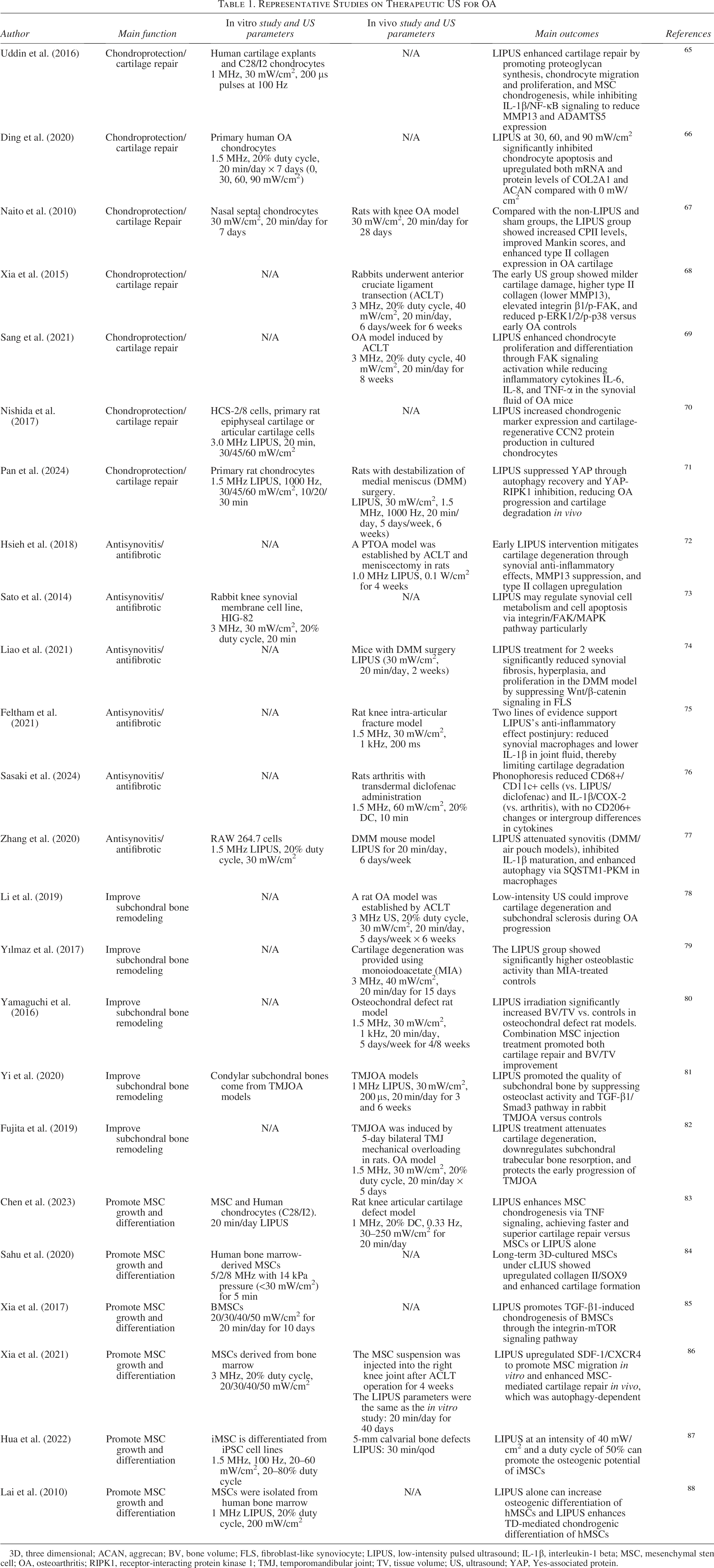
3D, three dimensional; ACAN, aggrecan; BV, bone volume; FLS, fibroblast-like synoviocyte; LIPUS, low-intensity pulsed ultrasound; IL-1β, interleukin-1 beta; MSC, mesenchymal stem cell; OA, osteoarthritis; RIPK1, receptor-interacting protein kinase 1; TMJ, temporomandibular joint; TV, tissue volume; US, ultrasound; YAP, Yes-associated protein.
Effects on chondrocytes
The degeneration of articular cartilage is considered a central feature of OA pathogenesis, with chondrocytes maintaining cartilage homeostasis by regulating ECM metabolism. 89 As mechanosensitive cells, chondrocytes respond to external mechanical signals. 61
Extensive preclinical studies have confirmed the protective effects of LIPUS on cartilage, including preventing chondrocyte degeneration and promoting matrix synthesis61,90 (Fig. 1). LIPUS promotes chondrocyte proliferation, increases glycosaminoglycan (GAG) content, and downregulates matrix metalloproteinase (MMP)-1 expression, thus inhibiting cartilage degradation.91,92 Additionally, LIPUS mitigates interleukin-1 beta (IL-1β)-induced cartilage damage by boosting ECM proteins, including type II collagen (Col II) and aggrecan (ACAN), while facilitating chondrocyte proliferation, migration, and differentiation. 65 Ding et al. 66 found that LIPUS activates SRY-Box transcription factor 9 (SOX9), a transcription factor that enhances ECM synthesis and secretion, while reducing apoptosis in OA chondrocytes. In vivo studies using rat OA models have confirmed these findings, showing that LIPUS stimulates ECM synthesis of Col II and ACAN. 67 These results highlight the therapeutic potential of LIPUS in modulating ECM metabolism and protecting cartilage in OA.
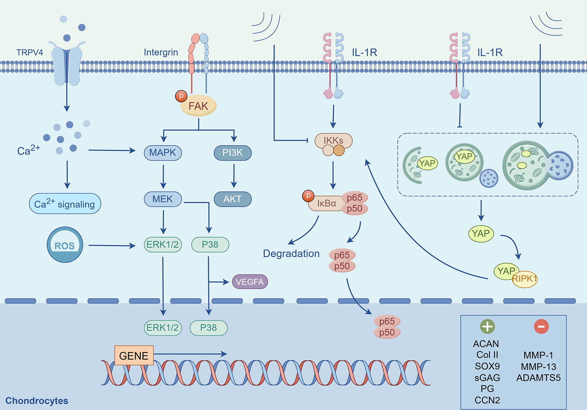
LIPUS regulates chondrocyte function through multiple pathways. Specifically, LIPUS promotes the expression of SOX9, Col II, and ACAN in OA chondrocytes by activating mechanosensitive integrin/FAK pathways and modulating the MEK/ERK and PI3K/AKT signaling cascades. Additionally, LIPUS may hinder chondrocyte degradation in OA by inhibiting p38 phosphorylation. The activation of TRPV4 ion channels further facilitates calcium influx and the subsequent activation of the MEK/ERK pathway. LIPUS also enhances the ERK pathway through the generation of ROS. Furthermore, LIPUS regulates chondrocyte catabolism and inflammation by inhibiting IL-1β and the downstream binding of the NF-κB pathway. It has also been shown to mitigate autophagy impairment induced by IL-1β, inhibit the interaction between YAP and PIPK1, and affect downstream NF-κB signaling, thereby exerting its biological regulatory functions. ACAN, aggrecan; ERK, extracellular signal-regulated kinase; IL-1β, interleukin-1 beta; LIPUS, low-intensity pulsed ultrasound; OA, osteoarthritis; TRPV4, transient receptor potential vanilloid 4.
The integrin/focal adhesion kinase (FAK)/mitogen-activated protein kinase (MAPK) signaling pathway is critical in mediating LIPUS-induced cartilage repair. Xia et al. 68 showed in a rabbit OA model that LIPUS increases integrin β1 expression, inducing FAK phosphorylation and subsequent extracellular signal-regulated kinases 1 and 2 (ERK1/2) activation, which promotes Col II synthesis. Further studies revealed that LIPUS upregulated Col II and integrin β1 expression in both normal and OA chondrocytes while specifically suppressing p38 phosphorylation in OA chondrocytes, highlighting LIPUS’s unique regulatory effect under pathological conditions. 93 This inhibitory effect was further confirmed in another study, where LIPUS slowed vascular endothelial growth factor A-induced cartilage degradation and synovitis by inhibiting p38 activity. 94 LIPUS also enhanced chondrocyte proliferation and differentiation, alleviating synovial inflammation in OA mice via FAK signaling activation.69,95 Moreover, LIPUS influences the integrin-FAK-phosphatidylinositol 3-kinase (PI3K)/protein kinase B (AKT) pathway, promoting ECM synthesis of ACAN and Col II in OA chondrocytes. 96 In this context, integrins, as mechanosensitive receptors on the cell surface, perceive external mechanical signals and convert them into biochemical signals, 97 a process underlying LIPUS-mediated therapeutic effects.
LIPUS elevated ROS production and activated ERK1/2 phosphorylation, leading to upregulation of cartilage-regenerative genes. 98 Research has shown that LIPUS enhances calcium influx into chondrocytes via the transient receptor potential vanilloid 4 (TRPV4) channel, subsequently activating p38 and ERK1/2 phosphorylation to enhance cellular communication network factor 2 (CCN2) expression, a key cartilage regeneration factor. 70 Similarly, LIPUS stimulated CCN2 expression through the ERK1/2 pathway in human medial meniscus cells, aiding meniscus repair.99,100 These findings suggest that LIPUS exerts distinct pathway-specific effects in different joint disease models.
LIPUS regulates multiple signaling pathways beyond MAPK to regulate chondrocyte growth and differentiation. It suppresses IL-1β-induced cartilage degradation by inhibiting nuclear factor kappa-light-chain-enhancer of activated B cells (NF-κB)-p65 and inhibitor of kappa B alpha (IκBα) phosphorylation, while enhancing chondrocyte migration, proliferation, and differentiation. 65 LIPUS also restores autophagy, a process essential for maintaining intracellular homeostasis, and delays OA progression by inhibiting the interaction between Yes-associated protein (YAP) and receptor-interacting protein kinase 1, reducing YAP expression and downstream NF-κB signaling. 71
In summary, the chondroprotective effects of LIPUS are mediated through multiple signaling pathways, including MAPK, NF-κB, and integrin-associated mechanotransduction, which regulate chondrocyte proliferation, differentiation, autophagy, and the balance between anabolic and catabolic processes.
Synovial tissue and synoviocytes
The synovium maintains joint homeostasis through complex immune and inflammatory interactions with cartilage. 101 Synoviocytes, particularly fibroblast-like synoviocytes (FLSs) and macrophages, play a central role in synovial inflammation, proliferation, and remodeling. 102
Increasing evidence suggests that US effectively suppresses synovial inflammation in both animal models and cellular studies of synovitis 16 (Fig. 2). In collagen-induced arthritis rats, US treatment significantly alleviated synovial hyperplasia after 4–6 weeks. 103 Early LIPUS intervention decreases synovial inflammation and MMP13 expression while enhancing Col II synthesis, thereby slowing cartilage degeneration.72,104 LIPUS exerts its anti-inflammatory and protective effects by activating ERK1/2 and p38 phosphorylation through the integrin/FAK/MAPK pathway. 73 Additionally, LIPUS modulates synovial function by upregulating hyaluronan synthases (HAS2 and HAS3) and inhibiting hyaluronidase (HYAL2) activity, leading to the accumulation of high-molecular-weight hyaluronan for joint lubrication. 105 The abnormal activation and functional changes of FLS are considered key factors in the onset and progression of OA. Studies indicated that LIPUS inhibits pathological FLS activation in OA by suppressing Wnt/β-catenin signaling activity, thereby preventing synovial fibrosis and hyperplasia. 74
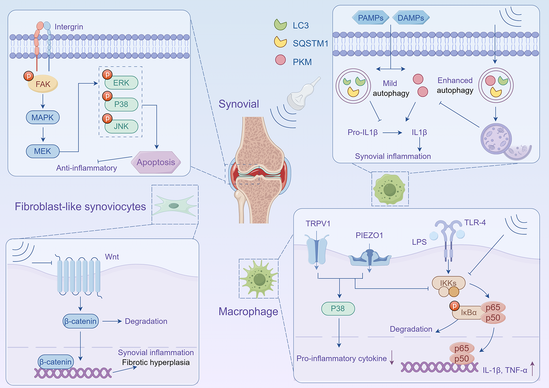
LIPUS combats synovitis and fibrosis through multiple pathways. LIPUS regulates the apoptosis and survival of synovial cells via the integrin/FAK/MAPK pathway. LIPUS inhibits synovial fibrosis by suppressing the Wnt/β-catenin signaling pathway in FLS. Furthermore, LIPUS alleviates synovial inflammation by enhancing autophagy levels in macrophages and inhibiting the maturation of IL-1β. The stimulation of LIPUS also activates the mechanosensitive ion channels PIEZO1 and TRPV1, which suppress NF-κB nuclear translocation, subsequently reducing the release of proinflammatory cytokines. FLS, fibroblast-like synoviocyte.
Macrophages are key immune cells within the synovium, controlling joint inflammation and tissue homeostasis. Studies have shown that pulsed US at 3.0 W/cm2 for 5 min significantly reduces inflammatory factor expression in lipopolysaccharide (LPS)-induced macrophages. 106 In post-traumatic OA rats, LIPUS reduced CD68+ macrophages in the synovium and lowered IL-1β levels in joint fluid, demonstrating its anti-inflammatory effects. 75 When combined with diclofenac, LIPUS synergistically alleviated joint inflammation and pain, potentially through M1 macrophages suppression. 76 LIPUS also increased the number of Lubricin-positive cells in OA models, suggesting it mitigates cartilage degradation by reducing early OA inflammation. 107 LPS-induced macrophages showed p65 nuclear translocation, which LIPUS inhibited by activating mechanosensitive ion channels PIEZO1 and TRPV1, reducing proinflammatory cytokine release through the p38 MAPK pathway. Importantly, blocking PIEZO1 and TRPV1 significantly diminished LIPUS’s anti-inflammatory effects, highlighting their central role in LIPUS-mediated inhibition of NF-κB signaling. 108 Similarly, LIPUS attenuated LPS-induced inflammation in U937 cells by preventing IκBα degradation and p65 nuclear translocation. 109
Notably, LIPUS promoted autophagy, facilitating the formation of sequestosome 1 - pyruvate kinase M (SQSTM1)-pyruvate kinase muscle isozyme (PKM) complexes and reducing PKM2 levels in LPS-adenosine triphosphate (ATP)-treated macrophages. This process suppressed mature IL-1β production, ultimately alleviated synovial inflammation and gait abnormalities in OA animal models. 77 These findings collectively demonstrate LIPUS’s multifaceted anti-inflammatory actions through macrophage modulation.
Subchondral bone
As OA progresses, subchondral bone loss worsens, disrupting the balance between osteoblasts and osteoclasts, which contributes significantly to OA onset and progression. 110
LIPUS exerts protective effects on subchondral bone remodeling in OA. One study examined the effects of LIPUS on joint cartilage and subchondral bone changes during OA progression under normal and functional disuse conditions. The results showed that LIPUS not only alleviated cartilage degeneration but also improved subchondral bone sclerosis during OA progression, with more pronounced effects observed under normal joint use conditions. 78 In monoiodoacetate (a glycolysis inhibitor that induces chondrocyte apoptosis)-induced rats, LIPUS enhanced osteoblast activity and bone mineral density, facilitating concurrent cartilage and subchondral bone regeneration. 79 Moreover, combining LIPUS with mesenchymal stem cell (MSC) therapy significantly improved cartilage repair and enhanced the bone volume/tissue volume ratio. 80
Osteoblasts and osteoclasts are known to be sensitive to mechanical stimulation. 111 Early LIPUS intervention attenuated knee OA progression by preserving tissue integrity, improving subchondral bone microstructure, and reducing both pain perception and diminishing sensory innervation. Moreover, daily LIPUS treatment inhibited osteoclastogenesis, potentially through neuromodulatory mechanisms. 63 In temporomandibular joint (TMJ) OA models, LIPUS improved subchondral bone quality by dual inhibition of osteoclast activity and the transforming growth factor (TGF)-β1/Smad3 signaling. 81 Further studies showed that LIPUS inhibits IL-6 expression and the TGF-β1/Smad3 pathway in subchondral bone, aiding TMJOA treatment. 112 Additionally, LIPUS counteracts mechanical overload-induced TMJOA by decreasing osteoclast numbers, restoring proteoglycan expression, and preventing cartilage degradation. 82
Collectively, these findings suggest that LIPUS modulates osteoblast–osteoclast interactions, playing a key role in maintaining bone homeostasis and remodeling subchondral bone.
MSCs
MSCs are multipotent cells capable of differentiating into various cell types, including chondrocytes and osteoblasts. US stimulation, through its mechanical, cavitation, and thermal effects, significantly enhances MSC proliferation, differentiation, and migration. 113
US parameters can direct MSC differentiation toward specific cell types. It promotes chondrogenic differentiation by enhancing TGF-β signaling, demonstrated by elevated proteoglycan production (3-fold increase) and intensified alcian blue staining. 114 LIPUS also suppresses the TNF signaling pathway, promoting MSC chondrogenesis and optimizing the anti-inflammatory microenvironment for chondrocytes. In vivo studies confirm these findings, showing superior cartilage repair in rat knee defects when combining LIPUS with MSC therapy versus controls. 83 Continuous LIPUS induces increased SOX9 expression in MSCs, a key transcription factor for chondrogenesis, through ERK1/2 phosphorylation. Prolonged continuous low-intensity US stimulation also increases collagen II and chondroitin sulfate in MSCs cultured in hydrogels. 84 As a crucial degradative process for maintaining intracellular homeostasis, autophagy is modulated by LIPUS and contributes to MSC chondrogenic differentiation. Specifically, LIPUS exerts dual effects through the integrin-mammalian target of rapamycin (mTOR) signaling pathway: it upregulates integrin and p-mTOR expression to suppress autophagy and promote bone marrow mesenchymal stem cell (BMSC) chondrogenesis, 115 while simultaneously mediating TGF-β1-induced chondrogenesis in BMSCs, an effect that can be reversed by specific inhibitors. 85 In the anterior cruciate ligament transection-induced OA model, LIPUS combined with MSC therapy significantly enhanced cell migration capacity and promoted cartilage regeneration through autophagy pathway activation. 86 These findings suggest that LIPUS-mediated autophagy regulation exhibits stage-specific characteristics, likely dependent on the distinct physiological states of cells.
US also positively influences MSC osteogenic differentiation. Studies have shown that LIPUS stimulates MSC osteogenesis via the Wnt/β-catenin signaling pathway, involving hypoxia-inducible factor 1-alpha (HIF-1α) and ras homolog family member A (RhoA) activation.87,116 Moreover, LIPUS enhances osteogenic differentiation by modulating specific miRNAs, such as miR-31-5p and miR-675-5p.116,117 When MSCs are treated with dexamethasone/TGF-β1 (TD) or BMP-2, LIPUS not only promoted osteogenic differentiation but also amplified the chondrogenic effects induced by TD. 88 These findings highlight the therapeutic potential of LIPUS in OA and other bone-related diseases by promoting MSC proliferation and differentiation.
Synergistic Effects of Therapeutic US and Biomaterials
Biomaterials, including natural, synthetic, inorganic, and metallic types, offer distinct therapeutic benefits (Fig. 3). The synergistic application of therapeutic US and biomaterials is vital for tissue repair, drug delivery, and microenvironmental modulation. US accelerates tissue repair by stimulating cellular proliferation, whereas biomaterials provide a stable microenvironment to enhance healing. The combination approach provides new possibilities for the treatment of OA, with this review focusing on these key mechanisms (Fig. 4).
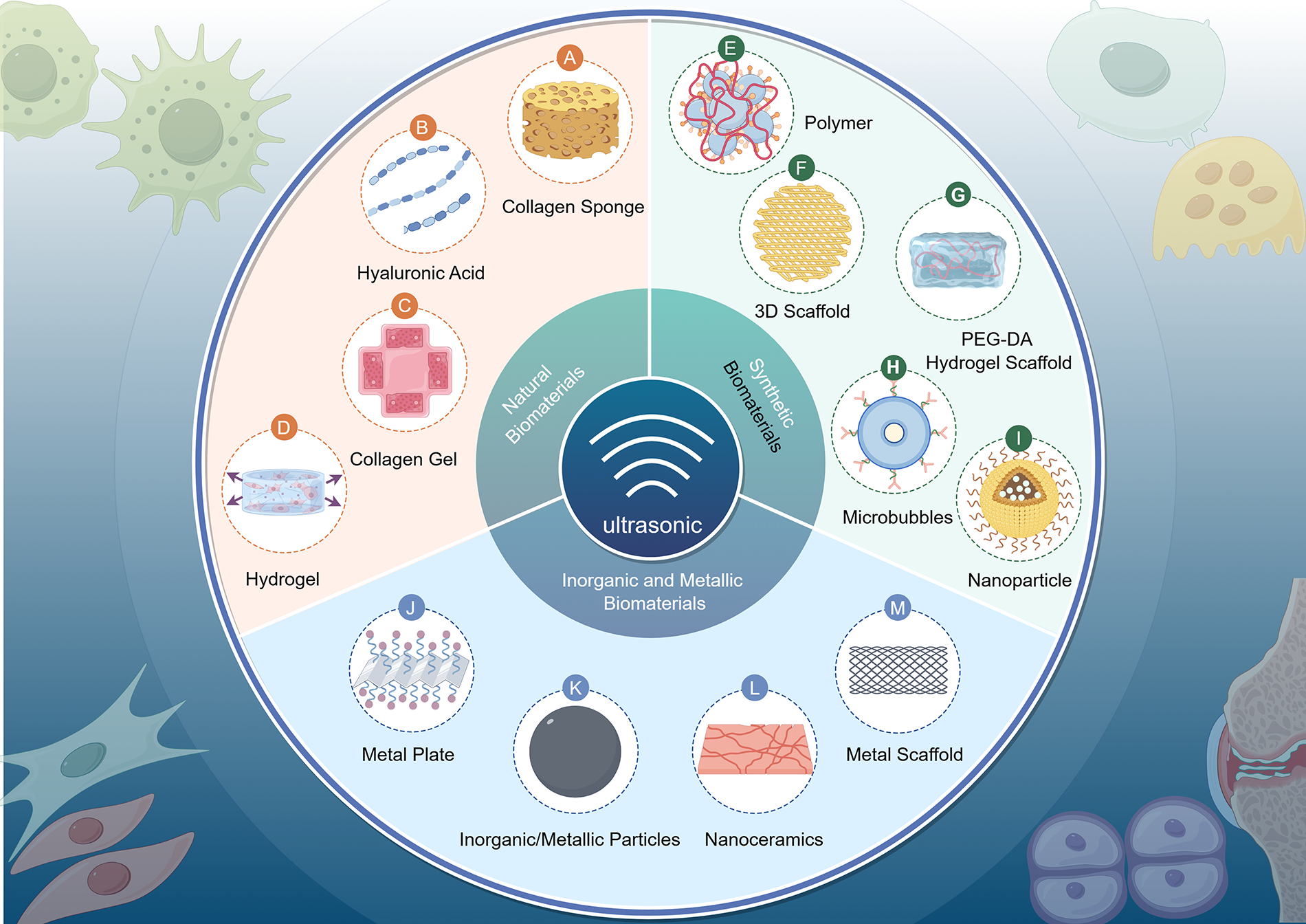
Various types of biomaterials can synergistically collaborate with the mechanical forces of US to exert biological effects that regulate cell behavior. US, ultrasound.
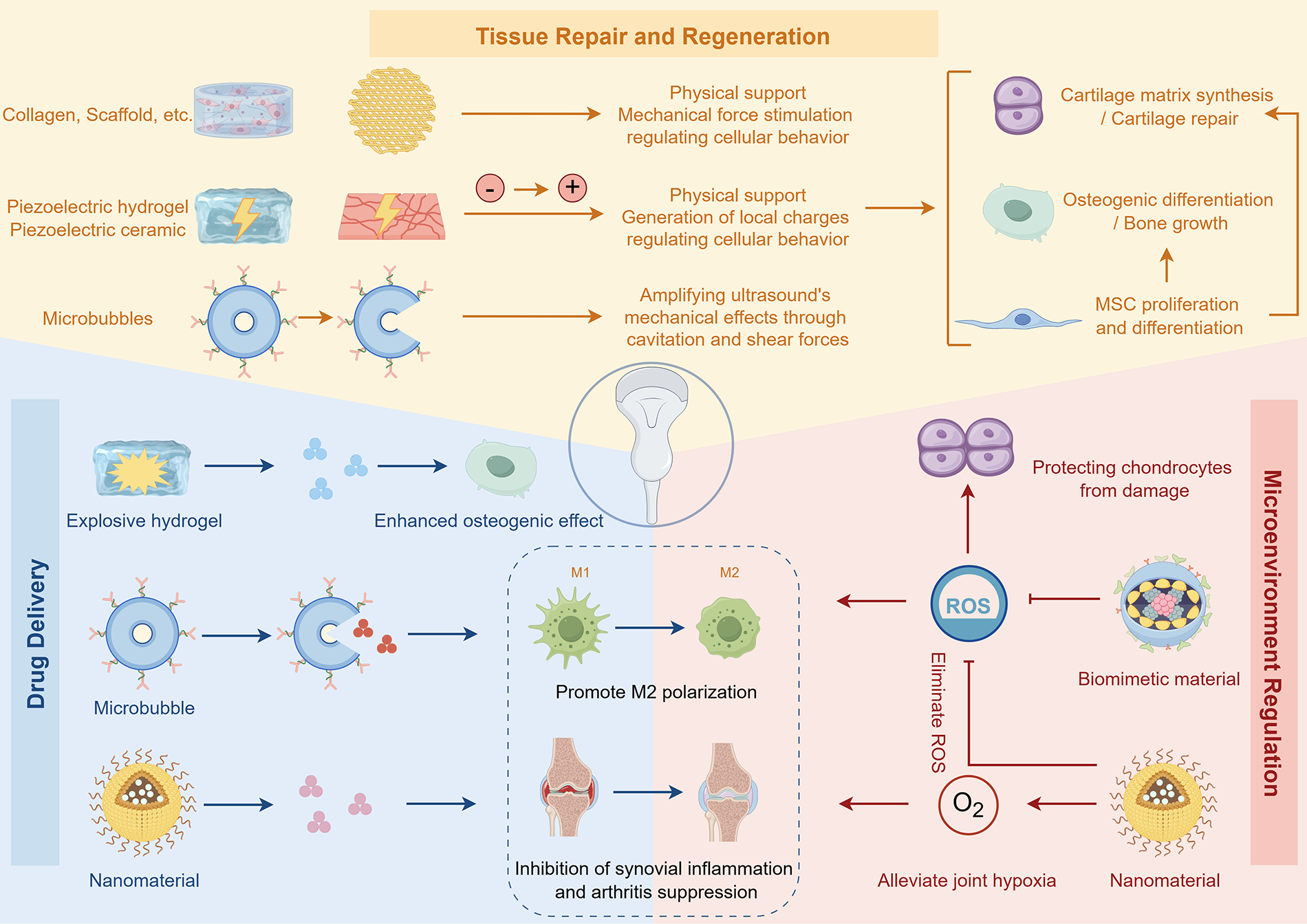
Biomaterials, particularly with US, have witnessed significant advancements in tissue engineering and regenerative medicine and are now widely employed for joint repair. US can modulate the physical properties of biomaterials, promote tissue repair and regeneration, enhance drug delivery, and regulate the local microenvironment. The synergistic effect of US and biomaterials promotes cartilage repair, reduces synovial inflammation, regulates subchondral bone remodeling and bone homeostasis, and ultimately promotes OA tissue repair. OA, osteoarthritis; US, ultrasound.
Effects on chondrocytes
Biomaterials such as collagen and HA, known for their biocompatibility and ability to mimic the natural ECM, are widely used in cartilage tissue engineering (Table 2). The synergistic interaction between US and biomaterials promotes cartilage repair primarily through mechanical stress. US-generated acoustic radiation forces and shear stresses directly stimulate chondrocytes while inducing microstrains in biomaterials, thereby mimicking the mechanical microenvironment of natural cartilage. Furthermore, cavitation effects enhance mass transport within the materials. Numerous experimental studies have confirmed this collaborative repair mechanism. For instance, Guo et al. 130 demonstrated that LIPUS treatment significantly enhanced repair outcomes in rabbit articular cartilage defects using an allogeneic chondrocyte-calcium alginate gel composite, yielding smoother cartilage tissue, improved tissue integration, and elevated collagen II expression. Similarly, another study indicated that low-intensity US enhanced the viability of human articular chondrocytes cultured in alginate while increasing the expression of Col II and proteoglycan. 131 In contrast, some studies reported limited effects of LIPUS on cell viability under specific conditions. Specifically, LIPUS stimulated GAG synthesis in monolayer or agarose-cultured chondrocytes by day 2, but this effect dissipated by day 5, indicating that the potential for pulsed low-intensity US-stimulated matrix synthesis may be limited in cartilage tissue engineering. 132
US Combined with Biomaterials for Cartilage Repair
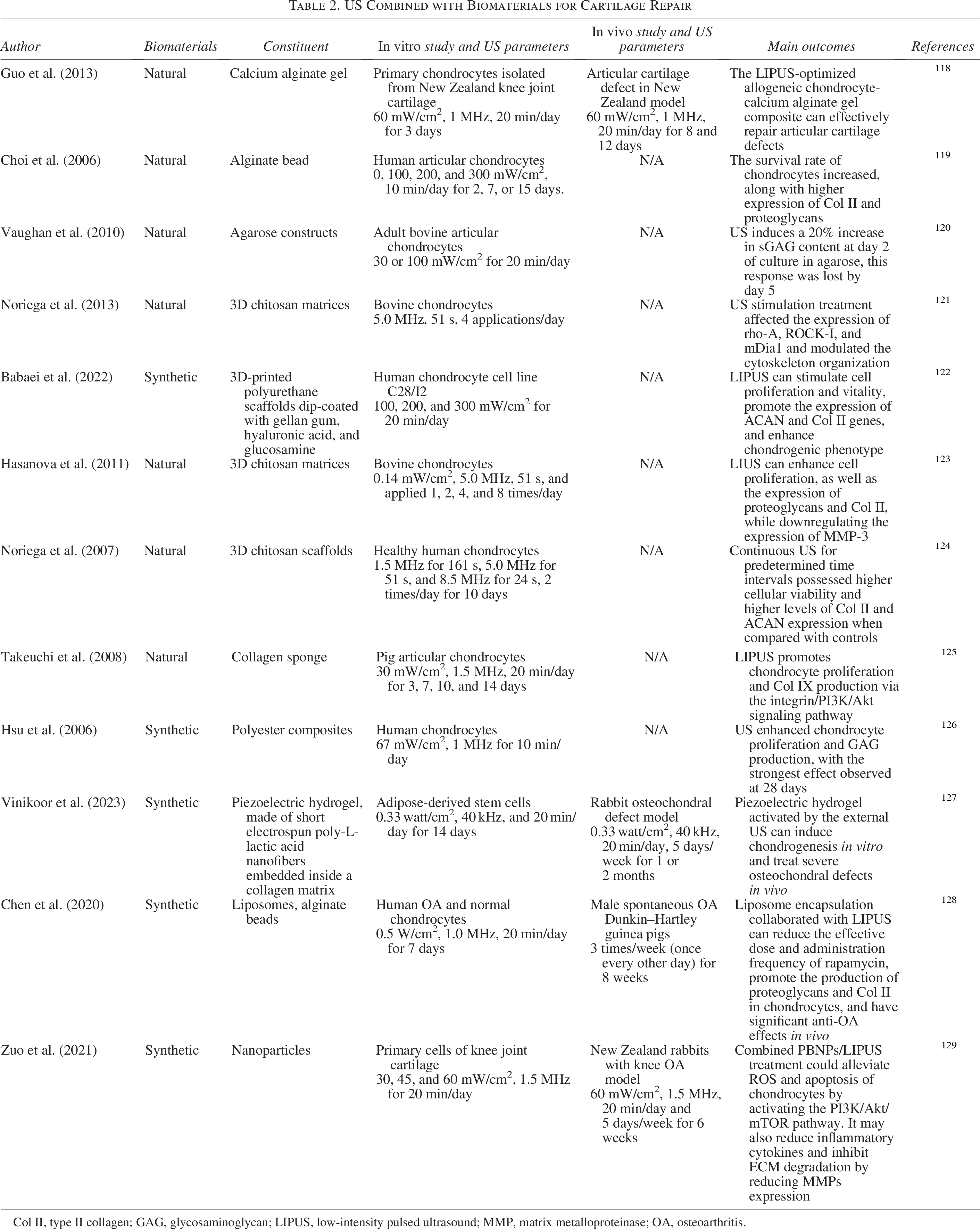
Col II, type II collagen; GAG, glycosaminoglycan; LIPUS, low-intensity pulsed ultrasound; MMP, matrix metalloproteinase; OA, osteoarthritis.
Recent investigations have increasingly focused on US’s effects on chondrocytes within 3D scaffolds. Low-intensity diffuse US stimulation was found to modulate cytoskeletal organization in chondrocytes seeded in 3D chitosan scaffolds. 133 LIPUS treatment on chondrocytes seeded in 3D-printed polyurethane scaffolds coated with gellan gum, HA, and glucosamine promoted chondrocyte proliferation, viability, and expression of cartilage phenotype-related genes. 134 Similarly, low-intensity US stimulation of bovine chondrocytes in 3D chitosan matrices enhanced cell proliferation and the expression of proteoglycan and Col II. 135 Continuous US stimulation in 3D chitosan scaffolds significantly enhanced chondrocyte viability and matrix synthesis, particularly increasing Col II and proteoglycan expression. 136 Mechanistically, LIPUS promotes chondrocyte proliferation and Col IX production in 3D scaffolds via the integrin/PI3K/Akt pathway. 137 Furthermore, Col II-modified polyester composite scaffolds combined with pulsed US enhanced chondrogenic potential of cultured chondrocytes. 138 Functional biomaterials, such as piezoelectric hydrogels, have revealed that US-induced localized electrical charges can stimulate stem cell migration, endogenous growth factor production, and chondrocyte marker expression. When injected into a rabbit osteochondral defect model, piezoelectric hydrogels, under US stimulation, facilitated cartilage healing. 139
The cavitation effect of US enhances cell membrane permeability, making it particularly effective for anti-inflammatory treatments. Studies show that combining LIPUS with liposome-encapsulated rapamycin in chondrocytes within alginate beads significantly increased proteoglycan and Col II production while suppressing IL-6 expression. Liposomes prolonged rapamycin’s efficacy, reduced adverse effects, and allowed LIPUS to lower the required drug dose and dosing frequency in OA treatment. 140 Furthermore, LIPUS improved the ROS scavenging ability of Prussian blue nanoparticles, reducing ROS levels, apoptosis, and MMP expression through activation of the PI3K/Akt/mTOR pathway, thereby protecting articular cartilage. 141 These findings highlight US’s dual capacity to modulate the cellular microenvironment while offering significant therapeutic benefits. In summary, US combined with biomaterials promotes chondrocyte proliferation, matrix synthesis, enhances drug delivery, and protects cartilage, demonstrating its potential in OA treatment.
Effects on synovial tissue and cells
The combined effects of US and biomaterials on synovial tissue have been primarily reflected in anti-inflammatory repair and drug delivery mechanisms. US enhances drug release and cellular uptake through cavitation and mechanical effects, enabling precise control of local drug concentration while promoting intracellular delivery via cell membrane permeabilization. 142 US with MBs improved skin permeability, enhancing diclofenac sodium gel delivery, which reduced synovial neovascularization and inflammation in arthritic regions. 143 US-driven sonodynamic therapy effectively killed activated synovitis cells while amplifying the therapeutic efficacy of nanozyme recombinant human serum albumin conjugated with SPX (Rh/SPX-HSA), further suppressing synovial inflammation. 58 Additionally, US-triggered MB destruction facilitated drug release from nanocarriers in the synovial cavity, reducing joint swelling, bone erosion, and inflammation in collagen-induced arthritis models. 144
Macrophages, as key immune cells in OA joints, drive cartilage degradation and accelerate OA progression through M1/M2 polarization imbalance and proinflammatory factor secretion. 145 US combined with biomaterials promoted macrophages polarization, cytokine secretion, and immune microenvironment improvement. US-stimulated release of biomimetic peptide nanofibers from hydrogels activated mitochondrial glycolysis, attenuated ROS production, and enhanced M2 macrophage polarization. 146 Piezoelectric biomaterials such as BaTiO3/collagen membranes, when activated by LIPUS, generated synergistic electrical and mechanical stimuli that enhanced Ca2+ influx via PIEZO1 channels, promoting bone regeneration. 147 Additionally, US-responsive IL-4-encapsulated microdroplets (MDs) not only suppressed inflammatory phenotypes in macrophages and enhanced M2 polarization but also promoted osteogenic differentiation in hBMSCs. 148
In summary, integrating US with biomaterials facilitates macrophage polarization, accelerates tissue regeneration, improves drug delivery efficiency, and modulates the microenvironment, effectively alleviating synovial inflammation.
Effects on subchondral bone
The pathological changes in subchondral bone in OA are closely associated with disrupted bone homeostasis. US combined with biomaterials demonstrates significant effects in regulating bone homeostasis and promoting bone regeneration. US induces hydrogel deformation, providing mechanical stimulation that promotes osteoblast differentiation. 149 Studies have shown that osteoblasts encapsulated in hydrogels with optimal stiffness exhibit upregulation of mineralization markers such as calcium, cyclooxygenase-2 (COX-2), and prostaglandin E2 (PGE2) under US stimulation, thereby enhancing mineralized tissue formation. In vivo experiments using nucleotide-binding oligomerization domain (NOD) scid gamma mice further confirmed that BMSC-laden optimized hydrogels with 4-week US treatment significantly promoted calvarial defect repair through synergistic cell delivery and mechanostimulus-induced osteogenesis. 150 Furthermore, US-responsive biomimetic hydrogel promotes BMSC osteogenic differentiation by inducing M2 macrophage secretion of BMP-2 and insulin-like growth factor I. In murine femoral defect models, these hydrogels significantly enhanced BMP-2 and type I collagen expression, demonstrating optimal bone healing efficacy. 146 Additional studies showed that LIPUS-generated mechanical stimulation further increased COX-2 and PGE2 expression in osteoblasts, elevating osteogenic markers including alkaline phosphatase and osteocalcin. 151 3D culture systems demonstrated superior osteogenic potential, with US-treated tricalcium phosphate scaffolds showing enhanced osteoblast adhesion and elevated phosphorylation of ERK1/2 and p38 phosphorylation compared with 2D cultures. 152
Piezoelectric biomaterials, capable of transducing mechanical stimuli into bioelectrical signals, work synergistically with LIPUS to promote osteoblast proliferation and differentiation. 153 For example, BaTiO3-coated scaffolds under intermittent LIPUS stimulation generated continuous electrical signals that activated mitochondrial function and cytoskeletal reorganization, improving osteoblast function. This combination of mechanical and electrical stimulation upregulated osteogenesis-related genes, particularly BMP-2, and reduced apoptosis, promoting osteoblast activity. 154
The combination of drug delivery and microenvironment modulation demonstrates significant potential in regulating bone homeostasis. US-responsive MDs, such as nuclear factor of activated T-cells cytoplasmic 1-polylactic acid (NFATc1-PLA)-exosome scaffolds, enhanced osteogenic differentiation of hBMSCs while modulating cytokine expression. 155 MDs-IL4 promoted macrophage polarization and further enhanced osteogenic differentiation. 148 Remarkably, these MDs exhibit temporally controlled structural transformations following US stimulation, with MDs-NFATc1 treatment significantly reducing osteoclast numbers, indicating a strong effect in inhibiting osteoclastogenesis. 156 Further studies have demonstrated that US-controlled “explosive” hydrogels loaded with resveratrol nanobubbles release drugs under low-intensity focused US, enhancing osteogenesis and maintaining bone microenvironmental homeostasis. 157 This innovative approach combining precise drug delivery with US-mediated modulation represents a promising therapeutic paradigm for regulating bone homeostasis.
Effects on MSCs
The combination of various hydrogel materials and US has shown notable effects in promoting chondrogenic differentiation of MSCs. In studies where rabbit MSCs were cultured in fibrin-HA or alginate hydrogels and treated with LIPUS in a chondrogenic medium, fibrin-HA hydrogels showed greater efficacy in promoting chondrogenesis, as indicated by increased production of sulfated GAGs and collagen, likely due to their superior mechanical properties. LIPUS further enhanced MSC chondrogenic differentiation in these hydrogels. 118
Similarly, scaffolds combined with LIPUS have promoted hMSC proliferation and chondrogenic differentiation. For instance, hMSCs seeded on a 3D-printed polyethylene glycol-diacrylate hydrogel scaffold showed increased proliferation and significantly higher levels of chondrogenic markers, including GAGs and Col II, following LIPUS treatment. 119 Further studies revealed that when lipid-coated, perfluorobutane-filled MBs were introduced, LIPUS treatment of hMSCs on this 3D scaffold led to a 40% increase in proliferation (compared with 18% with LIPUS alone). After 3-week chondrogenic differentiation, GAG production increased by 17% (vs. 5% increase with LIPUS alone), whereas Col II production rose by 78% (vs. 44% increase with LIPUS alone). 120 Similarly, in vivo studies using nonporous polyglycolic acid (PGA) scaffolds implanted in mice also showed increased chondrogenic biomarkers following LIPUS treatment.121,122 Furthermore, the combination of LIPUS and β-tricalcium phosphate (β-TCP) scaffolds synergistically promotes MSC proliferation and osteogenic differentiation. In experiments where BMSCs/β-TCP composites were subcutaneously implanted in rats, the LIPUS-treated group demonstrated significantly elevated alkaline phosphatase activity, osteocalcin levels, and improved angiogenesis and bone formation. 123
US combined with MBs has been shown to induce localized shear forces and controlled mechanical stress within cells, amplifying the mechanical effects of LIPUS. Nanoliposomes have been shown to act as nanomechanical force generators on cell membranes, promoting osteogenesis and bone formation in BMSCs through mechanisms involving TRPM7 ion channels and intracellular calcium oscillations. 124 Furthermore, 3D-printed scaffolds with high porosity, when combined with LIPUS, significantly promoted MSC proliferation, alkaline phosphatase activity, calcium deposition, and protein content. 125 Collectively, these findings highlight the potential of LIPUS combined with hydrogel scaffolds to enhance both chondrogenic and osteogenic differentiation of MSCs in vitro and in vivo.
Discussion
Numerous in vitro studies have shown that US can modulate ECM metabolism in OA joints and influence the microenvironment of synovium, cartilage, and subchondral bone.126,127 US also enhances the proliferation and differentiation of MSCs derived from various tissues, including bone marrow, adipose tissue, and the umbilical cord. 113 However, variations in experimental parameters, such as well thickness, gel layer, and medium volume, may introduce bias by affecting wave transmission and cellular responses. 158 Furthermore, temperature increases in the culture system may influence biological effects.18,61 Detailed analyses have revealed that at an US intensity of 30 mW/cm2, systems using coupling gel to connect the transducer to the culture plate exhibited a temperature increase of 3°C, whereas immersion of the transducer in water resulted in only a 0.2°C temperature change. This difference stems from the poor heat transfer performance of air medium leading to heat accumulation.128,158 Therefore, standardized US configuration calibration and temperature control measures are strongly recommended, particularly for nonimmersion systems.
The therapeutic effects of US vary between preclinical and clinical studies. US primarily shows positive effects in OA animal models, but these models are mostly limited to a single disease stage. There is a need to assess animal models at different disease progression stages to better mimic human OA development. Particularly lacking are comparative studies between young and aged models, whereas OA mainly occurs in the elderly population. Future studies should systematically compare the therapeutic effects of LIPUS in animal models of different ages, which is critical for clinical translation. The choice of appropriate animal models is critical, with species such as pigs, horses, and sheep offering closer anatomical resemblance to humans in terms of cartilage thickness and joint structure. 129 Despite promising results, US has not yet been incorporated into guidelines for knee OA management.159,160 This underscores the necessity for large-scale, multicenter clinical trials with stringent control over variables such as US intensity, frequency, and patient demographics to enhance the robustness of findings.
US demonstrates significant tissue penetration capability, but its therapeutic effects may vary significantly across different tissue types due to the involvement of multiple parameters, including frequency, intensity, duty cycle, and duration of intervention. 161 Compared with the mechanical forces required for bone regeneration, the mechanical stimulation needed for soft tissue repair is relatively weaker. In tissue engineering applications, various natural and synthetic scaffold materials have been developed to enhance US-mediated mechanical stimulation. For instance, MBs significantly amplify US cavitation effects to achieve controlled drug release, whereas hydrogels and scaffold materials provide cells with excellent structural support and an ideal microenvironment, thereby optimizing the mechanobiological effects of US. However, research on US parameters tailored to different material properties remains relatively scarce.
Effective OA treatment requires addressing joint cartilage, synovial inflammation, and subchondral bone remodeling. While most research has focused on bone tissue engineering, soft tissue repair also holds significant potential, including tendons, skeletal muscle, ligaments, and cartilage. 162 Developing novel US-responsive biomaterials requires a thorough understanding of US mechanisms, such as mechanotransduction, 163 cavitation effects, and cellular signaling cascades that determine biological responses. Further research is needed to explore the synergistic mechanisms between US and biomaterials.
In clinical translation, US, especially LIPUS, offers multiple advantages including localized application, noninvasiveness, ease of operation, and significant therapeutic benefits. As research progresses, LIPUS can target unmet clinical needs in the orthopedic field and is expected to significantly enhance OA management for numerous patients worldwide. Most importantly, LIPUS has shown unique synergistic therapeutic potential, but its efficacy and safety still require verification through more large-scale clinical trials.
Conclusion
Future research should focus on developing tunable US parameters and biomaterials for the treatment of OA across various pathological stages. This optimization strategy aims to enhance synergistic therapeutic mechanisms while expanding potential clinical applications. A deeper understanding of the mechanisms by which US operates, as well as the design requirements for biomaterials, will facilitate the provision of more personalized and precise therapeutic options for complex diseases.
Authors’ Contributions
W.H.: Writing original draft and literature search. X.H., C.P., and X.S.: Editing and revisions. T.X.: Conceptualization and revisions of the article. All authors have read and approved the final version of the article.
Footnotes
Funding Information
This study was supported by National Natural Science Foundation of China (grant nos. 82072556 and 82272610). The authors thank Figdraw for the assistance in creating schematic diagram.
Disclosure Statement
The authors have declared that no competing interest exists.
Declaration of Generative AI and AI-Assisted Technologies in the Writing Process
The authors declare that they did not use AI-assisted technologies to create this article.
Data Availability
The raw data supporting the conclusions of this article will be made available by the authors without reservation and can be obtained from the corresponding author.