
Abstract
The main objective of this research is to systematically summarize the characteristics of keratin-based materials and their current applications in the tissue engineering field, with a particular emphasis on highlighting their unique advantages over other traditional protein-based materials (such as collagen and silk fibroin). An electronic literature search of PubMed, Web of Science, and Scopus was conducted, identifying publications related to keratin-based materials and their application in tissue engineering. The majority of literature was published between 2015 and 2025. The structure of keratins, which is rich in disulfide bonds, gives it unique advantages in the field of tissue engineering, such as sustainability, versatility, and controllable degradability. Future research in this area could focus on improving the brittleness of keratin, developing more stable extracting sources, such as marine-derived sources, and conducting long-term clinical trials.
Impact Statement
Keratin-based materials have great potential in tissue regeneration and repair. This article reviews the research progress of keratin-based materials in recent years and summarizes the advantages of keratin-based products in tissue engineering fields, which contributes to the development of tissue engineering scaffolds.
Introduction
Biocompatible materials represent a critical class of natural or synthetic materials engineered to interface with biological systems for the evaluation, support, or replacement of tissues, organs, or bodily functions. 1 They have played an important role in biomedical fields since inception, enabling innovations such as tissue-mimicking implants, regenerative medicine platforms, and drug delivery systems. 2
Proteins are macromolecules capable of performing a wide range of biological functions. Their low cytotoxicity ensures minimal harm to living cells when integrated into biological systems, while their excellent biocompatibility allows interaction with tissues without triggering severe immune responses. Moreover, their biodegradability enables them to be broken down by the body’s natural enzymes into nontoxic by-products, avoiding long-term accumulation and potential complications. Over the past three decades, they have been widely used as biocompatible materials. 3 Collagen is extensively used in wound dressings due to its ability to promote cell adhesion and tissue regeneration. 4 Gelatin finds applications in drug delivery systems as a biodegradable matrix that controls the release of encapsulated drugs. 5 Silk fibroin is valued for its exceptional mechanical strength and biocompatibility, making it an ideal material for scaffolds in tissue engineering, such as for bone or skin regeneration. 6
Among different proteins, keratins stand out due to their high stability and adjustability. Besides, keratins can be easily obtained at a low cost. 7 In recent years, keratin-based materials have gained increasing attention. Many studies have reviewed keratins in terms of their extraction, properties, and biological applications.8–10 However, these studies have not placed keratins in the broader category of protein-based materials for horizontal comparison with systematically illustrate the performance and application advantages of keratin-based materials. Meanwhile, they may not incorporate the latest relevant research.
This article aims to summarize the current state of biomedical applications of keratin-based materials, shedding light on their advantages over other biocompatible materials, such as their biocompatibility, biodegradability, and cost-effectiveness. Additionally, this article tries to explore the prospects for their future development, including the expansion of application areas and the challenges and opportunities in their clinical translation.
Methods
This review conducted a comprehensive literature search to identify publications related to keratin-based materials and their application in tissue engineering. Databases searched included PubMed, Web of Science, and Scopus, with the majority of literature published between 2015 and 2025. Search terms comprised a combination of controlled vocabulary and free-text keywords, such as “keratin,” “tissue engineering,” “tissue regeneration,” “bone regeneration,” “vascular regeneration,” “nerve regeneration,” and “biocompatible materials.” Following deduplication and screening of all retrieved results based on their quality, approximately 115 articles relevant to this review were ultimately selected (Fig. 1).
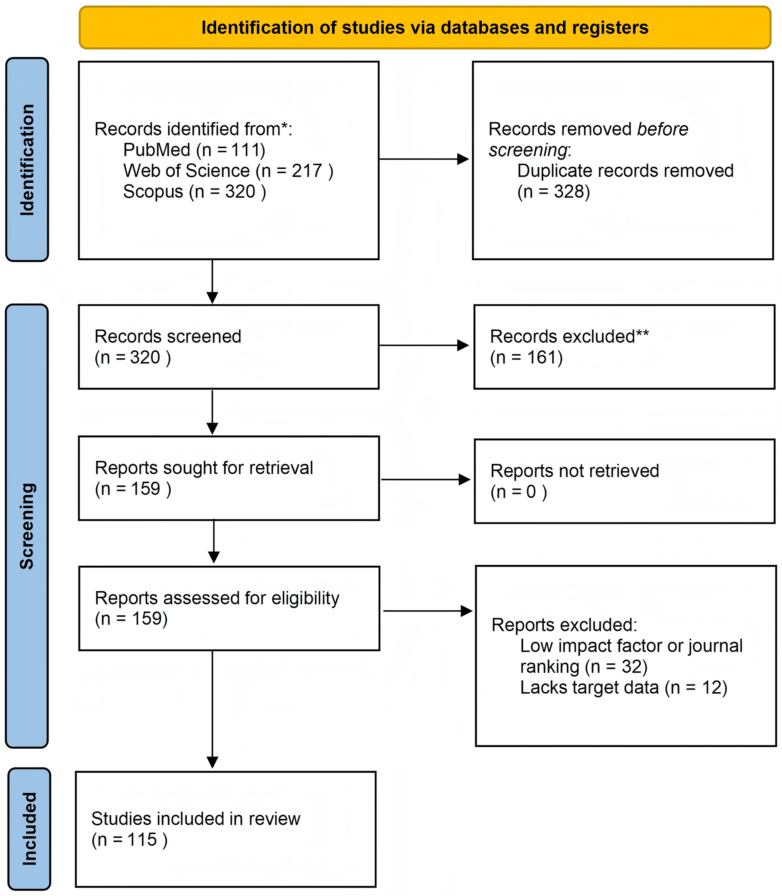
PRISMA 2020 flow diagram showing selection of studies. PRISMA, Preferred Reporting Items for Systematic Reviews and Meta-Analyses.
The structure and classification of keratins
Keratins are an insoluble protein primarily distributed in the epithelial tissues of humans and animals. It is ubiquitously sourced from abundant agro-industrial waste streams and natural sources, including human hair, wool, feathers, nails, horns, and hooves. They can be extracted from human hair, wool, feathers, beaks, hooks, nails, horns, scales, and claws. 9 In biological organisms, keratins are synthesized by epithelial cells and belong to the family of intermediate filament proteins. They exhibit a complex hierarchical structure, which can be categorized into polypeptide chain structure, filament matrix structure, lamellar structure, and “sandwich” structure, from the microscopic to the macroscopic scale. 11 Typically, the polypeptide chain structure refers to the sequence of amino acids that constitute keratins. The filament matrix structure, which is a nanoscale structure, refers to the filaments composed of multiple polypeptide chains arranged in an orderly manner and embedded in the amorphous matrix, forming the basic structure of keratins.
Based on the structural feature revealed by X-ray diffraction, keratins can be structurally classified into α-keratin, β-keratin, feathery keratin, and amorphous keratin. Since the diffraction image of feathery keratin is identical to that of β-keratin, it is generally accepted that feathery keratin belongs to β-keratin. 2 Amorphous keratin refers to the amorphous matrix of keratin. Therefore, keratins are usually divided into two types, α and β, based on their ordered structure.
α-Keratin is predominantly found in mammals, with common sources including wool, hair, nails, hooves, and horns. The polypeptide chains are arranged in the form of a right-handed α-helix, and two right-handed α-helical polypeptide chains are connected by disulfide bonds to form a left-handed α-helical dimer. The dimer, cross-linked by disulfide bonds, is connected and staggered in parallel to form a prefilament with a diameter of 2 nm. Subsequently, two prefilaments arranged in parallel form fibrils, and four fibrils combine to form an intermediate filament with a diameter of 7 nm12,13 (Fig. 2). As keratinocytes approach maturity, sulfur-rich amorphous matrix proteins are produced between the intermediate filaments and then attached to form keratin. Among them, the intermediate filaments have a lower sulfur content, while the amorphous matrix proteins mainly consist of protein chains containing cysteine residues or a large number of glycine, tyrosine, and phenylalanine residues and have a higher sulfur content.
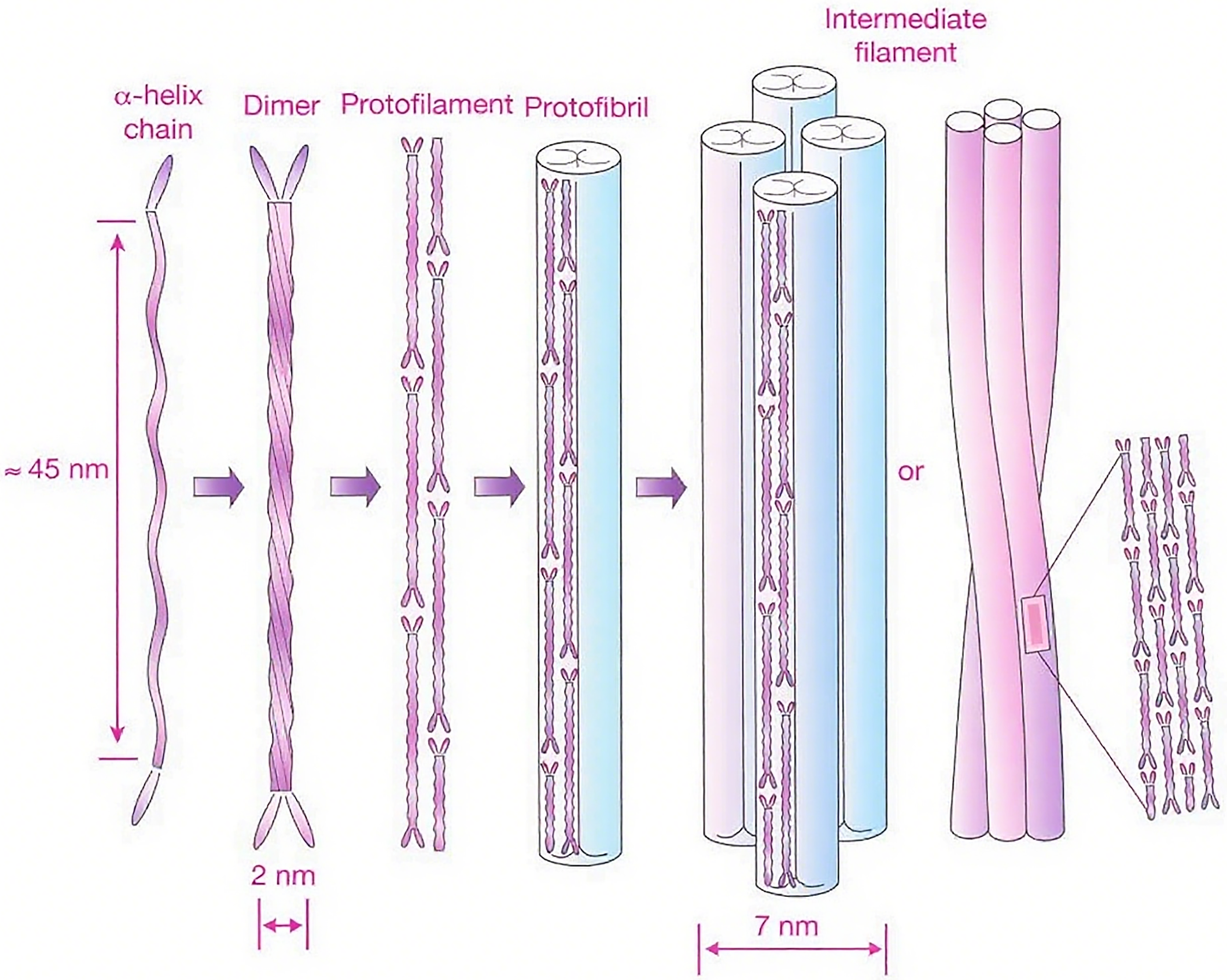
Intermediate filament structure of α-keratin.
β-Keratin is mainly present in the hard tissues of birds and lizards, such as feathers, beaks, claws, and scales. Unlike α-keratin, the keratin filaments and amorphous matrix proteins of β-keratin are not synthesized in an orderly manner. The polypeptide chain of β-keratin exists in a β-folded structure. Each polypeptide chain is folded into four transversely arranged peptide chains, which are connected by hydrogen bonds. These chains are then arranged in parallel or antiparallel to form β-folded sheets, with the antiparallel arrangement being more stable. The β-folded sheet is subsequently twisted to form a left-handed helix, in which two folded sheets are superimposed in reverse to form a β-keratin filament with a diameter of 4 nm, 14 which binds to the surrounding amorphous matrix protein (Fig. 3). In β-keratin, there is no significant difference in sulfur content between keratin filaments and matrix proteins.
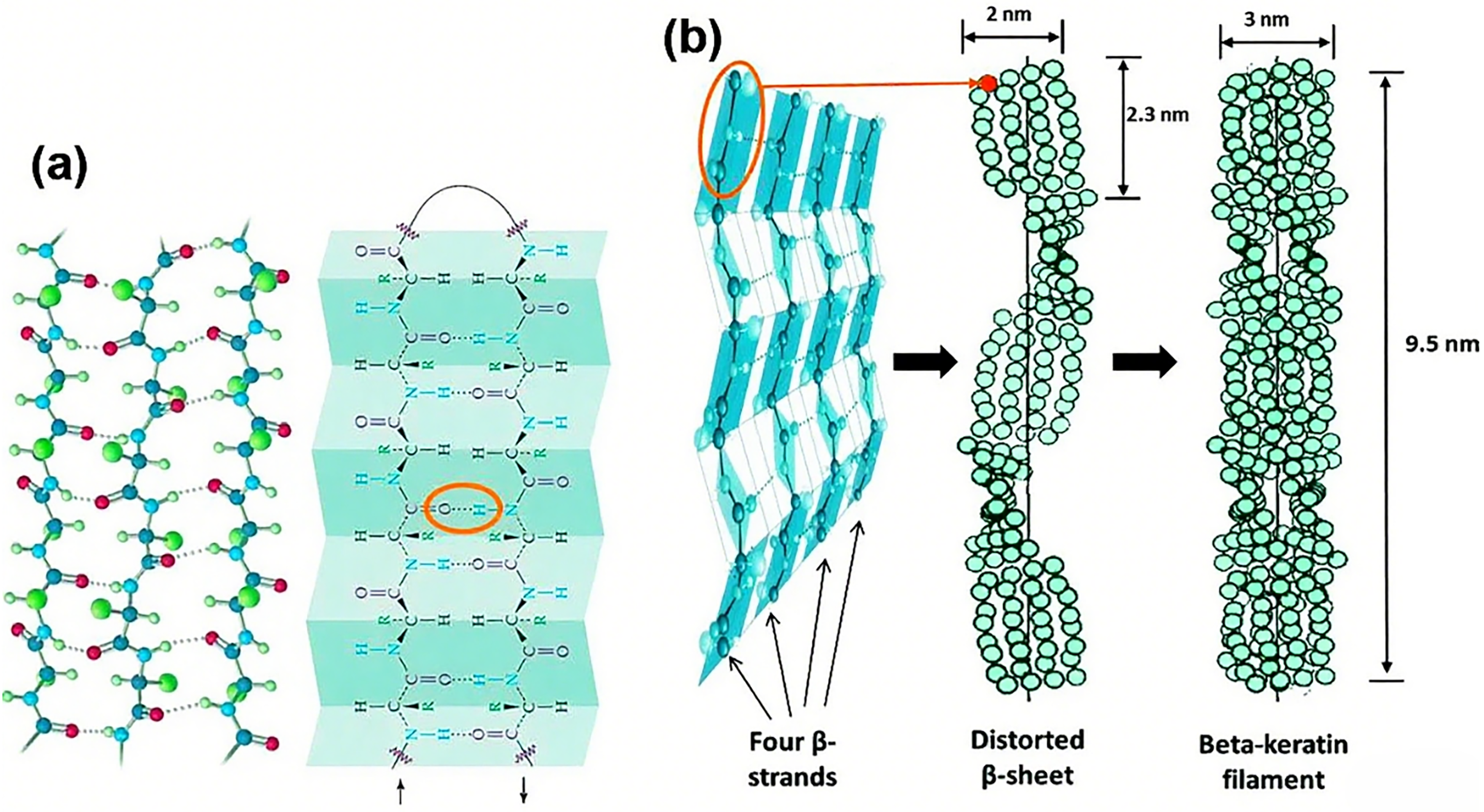
Structure of the β-keratin filaments:
Properties of keratin
Mechanical properties
The most prominent advantage of keratins is their excellent mechanical strength. In general, keratins exhibit higher hardness and lower solubility than other proteins due to their larger number of disulfide bonds. The mechanical properties of different keratins are closely related to the types and content of amino acids. Keratins with more cysteine residues are harder because of their higher disulfide bond density. 10
When humidity or moisture content increases, the stiffness, strength, and hardness of keratin-based materials decrease. This high moisture sensitivity can be modified. For instance, Wu et al. prepared feather keratin/polyvinyl alcohol/tris(hydroxymethyl) aminomethane film using transglutaminase (TG), CaCl2, and genipin as cross-linking agents. All three cross-linkers were found water resistance of the blend films, in addition to enhancing the mechanical properties. The elongation at break increased from 10.83% to 31%, 36.2%, and 50.5% (corresponding to TG, CaCl2, and genipin cross-linking, respectively), while the tensile strength increased from 9.58 MPa to 12.34 MPa, 12.03 MPa, and 11.04 MPa (corresponding to TG, CaCl2, and genipin cross-linking, respectively). 15 α-Keratin has higher hardness and lower tensile properties due to the high sulfur content of its amorphous matrix. Under wet conditions, its breaking strain is approximately 45%, and Young’s modulus is around 2000 MPa. 16
Although keratins possess higher strength, they are also more brittle. 17 Therefore, other substances are often added to modify keratin-based materials (Table 1). For example, Wei et al. added glycerol to the keratin/gelatin/curcumin composite films, and the tensile strength of the film increased from 12.45 to 13.73 MPa, and the elongation at break increased from 4.6% to 56.7%. 23 Ding et al. added citric acid to an FK/PVA/PEO nanofiber membrane and found that the tensile strength of citric-treated membranes was about 4.5 times higher than that of the control group. 21
The Mechanical Properties of Keratin-Based Materials After Adding Different Modifying Agents or Using Different Modification Methods

Better mechanical properties make it possible for keratin-based materials to maintain certain shape and withstand the stress in vivo, thus achieving more satisfying results in clinical applications.
Biocompatibility and biodegradability
For a tissue engineering material, excellent biocompatibility and biodegradability are essential, and keratins not only meet but also excel in these criteria.
Biocompatibility refers to the ability of a material to interact with cells without producing toxic side effects. Collagen and gelatin are typical protein materials commonly used in biomedical engineering. Keratins have poorer biocompatibility compared with collagen, but their biocompatibility is comparable with that of gelatin, 18 making it a potential candidate for human body applications.
The biocompatibility of keratin is demonstrated by its capacity to support the proliferation of a wide range of cells, 19 mainly due to the presence of cell-binding motifs such as arginine–glycine–aspartate, leucine–aspartate–valine, leucine–aspartate, and glutamate–aspartate, 9 which are capable of forming fibronectin-like cell-binding domains. 7 Studies have shown that keratins promote fibroblast proliferation mainly because it activates the integrin–extracellular regulated protein kinases (ERK) pathway and upregulates the synthesis of collagen I/III. 23 Moay et al. fabricated a sponge from keratin and alginate that supports the proliferation of human dermal fibroblasts. 20
Lin et al. found that human hair keratin scaffold can increase the proliferation rate of MG63 osteoblasts by 30 − 50%, 22 and in a further study, they found that the keratin/chitosan membrane can promote the osteogenic differentiation of human adipose stem cells. 24
Keratins have also been revealed to support the growth and adhesion of several cell types such as hepatocytes, 25 mesenchymal stem cells, 26 Schwann cells, 27 and dental pulp stem cells 28 in both two-dimensional (2D) and 3D environments (Table 2). This high biocompatibility allows keratin-based materials to be safely used in vivo and able to repair a wide range of tissue defects.
Biocompatibility and Application Fields of Keratins

Biodegradation refers to the process by which biodegradable polymer materials are catabolized into nontoxic small molecules in the human body. Keratins can be catalyzed by trypsin in humans, and a recent study demonstrated that approximately 50% of keratin hydrogel materials were degraded after 42 days of cell culture in vitro. 36
This enables keratin-based materials to degrade in vivo, so no invasive methods are needed to take the material out of the human body.
Hemostasis ability
In clinical applications, keratin has been proven to possess hemostatic effects and can accelerate blood clotting. The possible mechanism is that keratin-based materials can mimic the extracellular matrix to mediate platelet adhesion through glutamate–aspartate–serine/leucine–aspartate–valine sites, providing a catalytic interface for the coagulation cascade and thereby promoting primary coagulation. 5 Furthermore, keratin accelerates the polymerization of fibrinogen to fibrin, 14 thus accelerating secondary coagulation.
Van Dyke’s group utilized human hair-derived hydrogels as a hemostatic material and found their effects comparable with those of commercial hemostatic agents (Hemcon® and Quikclot®) in a lethal liver transection rabbit model. 43 Luo et al. prepared keratin nanoparticles via the emulsion-diffusion method, which demonstrated excellent hemostatic efficacy. In a liver puncture experiment, the application of these nanoparticles resulted in an approximately 400% reduction in blood loss. Additionally, the keratin nanoparticles achieved hemostasis over 150% and 250% faster compared with controls. 44
Excellent hemostasis ability enables keratin to perform well in wound healing and work as a hemostatic product in clinical applications.
Antimicrobial ability
In addition to the aforementioned prominent characteristics, keratin-based materials also possess a minor antimicrobial function. Feather keratin, which has fewer α-helices and a random structure, exhibits higher antibacterial activity than wool keratin with more α-helices and an ordered structure.9,45 Therefore, it is speculated that the antibacterial ability of keratin is related to its secondary structure. Compared with α-keratin formed by α-helices, the lamellar conformation of β-keratin binds to lipopolysaccharide on the bacterial surface more easily and induces changes in membrane permeability. 46 Current keratin-based materials have inhibitory effects on both Gram-positive bacteria, like Staphylococcus aureus, and Gram-negative bacteria, like Escherichia coli (Table 3). 47 Processing keratins into peptides or nanoparticles can enhance their antimicrobial ability. 52
Antimicrobial Ability of Different Types of Keratins

Antimicrobial properties can inhibit the occurrence of infection, promote wound healing, and tissue recovery.
Drawbacks
Despite its versatile properties, keratins also have some inherent limitations. The tensile strength of keratin-based materials is generally low, and their mechanical integrity is rapidly compromised under stress or environmental changes. Additionally, their mechanical properties are highly sensitive to environmental humidity, with stiffness significantly decreasing in moist conditions, making it difficult to maintain consistent performance across varying environments. 53 Compared with other biomaterials such as silk fibroin, keratin may exhibit relatively limited functionality in inducing high cell migration and promoting the expression of genes related to wound healing.
Overall, the various properties of keratin remain quite competitive compared with other protein-based materials (Table 4).54–76
Comparison Between Common Protein Materials

Biomedical applications of keratins in tissue engineering
When the structure and properties of keratins are considered together, it proves to be a highly suitable tissue engineering material.
Tissue engineering induces the migration and differentiation of tissue cells through natural or synthetic biomaterials, which can restore the structure and functions of organs or tissues and avoid the immune problems caused by transplantation. 8 The biocompatibility, biodegradability, nontoxicity, and nonimmunogenicity of keratin make it an ideal tissue engineering material. It has been widely used in the regeneration of bone and cartilage tissue, nerve tissue, and vascular tissue. 24
Skin tissue
The skin serves as the natural barrier of the human body, defending against external bacteria. However, when the skin is compromised due to trauma, burns, surgery, or chronic diseases, bacteria can invade the body and cause inflammation. This process can be mitigated by using dressings to promote wound healing. Given that keratin possesses properties such as hemostasis, antibacterial activity, and anti-inflammatory effects, 77 it is an ideal wound dressing material for normal and chronic wounds like diabety 78 or radiated 79 wounds. Naturally derived keratin, such as the molt of the spot-bellied pit viper and human hair, has been shown to promote granulation tissue formation, re-epithelialization, neovascularization, and hair follicle development in the early stages of wound healing.80–83 In current studies, keratin composites are prepared as membrane- or hydrogel-based materials to enhance the hemostatic and antimicrobial properties of the material. 84
Membrane-based materials
Membrane is a common form of keratin wound dressing. A biofilm made from horn-derived keratin and chitosan supports the attachment of fibroblasts and acts as a carrier for the release of the antimicrobial drug mupirocin, improving the bioavailability of the drug at the wound site, increasing the absorption of exudate, and reducing patient pain by decreasing the frequency of dressing changes. 85
However, membranes fabricated by casting often have a lower porosity rate, leading to lower water vapor transmission rates. This may be overcome by mixing keratin with other substances, like chitosan, which has a higher water vapor transmission rate. Ganesan prepared a cast keratin film with silk fibroin and chitosan. The water vapor transmission rate of the composite membrane was about 1800 g/m2/day, comparable with the commercial wound dressing. 86
Another way of increasing the water vapor transmission rates is to fabricate the membrane using keratin nanofiber, which can be achieved through electrospinning. 47 Nanofiber electrospinning technology can produce nanofiber membrane materials with higher porosity, thus enhancing water vapor permeability and the absorption of wound exudate.87–90 The morphology of the nanofiber structure is similar to that of collagen fibers or elastin fibers in the natural extracellular matrix, thereby promoting the adhesion and migration of fibroblasts and keratinocytes and ultimately contributing to the regeneration and healing of the wound site. Ye et al. extracted high-molecular-weight keratin from wool through a multienzyme cascade, then electrospun it with 3-hydroxybutyrate-co-3-hydroxyvaleric acid (PHBV), and subsequently introduced silver ions into the nanofibers through in situ bioreduction to form polymer keratin/PHBV/silver nanoparticle nanofiber membranes. 91 The high molecular weight of keratin (120 kD) confers good strength to the material, with its Young’s modulus and tensile strength exceeding that of the human stratum corneum.
New technologies can improve the performance of keratin membranes and expand their application. Navarro et al. prepared membrane from keratin photosensitive resin and tyrosine in oxidized keratin through cross-linking and 3D printing and were able to regulate the substance transportation of the membrane by altering the cross-linking degree. The prepared membrane can enable growth factors from the subcutaneous fat layer to penetrate into the dermal membrane and thereby promote wound healing. 92
Hydrogel-based materials
Hydrogel is a material with a biological network structure formed by the cross-linking and polymerization of hydrophilic macromolecules. Due to their high water content (70 − 90%), hydrogels can provide a favorable environment for cell expansion. Unlike casted membrane materials, hydrogels have a porous structure and thus have better water and gas permeability, as well as higher absorption of exudate. In situ cross-linked keratin hydrogels prepared from human hair-derived keratin can effectively promote keratinocyte migration and epithelial–mesenchymal transition, thereby facilitating wound healing. 93 Feather-derived keratin hydrogels exhibit similar wound healing effects to human hair-derived keratin hydrogels 94 and even have higher fibroblast proliferation rates and better mechanical strength. 95
However, low mechanical properties restrict the application of hydrogels. This limitation can be improved to some extent by cross-linking with other substances and improving the preparation process.19,96 Zhu et al. used tannic acid as a cross-linker to modify keratin/sodium alginate/carboxymethyl chitosan hydrogels. The elastic modulus of the modified hydrogel was 589.74 kPa, the toughness was 211.74 kJ/m3, and the elongation at break was 75.39%, which was similar to the human skin modulus. 97 Wang et al. modified the structure and properties of keratin hydrogels by controlling the number of freeze–thaw cycles. The compressive stress of RHK hydrogel (FT9) reached 14.927 kPa when the compressive strain was 53%. 98
Bone tissue
Trauma, surgery, and systemic diseases can lead to bone defects. Among the methods of repairing bone defects, natural bone grafting is costly and can cause damage to the donor site. Synthetic bone scaffolds are nondegradable and lack osteogenicity. Therefore, bone tissue engineering scaffold materials combined with bioactive factors have become an effective way to repair bone defects. 9 Keratin is an ideal bone tissue engineering material due to its stable structure and ease of modification. At present, most studies have prepared composite materials with other polymers to improve the mechanical and biological properties of keratin, thereby achieving better regeneration effects.99–102
Hydroxyapatite (HA) is one of the constituent substances of human bone tissue, and thus it is often used in bone tissue repairing materials.102–106 Sarrami et al. prepared a nanocomposite scaffold by adding nano-HA to a polyhydroxybutyrate-keratin scaffold, which significantly enhanced the tensile strength and Young’s modulus of the scaffold. It also improved the hydrophilicity of the material, slowed down the weight loss and pH change of the scaffold during degradation, and enabled the scaffold to deposit a calcium phosphate layer in simulated body fluid. 103 Feroz et al. prepared a bilayer keratin/HA membrane containing two different concentrations of HA to mimic the hierarchical structure of alveolar bone, which was shown to have good osteogenic properties. 104
Montmorillonite is a silicate with good biocompatibility, which can be used as a carrier for ion-exchange drugs 107 and can also improve the mechanical properties and biological activity of hydrogels. 108 Studies have shown that montmorillonite can upregulate the expression of the bone morphogenetic protein/Smad proteins (BMP/SMAD) signaling pathway, ultimately enhancing the expression of osteogenic genes to promote osteogenesis. 109 Ke et al. prepared feather keratin–montmorillonite nanocomposite hydrogels, and the addition of montmorillonite significantly improved the mechanical properties of the hydrogels. It also promoted the osteogenic differentiation of bone marrow mesenchymal stem cells by activating the BMP-2/RUNX2/p-SMAD pathway, ultimately repairing tissue defects. 13
Nerve tissue
The longitudinal microfilament structure of keratin can provide an adhesion mediator, growth space, and orientation for Schwann cells. Additionally, it can promote the secretion of neurotrophic factors, regulate the expression of inflammatory factors in macrophages, and facilitate the elongation of Schwann cell axons to repair peripheral nerve tissue. 110 Qin et al. used human hair keratin as a conduit and carried IL-1β-activated Schwann cells on it to construct an artificial nerve graft. This graft successfully restored the function of the sciatic nerve in mice, with better outcomes than direct suturing. The study proposed that the artificial nerve may promote nerve repair by upregulating the fibroblast growth factor 2 (FGF2) and transforming growth factor-β (TGF-β) signaling pathways. 37
Vascular tissue
In vascular tissue engineering, tubular biodegradable materials are inserted into the body to induce the migration and growth of vascular endothelial cells, forming new blood vessels and replacing diseased ones. Keratin-based materials are ideal biomaterials due to their rich content of cell-binding motifs, which can mimic the binding of tissues to receptors of various cell surface integrins, thereby promoting cell migration, adhesion, and growth. To prevent thrombosis and maintain a higher graft patency rate, keratin-based materials are often combined with anticoagulant drugs.41,111–113 Wan et al. co-electrospun poly(ε-caprolactone) (PCL) with keratin and covalently conjugated heparin to create heparinized PCL/keratin pads. These pads can catalyze the release of NO from NO donors in the blood, promoting endothelial cell growth, reducing smooth muscle cell proliferation and platelet adhesion, and thus enhancing the adhesion and growth of human umbilical vein endothelial cells. 111 The addition of copper 114 or silver nanoparticles 115 to PCL/keratin pads can improve the antimicrobial properties of the material.
Advantages of keratin-based materials in biomedical applications
Mechanical strength and stability
Strong intermolecular interactions, including disulfide bonds, hydrogen bonds, ionic bonds, and hydrophobic interactions, render keratin-based materials more rigid than other biopolymeric materials such as collagen and gelatin. The elastic modulus of hard α-keratin (e.g., human hair, wool) can reach 2.3–4.5 GPa in the dry state, and the modulus of soft α-keratin (e.g., epidermis) can also reach 2.0 GPa. 116 On the other hand, pure collagen has weak mechanical properties, typically with a tensile strength of 0.19–0.44 MPa (e.g., porcine collagen films) and a Young’s modulus of 1.88–2.86 MPa. 117 And in some studies, the Young’s modulus of gelatin was reported about 40 kPa.118,119 With high elastic modulus, keratin-based materials can meet the need for mechanical support in tissue regeneration of bone, blood vessels, nerves, and so on.
In terms of biodegradation, keratin demonstrates superior stability compared with the majority of other biopolymers. Keratin is insoluble in water, weak acids, weak alkalis and organic solvents under physiological conditions and remains stable in the presence of pepsin and trypsin. 120 This resistance derives from its high content of disulfide bonds, which hinders the exposure of the binding site of the enzyme. 5 It is the only natural biomaterial that is not a substrate targeted by specific tissue transformation-related enzymes, 121 which confers long-term stability and degradation controllability after implantation.
Studies have shown that under the same conditions, keratin degrades slower than collagen. 122 Keratin hydrogels need 3–4 weeks to fully degrade in vivo, 123 whereas pure collagen is nearly completely degraded (about 100%) within 15 h in the presence of collagenase. 124 And in the presence of enzymes, gelatin degrades 80 − 90% within 72–96 h. 125
This slow degradation rate facilitates in situ regeneration of organs and tissues. It shows significant advantages in medical scenarios that require long-term functional maintenance such as bone regeneration, wound healing, and drug delivery.
Functional versatility and modifiability
Compared with other protein materials, keratin is rich in carboxyl, amino, and disulfide bonds. The presence of carboxyl and amino groups enables keratin to react with acids.
The presence of carboxyl, amino, and disulfide bonds also provides sites for the coupling, functionalization, and chemical modification of keratins, making it possible for the creation of versatile functional composites. Han et al. prepared hydrogels for drug delivery using iodoacetamide as an alkylating agent for keratin alkylation. 126 Different levels of alkylation lead to different viscoelastic properties and erosion time, matching the diverse needs of drug delivery. Cal et al. prepared a boron–silicon–collagen–human hair keratin hydrogel with collagen and human hair keratin treated with tetraethoxysilane to promote osteogenesis. 127
Intrinsic biological activity
Despite the cell-binding motifs promoting cell proliferation and adhesion, keratin also plays a role in regulating the immune system. Evidence proved that keratin biomaterials could induce macrophage transformation to an anti-inflammatory phenotype (M2 type), reduce the release of proinflammatory factors (e.g., TNF-α, IL-6), and increase the expression of anti-inflammatory factors (e.g., IL-10, TGF-β), thus reducing inflammation and promoting tissue healing. 128
Additionally, the abundant cysteine residues and disulfide bonds in keratin are easily oxidized, endowing keratin with reducing properties. This allows keratin to scavenge free radicals, resist oxidation, and avoid inflammatory reactions and cell necrosis caused by free radical accumulation. 19 Keratin particles extracted from chicken feathers have demonstrated strong anticancer activity against HeLa and SK-OV-3 cell lines, as well as strong free radical scavenging activity against 2,2-diphenyl-1-picrylhydrazyl and 3-ethylbenzothiazoline-6-sulfonic acid. 129 This suggests that keratin-based materials have potential applications in the antitumor field.
Keratin is also present on the surface of natural cells and may be involved in receptor-mediated endocytosis and immune evasion in cancer cells. These cell surface keratins can be targeted by chemotherapy drugs, 130 making the application of keratin in the antitumor field feasible.
Wide and sustainable sources
Keratin is mainly derived from animal by-products such as hair, feathers, nails, and ungulates. 131 For example, agricultural and slaughterhouse wastes such as chicken feathers and wool can be used as low-cost raw materials to reduce environmental pollution. In contrast, collagen is mostly extracted from animal skin or bone, and chitosan relies on crustacean shells, which are subject to resource competition and ethical controversy.
Recently, marine-derived biomaterials have attracted increasing attention. A large number of protein-based materials can be extracted from fish and fishery processing wastes (such as fish scales and skins), featuring high resource utilization efficiency. Marine-sourced keratin exhibits excellent properties 132 ; dry hagfish threads show high initial stiffness of 3.6 GPa and a high tensile stress of 530 MPa while wet threads exhibit stiffness of 6 MPa and tensile strength of 180 MPa.116,133
Despite the abundance of sources, the extraction of keratin is still a conundrum. Keratin is extracted from hair or feathers, which carries the risk of residual animal-derived pathogens. Moreover, chemical extraction processes (e.g., the reduction method) may introduce defects associated with cytotoxic reagents. Enzymes derived from marine bacteria may serve as the key to breakthroughs in keratin extraction technology. The high-salinity, low-temperature, and alkaline environment of the ocean enables enzymes derived from marine bacteria to have a wide range of temperature and pH, and salt tolerance.134–145 Like marine bacterial dextranases 137 and marine collagenase 138 show high catalytic performance and stability, studies on marine keratinase139–142 proved marine bacterial keratinase an effective and eco-friendly way to extract keratin from keratinous sources. The continuous discovery of keratinases in marine bacteria represents a direction for the efficient extraction and large-scale production of keratin in the future.
Currently, most studies focus on the extraction of marine-sourced collagen and collagenase,132,143 while relatively few studies focus on keratins and their extraction. The abundant marine resources can significantly increase keratin’s yield, laying a foundation for the large-scale production of keratin-based products.
Conclusion and Outlook
Keratin-based biomaterials possess a set of distinctive advantages that set them apart from conventional protein-derived materials. These include their unique chemical profile characterized by high sulfur content, enhanced structural stability, exceptional functional versatility paired with tunable modifiability, inherent biological activity, and an environmentally sustainable sourcing pathway. Such multifaceted attributes render keratin biomaterials a highly compelling option in biomedical research and applications, driving their integration into diverse domains, including wound repair, controlled drug delivery, and tissue regeneration strategies.
However, several challenges remain. The most critical issue is its relatively high brittleness, which results in poor film-forming ability. This limits its applications to a certain extent. Currently, improvements are mainly achieved by adding cross-linking agents and modifying disulfide bonds, but there is still room for further enhancement, such as developing novel cross-linking agents, creating keratin-based composite materials, composite structures, and processing methods.144,145 Second, safer extraction methods with higher stability and greater efficiency are yet to be developed; marine organisms could be a priority for future research focus.
Last, most of the current studies are limited to cell experiments or small animal experiments (e.g., rat skull defect models) and lack long-term repair data from large animals (such as sheep and pigs), as well as evidence from multicenter clinical trials. To fully unlock their biomedical potential, future studies must address critical challenges spanning cost optimization, scalable manufacturing processes, and successful clinical translation.
These hurdles represent key milestones that need to be overcome to transition keratin-based technologies from experimental stages to practical health care solutions.
Authors’ Contributions
Y.-N.L.: Writing—original draft (lead); X.-Y.W.: Conceptualization (lead), supervision (equal), and writing—review and editing (equal); and P.Y.: Writing—review and editing (equal) and supervision (equal).
Footnotes
Funding Information
This research did not receive any specific grant from funding agencies in the public, commercial, or not-for-profit sectors.
Disclosure Statement
No competing financial interests exist.