



Abstract
Background:
Unidirectional displacement flow (UDF) ventilation systems in operating rooms are characterized by a uniformity of velocity ≥80% and protect patients and operating room personnel against exposure to hazardous substances. However, the air below the surgical lights and in the surrounding zone is turbulent, which impairs the ventilation system’s effect.
Aim:
We first used the recovery time (RT) as specified in International Organization for Standardization 14644 to determine the particle reduction capacity in the turbulent spaces of an operating room with a UDF system.
Methods:
The uniformity of velocity was analyzed by comfort-level probe grid measurements in the protected area below a hemispherical closed-shaped and a semi-open column-shaped surgical light (tilt angles: 0°/15°/30°) and in the surrounding zone of a research operating room. Thereafter, RTs were calculated.
Results:
At a supply air volume of 10,500 m3/h, the velocity, reported as average uniformity ± standard deviation, was uniform in the protected area without lights (95.8% ± 1.7%), but locally turbulent below the hemispherical closed-shaped (69.3% ± 14.6%), the semi-open column-shaped light (66.9% ± 10.9%), and in the surrounding zone (51.5% ± 17.6%). The RTs ranged between 1.1 and 1.7 min below the lights and 3.5 ± 0.28 min in the surrounding zone and depended exponentially on the volume flow rate.
Conclusions:
Compared to an RT of ≤20 min as required for operating rooms with mixed dilution flow, particles here were eliminated 12–18 times more quickly from below the surgical lights and 5.7 times from the surrounding zone. Thus, the effect of the lights was negligible and the UDF’s retained its strong protective effect.
Ventilation systems in hospitals are used to maximize infection prevention and control for employees, patients, and visitors. However, in operating rooms, the quality of the ventilation system is of particular importance for the following reasons. First, it is used to control and dilute airborne contaminants and protect the patient from airborne pathogens by significantly reducing their presence in the air, thereby reducing the risk of surgical site infections. Second, the ventilation system protects the operating room personnel against exposure to microorganisms, cells, blood fragments, anesthetic gases, and surgical smoke, which are proven health risks, and third, the ventilation system is needed to ensure the thermal comfort of the operating room personnel (Alp et al., 2006; Heinsohn & Jewett, 1993).
According to the internationally accepted standard International Organization for Standardization (ISO) 14644-4 and Deutsches Institut für Normung (DIN) 1946-4, the unidirectional displacement flow (UDF) ventilation system can satisfy all requirements of the air cleanliness for ISO classes 5 up to 2, while mixed diluting flow (MDF) ventilation is typical for the less clean ISO classes 6–8 (DIN 1946-4, 2018; ISO 14644-4, 2022). In addition, UDF systems are often preferred by the surgeons due to its high air velocity and the low supply air temperature (≤19°C), which prevent them from sweating, while others (e.g., anesthesiologists) often need additional cloths due to their comparatively lower metabolic rates. However, staff in operating rooms equipped with UDF systems are less impaired by draught effects than in operating rooms with MDF ventilation (ISO 7730, 2006; Khalia et al., 2020; Uscinowicz et al., 2015). Basically, UDF systems consist of an inner “protected area,” which is determined by an air outlet at the ceiling sized 10 m2 (3.2 m × 3.2 m; ISO 14644-4, 2022), and an outer “surrounding zone.” Sterile filtered cool air passes through textile fibers (laminarisator) in the outlet at the ceiling (≤3 m above floor level), leading to parallel airstreams (“laminar flow”) down to the patient and the operating room personnel (within the sterile field). Thereby, the air in the protected area is continuously displaced by the sterile supply air without dilution or mixing. The quality of the UDF system is primarily judged by its technical performance, expressed by the parameters “uniformity of velocity” and “average airflow velocity” in the protected area (further quantifications, such as the “degree of protection” or the “turbulence intensity” can be tested once the technical requirements are met). The uniformity of velocity is defined by ISO 14644-4 (2022). In addition, DIN 1946-4 defines “turbulent” as a degree of turbulence ≥20%. Thus, the uniformity of velocity (Uv) for the quantification “turbulent” can be calculated according to the formula: Uv = (100 − degree of turbulence) = 100 − (≥20) = <80%. Consequently, a uniformity of velocity ≥ 80% should be achieved to avoid turbulent conditions. An average airflow velocity of 0.285 m/s leads to 10,500-m3/h supply air passing through the protected area (size: 10.2 m2 and altitude: 3 m) within 10s, leading to 340 air exchanges per hour. Thus, biological particles, surgical smoke, microorganisms, and anesthetic gases are eliminated completely out of the protected area independent of the concentration of the hazardous substances. The supply air from the protected area flows into the surrounding zone, where most of the nonsterile personnel are staying. Moreover, it should be acknowledged that the rapid elimination of hazardous substances is fundamentally impaired below the surgical lights. Depending on their size, structure acerbity, surface temperature, and so on, the surgical lights transform the laminar flow into a more turbulent mixed flow (MDF) and limit the protective effect of the UDF system for the personnel as well as the patient locally in the center of the protected area (Aganovic et al., 2017; Refaie et al., 2017; Traversari et al., 2017; Zoon et al., 2010). From a metrological point of view and according to ISO 14644-3 the recovery time ([RT] or recovery rate, alternatively) must be used to validate the protective function in the turbulent local spaces underneath the surgical lights. The RT is defined as the time needed to reduce the particle concentration in the operating room by 99%. This is equal to a 100-fold particle reduction or a reduction by two log steps (log10 = 2, 100-fold) and the standard evaluation parameter for MDF conditions (DIN 1946-4, 2018; ISO 14644 -1, 2016; ISO 14644-4, 2022). This also applies to the surrounding zone of an operating room with UDF ventilation, as it has been shown that the air flow there is turbulent and thus should be evaluated based on the RT (Lans et al., 2022).
Previous studies have investigated the impact of surgical lights on the protective effect of UDF systems by means of air velocity, degree of turbulence (Aganovic et al., 2017; Chow et al., 2006), or other noninternationally standardized methods (Chow et al., 2006; Kai et al., 2019; McNeill et al., 2013; Refaie et al., 2017; Traversari et al., 2017; Zoon et al., 2010). However, these parameters do not permit a quantification of the UDF’s protective effect in the locally turbulent environment; for this reason, the results of the uniformity of velocity (calculated by the degree of turbulence) and subsequent testing of the RT were necessary. Moreover, simulations by computational fluid dynamics (Brohus et al., 2006; Cao et al., 2018; Liu et al., 2014) are not an acceptable analytical tool according to ISO 14644-3:2019 and therefore should not be used exclusively.
In this study, we used a new two-step procedure to measure the particle reduction capacity of an UDF system. First, the type of airflow was determined (unidirectional vs. turbulent), before measuring the RT according to ISO 14644 in the locally turbulent areas (below surgical lights, in the surrounding zone). To mimic the conditions in daily clinical practice, measurements were performed at heights of 1.55–1.75 m above floor level (representing the air contamination in the breathing zones of personnel within the sterile field) and with a supply air volume flow up to 10,500 m3/h. The UDF’s protective effect was measured for two surgical light shapes (hemispherical closed-shaped vs. semi-open column-shaped) and three different working angles (0°, 15°, and 30°).
Method
This study was conducted at the University of Applied Sciences, Giessen, Germany, between January and May 2023. The need for ethical approval was waived by the local Ethics board, as neither humans nor animals were investigated in this laboratory study.
Experimental Setting
All experiments were performed in a laboratory operating room, built to a 1:1 ratio with a typical total volume of 132 m3, which was equipped with an UDF ventilation system (Rox-Klimatechnik GmbH, Weitefeld, Germany, size of the laminarisator: 3.2 m × 3.2 m, height above floor level: 2.7 m), resulting in a protected area of 10 m2 (28 m3), installed in the geometric center of the room, and a supply air volume flow adjustable between 3,500 and 11,500 m3/h. An operating table (Operon-B 710, Berchtold GmbH & Co. KG, Germany) was placed in the middle of the protected area and set at a height of 0.9 m. Exhaust air slots were installed in all four corners of the surrounding zone.
Surgical Lights
The effect of surgical lights on the uniformity of velocity was evaluated. Therefore, two geometrically different surgical lights were successively investigated, one was a hemispherical closed-shaped light (Aurinio Wave FTC STD, Trilux AG, Spreitenbach, German) and the other was a semi-open column-shaped light (Aurinio L160, Trilux AG, Spreitenbach, Germany; Figure 1).

Surgical lights. (A) Hemispherical closed-shaped light with a maximum diameter of 800 mm and (B) semi-open column-shaped light with a maximum size of 700 × 800 nm. Both lights contained LED.
The surgical lights were fixed at the ceiling via cardanic arms (Acrobat 2000, Ondal Medical Systems GmbH, Hünfeld, German) and positioned at a height of 2.05 m. Their geometric center was adjusted 0.6 m from the center of the operating table. Measured tilt angles were 0°, 15°, and 30° with lights shining toward the center of the operating table. The lights were switched on with an illuminance of 75 klx according to standard DIN 1946-4. The maximum surface temperature was 22.7°C for Aurinio L 120 premium and 23.6°C for Aurinio L 160. The uniformity of velocity was measured as hereinafter described.
“Uniformity of Velocity” Measurements
According to ISO 14644-3 and DIN 1946-4, the “uniformity of velocity” and the air temperature were determined by grid measurements in the whole operating room (Figure 2; 22 rows and 21 columns), which consisted of the protected area (rows: 6–15 and columns: 7–16) and the surrounding zone (ISO 14644-4, 2022; ISO 14644-3, 2019).

Measuring grid to determine the uniformity of velocity. The horizontally aligned metal rod with the 12 comfort-level probes was shifted 20 times in parallel from left to right for 0.3 m and 21 times from top to bottom for 0.3 m. The inner black square represents the protected area and the outer square the surrounding zone.
Therefore, 12 comfort-level probes (Testo SE & Co. KGaA, Titisee-Neustadt, Germany) were adjusted at distances of 30 cm from each other on a movable horizontal metal rod and connected to three control units (Testo 350-M/XL Control Unit, Titisee-Neustadt, Germany) for documentation and data storage. The comfort level probes were calibrated with 10,500-m3/h supply air. The comfort-level probes were shifted 20 times in parallel from left to right for 0.3 m and 21 times from top to bottom for 0.3 m, resulting in 462 measuring positions.
First, the grid measurement was done in the whole operating room without the influence of the surgical lights (these were swung out of the measuring positions) at a height of 1.2 m above floor level for supply air flows of 9,700, 10,500, and 11,500 m3/h. Then, the grid measurement was repeated in the protected area with a supply air flow of 10,500 m3/h to evaluate the influence of both surgical lights in standard positions according to DIN 1946-4. The air velocity and the temperature in each position were measured every second for an interval of 3 min. Then, the uniformity of velocity was calculated according to the formula defined by ISO 14644-3 (ISO 14644-3, 2019):
Uniformity of velocity [%] = 1 − (standard deviation of air velocity [m/s]/average air velocity [m/s]).
All measurements were repeated on five different days.
Recovery Time
In those areas in the operating room that did not fulfill the requirement for uniformity of velocity (<80%), the RT was measured. Therefore, the particle level was determined below the geometric center of the surgical lights in the protected area and on different positions along each side of the operating room in the surrounding zone, while an ATM225 aerosol generator (Klotz, Bad Liebenzell, Germany) emitted aerosol particles originating from liquid diethyl hexyl sebacate in the air at a height of 2 m for 5 min. Particle emission positions were below the geometric center of the surgical lights in the protected area and on different positions along each side of the operating room in the surrounding zone (Figures 3 and 4).

Measuring positions below the surgical lights to determine the recovery time in the locally turbulent protected area, which is also the breathing zone of the operating room personnel. Particles were emitted from a particle generator at a height of 2 m in the geometric center of each tested surgical light. The development of the particle concentration was continuously monitored at heights of 1.75 and 1.55 m, respectively, by a particle measuring system, until a reduction of more than two log steps was reached. Experiments were performed with both surgical lights in three different tilt angles. PC = laptop; OPC = optical particle counter; VD = dilution system; OR-table = operating table; UDF = unidirectional displacement flow.
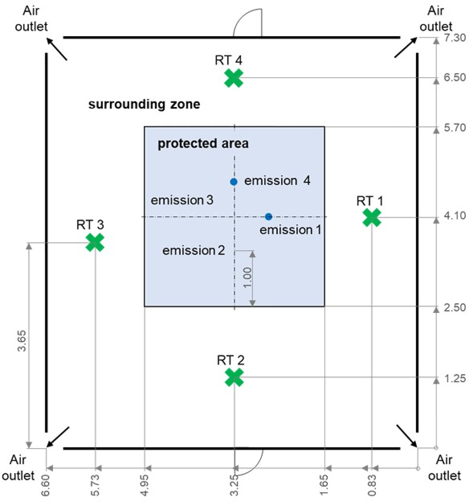
Particles were emitted from a particle generator at a height of 2 m successively at four different emission positions (Emission 1–4) in the protected area, each 1 m inward from the border of the inner outled. For recovery time (RT), calculations corresponding measuring positions (RTs 1–4) were defined in the surrounding area at a height of 1.65 m.
The increase of the particle concentration and its subsequent decrease were determined by a particle measuring system, consisting of the mobile AbakusTM optical particle counter and a 1:100 dilution system (Bad Liebenzell, Germany; software “Log and Show 3.1”). The optical particle counter had a measuring volume of 28.3 l/min (concentration limit: 35,000,000 particles sized 0.3 µm/m3) and 10-s measuring intervals. To avoid a systematic error due to the high measuring volume of the particle counter (high amounts of sucked particles with low remaining particles in the air and a consecutively false low RT), a bypass for sterile filtered air was installed. Therefore, a y-piece was placed in the antistatic hose between the particle measuring system and measurement position (Figure 3). A calibrated rotameter (adjustable range: 20–250 L/h) continuously monitored the restriction of the volume flow and volume of the particle counter to 2.8 L/min. The antistatic hose terminated on a tripod at heights of 1.55 and 1.75 m above floor level (below the geometric center of the surgical lights in the protected area). The RT was determined at these two heights below the lights to monitor the particle reduction of two different particle concentrations. In the surrounding area, the antistatic hose terminated on a tripod at a height of 1.65 m.
For calculating the RT according to ISO 14644, the particle concentration was determined when a particle reduction of more than two log steps was achieved (ISO 14644-3, 2019). The RTs for both surgical light shapes, all measuring positions, and in all heights above floor level in the surrounding zone were determined on five different days.
Statistics
For statistical analysis, the program IBM SPSS Version 20 (IBM Corp., Armonk, NY) was used. Variables were tested for normal distribution by using the Kolmogorov–Smirnov test with Lilliefors correction, before group differences were tested by either students t test or analysis of variance. The continuous variables are presented as mean (±standard deviation [SD]). A p value < .05 with an error probability < 5% (two-sided test) was considered statistically significant.
Results
The uniformity of velocity was first measured in the operating room at a height of 1.2 m without the influence of the surgical lights. Data revealed a uniformity of velocity of 95.8% ± 1.7% in the protected area, demonstrating the high homogeneity of the UDF in the center of the operating room, whereas it was 51.5% ± 17.6% in the surrounding zone, reflecting the turbulent conditions. Figure 5 shows the details of all measuring positions. The relationship between the uniformity of velocity and supply air volume flow for the protected zone as well as for the surrounding zone and its significances is demonstrated in Table 1.
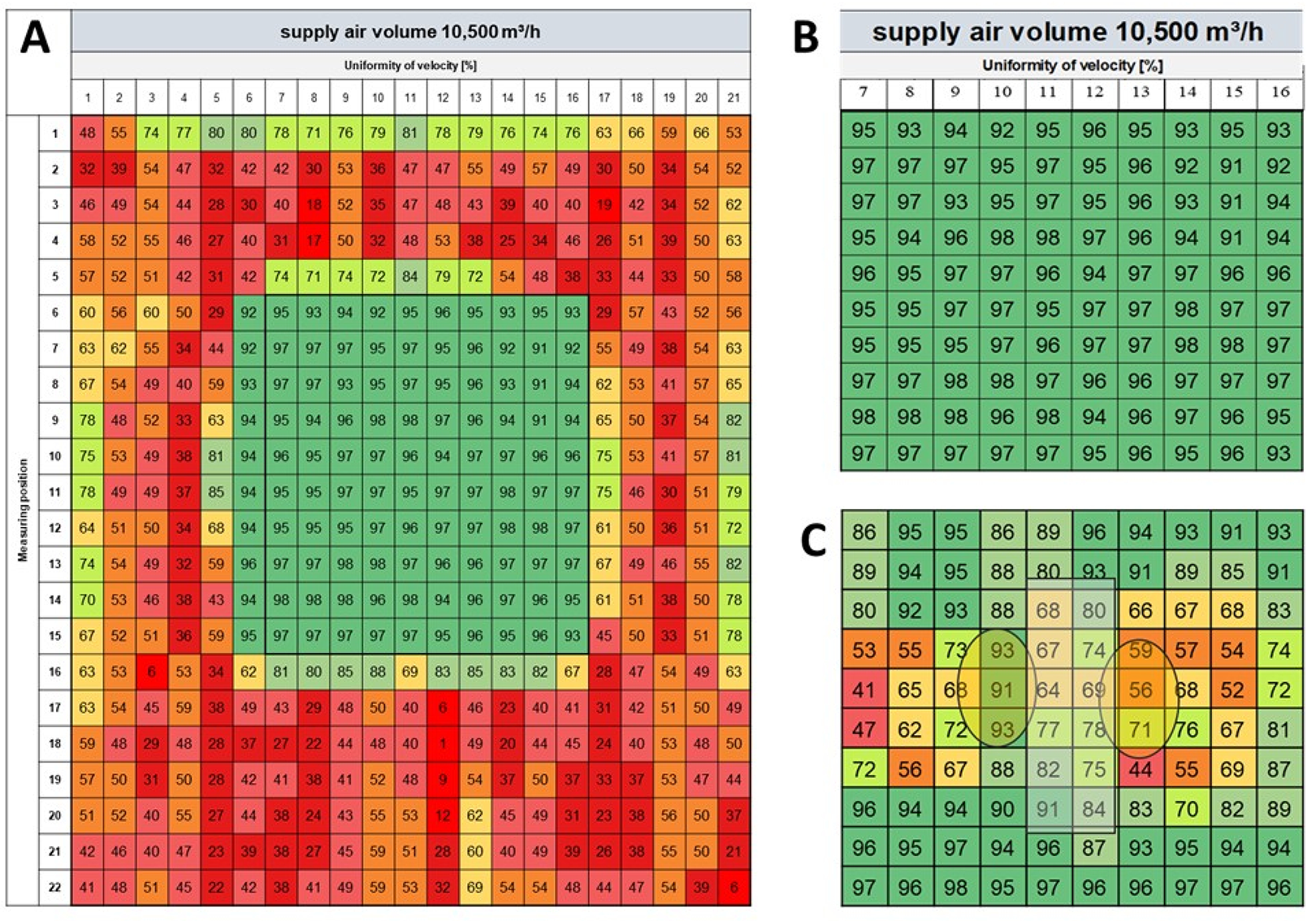
Uniformity of velocity (Uv) [%] detected in a grid (30 cm × 30 cm) at a supply air volume flow of 10,500 m3/h. (A) Operating room with lights swung out of the measuring positions. The velocity was always uniform in the protected area (dark green square in the center of the operating room (uniformity of velocity > 90%), but turbulent in the surrounding area. (B) Magnification from A for comparison with C. (C) protected area with surgical lights (yellow ovals) and operating table (gray rectangle). The surgical lights with a tilt angle of 30° caused a turbulent flow with a locally inhibited uniformity of velocity in the protected area. Data are given as mean (n = 5 per measuring position).
Relationship Between Uniformity of Velocity and Supply Air Volume Without the Impact of Surgical Lights.
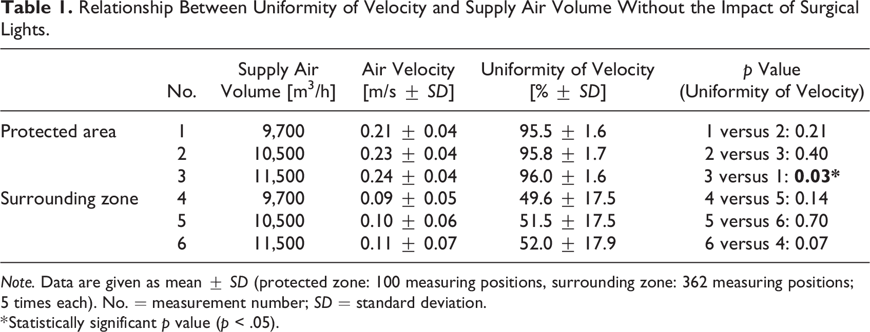
Note. Data are given as mean ± SD (protected zone: 100 measuring positions, surrounding zone: 362 measuring positions; 5 times each). No. = measurement number; SD = standard deviation.
* Statistically significant p value (p < .05).
Thereafter, the impact of the surgical lights on the uniformity of velocity in the protected area with a supply air volume of 10,500 m3/h was tested again at a height of 1.2 m and with the lights tilted to an angle of 30° according to DIN 1946-4. The lights led to a significantly lower uniformity of velocity in the protected zone and locally turbulent flow condition in several measuring positions (without lights: 95.8% ± 1.7% vs. with lights: 81.0 ± 14.7; p < .001). No statistical difference was seen for the uniformity of velocity between the hemispherical closed-shaped light (measuring position rows 7–11, columns 9–12: 69.3% ± 14.6%) and the semi-open column-shaped light (measuring position rows 12–16, columns 9–12: 66.9% ± 10.9%; p = .559; Figure 5).
As the air flow has been shown to be turbulent in the surrounding zone as well as below the surgical lights in the protected area, the RT was used here to evaluate the local impact on the UDF ventilation system. Therefore, the RT was measured 10 times below each surgical light in the protected area with a supply volume flow of 10,500 m3/h and in heights of 1.55 and 1.75 m above the floor level. The mean ± SD RT for the hemispherical closed-shaped light was 1.7 ± 0.2 min at a tilt angle of 0°. This time was reduced to 1.3 ± 0.1 min at the more common clinical working angles of 15° and 30°. The RTs of the semi-open column-shaped light were 1.2 ± 0.1 min for tilt angles of 0° and 15° and 1.1 ± 0.1 min for 30°. The RTs of the semi-open column-shaped light were significantly shorter for all three tilt angles than for the hemispherical closed-shaped light (p < .001). Differences between 1.55 and 1.75 m were not statistically significant for both lights and all tilt angles (hemispherical closed-shaped light: 0°: p = .473; 15°: p = .999, 30°: p = .080; semi-open column-shaped light: 0°: p = .999; 15°: p = .070, 30°: p = .999).
The mean RT in the surrounding zone was 3.5 ± 0.28 min without significant differences between the four measuring positions (RT 1: 3.5 ± 0.27; RT 2: 3.6 ± 0.31; RT 3: 3.6 ± 0.17; RT 4: 3.3 ± 0.24 min). However, the RT depended exponentially on the volume flow rate (Figure 6).
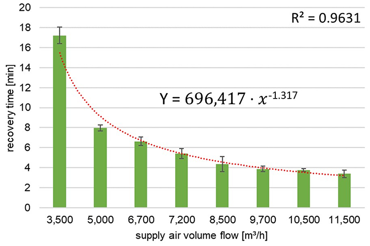
Relationship between recovery time (RT) and supply air volume flow in the surrounding area. A low supply air volume leads to a comparatively long RT. The function RT = 69,642·x -1.317 (x = supply air flow) allows the determination of the RT for every supply flow.
A supply air flow of 3,500 m3/h, for example, led to an RT of 17.2 ± 0.8 min, whereas it was 3.4 ± 0.4 min with a supply air flow of 11,500 m3/h (p < .001). Based on our measurements, we calculated the function Y = 696,417·x −1.317 (Y = RT and x = supply volume flow), which allows to determine the RT for every supply volume.
Discussion
Multiple studies have shown that UDF ventilation systems reduce particles, microorganisms, biological fragments, surgical smoke, and anesthetic gases in the air of operating rooms more effectively than MDF ventilation. This will therefore have a higher health protective effect for the personnel and may contribute to reducing the risk of surgical site infections for patients (Aganovic et al., 2017; Herzog-Niescery et al., 2015; Romano et al., 2020; Seipp et al., 2022). However, about a decade ago, studies pointed out that the velocity in operating rooms with UDF ventilation is not uniform below the surgical lights and assumed that the beneficial effect of the UDF system is significantly affected. In 2010, Zoon et al. investigated the effect of three different surgical light dummies (closed-shaped, semi-open-shaped, and open-shaped), which were built in a 1:1 ratio to functional operating room lights. They revealed a turbulent air flow below the lights, but the effect of the surgical light’s heat release, which is regularly observed in clinical practice, was disregarded and only a tilt angle of 0° (worst, but practically unusual position) was investigated (Zoon et al., 2010). The turbulent flow conditions below functional surgical lights were later confirmed by different research groups, but they either used bubble tests, computational fluid mechanics simulation, or measured the changes in air flow velocity, all of which are not favored according to ISO 14644 (Aganovic et al., 2017; Al-Waked, 2010; Refaie et al., 2017).
In this study, we demonstrated that the surgical lights significantly impair the uniformity of velocity in the protected area. Using the RT for the locally turbulent flow conditions, we quantified the particle reduction capacity of the UDF ventilation system below the surgical lights and within the personnels breathing zones (1.55–1.75 m). Data revealed mean RTs of 1.1–1.7 min for a supply air flow of 10,500 m3/h. This means that even though the air flow is locally turbulent below the surgical lights, the contaminated air (particles in this study) is eliminated approximately 12–18 times quicker compared to operating rooms with MDF ventilation, which are defined by an RT of up to 20 min according to DIN 1946-4. An explanation for this quick RT despite the turbulent conditions might be that the supply air enters the operating room through the laminarisator and thus above the surgical lights, around which the flow is directed. This 12–18 times more efficient elimination of particles is also consistent with results from Romano and colleagues, who reported a 13-times lower particle load in operating rooms with UDF ventilation systems compared to MDF ventilation (Romano et al., 2017).
In addition, the impact of the surgical lights’ tilt angles (0°, 15°, and 30°) on the RTs was evaluated. A significant difference was observed between the two lights for all three tilt angles, which was already assumed by others (Traversari, 2017). The 0°-position represented the worst condition for both lights in the UDF ventilation system, even though the significantly different RTs (1.7 ± 0.2 vs. 1.3 ± 0.1 min) appeared negligible. However, a 0°-position is not common during real surgery, and the RTs for tilt angles between 15°–30° resulted in a minimal, but significant difference of 0.1 min. Thus, we recommend a working angle of 30°. Although we are aware that the lights are moved frequently in clinical practice, it seems necessary to evaluate and qualify new developments of surgical lights under comparable standard conditions.
Additionally, the impact of the surgical lights’ parameters “shape” and maximum surface temperature on the uniformity of velocity was investigated.
We found that the uniformity of velocity was marginally lower (namely, more turbulent) and the RT significantly longer below the hemispherical closed-shaped light compared to the semi-open column-shaped one. A reason for this might be that the flat-shaped semi-open light may disrupt the airflow more than a rounded surgical light, whereas particles below the closed-shaped light rotate more, not influenced by the total downstream airflow, which changes toward a circumferential sideward direction. The significantly different RTs can be explained by the sensitive measuring method and by a more effective particle washout below the semi-open light. However, this difference (1.1 vs. 1.7 min) is negligible in clinical practice, as RTs up to 20 min are acceptable in operating rooms with mixed dilution flow. Thus, our results are basically in line with observations from Aganovic et al., who stated that the uniformity of velocity is lower below the hemispherical closed-shaped surgical light. This was due to its coherent surface compared to the semi-open column-shaped light, where the airflow passes downstream through the openings (Aganovic et al., 2017). However, they observed no difference in microbiological sampling, which was defined as a parameter of the UDF’s protective effect in their study. Nevertheless, it should be recognized that the results for surgical lights with other forms or surfaces might be different.
Another finding from this study relates to the turbulent air flow in the surrounding zone, outside of the sterile field where most of the staff (circulating practitioners, sometimes the anesthesiologist) stay. At first, we investigated the RT with 10,500-m3/h supply air at four different positions in the surrounding zone of an UDF system corresponding to DIN standard (laminarisator: 10.2 m2). The mean RT was 3.5 ± 0.28 min without significant differences between the measuring positions, which is justifiable due to the homogeneous distribution of supply air flowing out of the protected area, combined with exhaust air slots installed in all four corners of the operating room. This allowed us to determine the RTs of the surrounding zone at one representative measuring position only and to calculate the performance function that correlates the RT to the supply air volume. The RTs in the operating room with a size of 132 m3 ranged between 3.4 ± 0.4 min for 11,500 m3/h and 17.2 ± 0.8 min for 3,500 m3/h. This is in line with the results from Lans and colleagues, who published a research study about the protective effect of small UDF ventilation systems (laminarisator: 5.7–7.1 m) in the surrounding zone of operating rooms in the Netherlands. They determined an RT of 6 ± 1.2 min with small supply air flows of 5.593–8.690 m3/h, although the particle counter used (Lighthouse 3016 with a flow rate of 2.83 L/min) had a 10-times lower flow rate as required for clean room measurement according to ISO 14644-1 (Lans et al., 2022). Compared to our results, this means that UDF systems with small supply air flows reduce hazardous substances by about 3.3 times and UDF systems with a supply air flow of 10,500 m3/h about 5.7 times more effectively out of the surrounding zone than MDF ventilation with RTs up to 20 min. This clearly provides a significantly improved protective effect for the nonsterile dressed personnel in the periphery of the operating room. In addition, our results agree with the current findings of Wagner et al. (2021), who reported significantly lower particle concentrations in the protected area (operating and instrument tables) compared to the surrounding area in operating rooms equipped with UDF systems with single large diffusers and multiple diffuser arrays design. The lower particle concentrations can be explained by the “displacement effect” in the protected area, which transports the hazardous substances down to the floor due to the vertical unidirectional flow velocity of 0.28 m/s within < 10 s, before they spread into the larger volume of the surrounding area with its turbulent mixed flow.
The requirements for the protective effect of operating room ventilation systems are based on the maximum concentration of hazardous substances associated with a possible health risk. In 1982, Whyte et al. reported airborne germ concentrations of 413 CFU/m3 (Colony Forming Units) in operating rooms with MDF ventilation, whereas only 4.3 CFU/m3 in operating rooms with UDF ventilation (97-fold reduction). Thirty years later, 37.5–44.3 CFU/m3 were detected during orthopedic surgery in Sweden and in the Netherlands (Andersson et al., 2012; Mathijssen et al., 2016). Thus, the ventilation system should guarantee to reduce the maximum of 1,000 airborne bacteria per m3 (reduction by three log steps, log10 = 3). This can be achieved within 10s in operating rooms with unrestricted UDF ventilation (and in 1.7–2.6 min below the surgical lights), whereas it lasts 30 min in operating rooms with turbulent MDF ventilation.
However, the highest concentrations of hazardous substances result from the surgical smoke with maximum particle concentrations of >1E + 11 per m3 (Ragde et al., 2016; Romano et al., 2020). Here, a reduction capacity of six log steps (log10 = 6) by the ventilation system is needed. This can be achieved in the protected area of UDF ventilation systems within 10 s and in the locally turbulent spaces below the surgical lights in 3.3–5.1 min, but it lasts more than 60 min in operating rooms with turbulent MDF ventilation. Moreover, it should be noted that surgical smoke exposure due to high-frequency electrocautery is not a “single-spot-issue” but is repeated frequently during surgery. Long RTs such as in operating rooms with MDF ventilation then quickly lead to particle accumulation with consecutive high persisting concentrations of surgical smoke pollutants.
A limitation of this study is that we simulated clinical conditions in a laboratory setting. This allowed us to focus on the impact of the surgical lights, on the uniformity of velocity, as well as on the methodically correct determination of the RT by the elimination of other variables and confounding factors; however, daily routine in clinical practice is different. Thus, the lights are moved constantly from one side to another and are used in different angles; both personnel within and outside of the sterile field are moving in the surrounding zone. Doors are opened and closed frequently, and equipments—besides just the operating table—are placed throughout the operating room, which can hinder the supply air to flow directly to the exhaust air outlets. All these factors can influence the protective effect of the UDF system in the protective area as well as in the surrounding area temporary. Nevertheless, even if conditions are less optimal in practice, the UDF’s protective effect is still considerably higher than that of MDF ventilation.
Another possible limitation could result from the particle counter, which draws off the particles below the surgical lights to determine their concentration. As explained in the “Method” section, a high measuring volume may lead to a false low particle concentration in this area and consecutively to a false low RT. We are aware of this phenomenon and therefore installed a bypass of sterile air and thus reduced the succeeded volume. The resulting comparatively low measuring volume sucked in less particles; however, this did not impair the measurements due to the high particle concentration in the air. Moreover, we determined the particle concentration at two different heights below the lights (30 cm—corresponds to 1.75 m, and 50 cm—corresponds to 1.55 m) and obtained comparable results.
In summary, local turbulent spaces below the surgical lights and in the surrounding zone were of special interest and compared to the effectiveness achieved using MDF. The RT is the correct method to evaluate the protective effect of an operating room ventilation system in locally turbulent areas, even though an UDF system is installed. Data revealed a 12–18 times more efficient elimination of particles in the protected area below the lights and a 5.7 times more efficient elimination in the surrounding zone compared to standard MDF ventilation systems with an RT of 20 min. While surgical lights had significant effects on the uniformity of velocity, the RTs underneath the lights demonstrated a high protective effect, whereas the differences caused by various tilt angles (0°, 15°, and 30°) only slightly affected the RT (1.1–1.7 min). However, the UDF’s deterioration in performance due to the surgical lights is negligible and it nevertheless retains its strong protective effect.
Implications for Practice
The aim of this study was to determine the RT in the turbulent spaces of an operating room equipped with a UDF ventilation system according to ISO 14644.
The air below the surgical lights was locally turbulent, leading to a mean RT of 1.1–1.7 min.
No statistical difference was seen for the uniformity of velocity between the hemispherical-shaped light and the semi-open column-shaped light at a tilt angle of 30° according to DIN 1946-4.
The overall effect of the surgical lights on the particle reduction capacity of the UDF system was negligible.
The mean RT in the turbulent air of the surrounding zone was 3.5 min, which still is approximately 6 times quicker than a standard mixed dilution flow system.
Footnotes
Declaration of Conflicting Interests
The authors declared no potential conflicts of interest with respect to the research, authorship, and/or publication of this article.
Funding
The authors received no financial support for the research, authorship, and/or publication of this article.
