



Abstract
Purpose:
To investigate the impact of two different routes of administration on the lung and systemic exposure of drugs designed for local delivery to the lung.
Methods:
In our comparative studies, similar lung doses of three different drugs were administered to rodents by both intratracheal instillation and inhaled delivery.
Results:
An obvious but unexpected difference in the exposure was observed. Immediately after the dose, the initial plasma concentration was much higher whereas the initial fraction of the lung dose in the lung was clearly lower for the instillation compared with the inhaled delivery. There was also a difference in the tolerability for one of the drugs when the same lung dose was administered and the inhaled dose was, in contrast to the instilled dose, tolerated by the mice.
Conclusions:
A plausible explanation for the observed but contrary to expected difference is that the drugs leaked from the lung into the systemic circulation already during the instillation procedure due to its invasive nature.
Introduction
Intratracheal instillation (Fig. 1) is an invasive, non-physiological bolus administration, and there are several comparative studies1–7 describing the considerable difference in the lung distribution of the dosed material. In these comparative studies the lung distribution was measured with several different methods, that is, gamma scintillation of radiocolloids, 1 autoradiography of the optical density of dust, 2 gamma scintillation imaging of radiocolloids, 3 immunohistochemistry staining of a ricin-specific antibody, 4 imaging mass spectrometry of tiotropium, 5 positron emission tomography imaging of a radiotracer, 6 and light sheet fluorescence microscopy of a melamine resin fluorophore. 7 A consistent major disparity in the lung distribution between the intratracheal instillation and the inhaled delivery was reported. The instilled bolus dose resulted in an inhomogeneous distribution concentrated around central airways that failed to reach the peripheral regions, and with a large inter-individual variability, where sometimes entire lung lobes were missed. The inhaled delivery, in contrast, resulted in a homogenous distribution in the entire lung, also reaching the peripheral regions. It is likely that such a large difference in the lung distribution will influence the clearance as well as the degree of systemic absorption, 8 and the latter is supported by a reported tenfold higher bioavailability of inhaled compared with instilled insulin. 3 Other studies that focused solely on inhaled delivery9–11 have also concluded that the dosed test material was uniformly distributed in the entire lung.
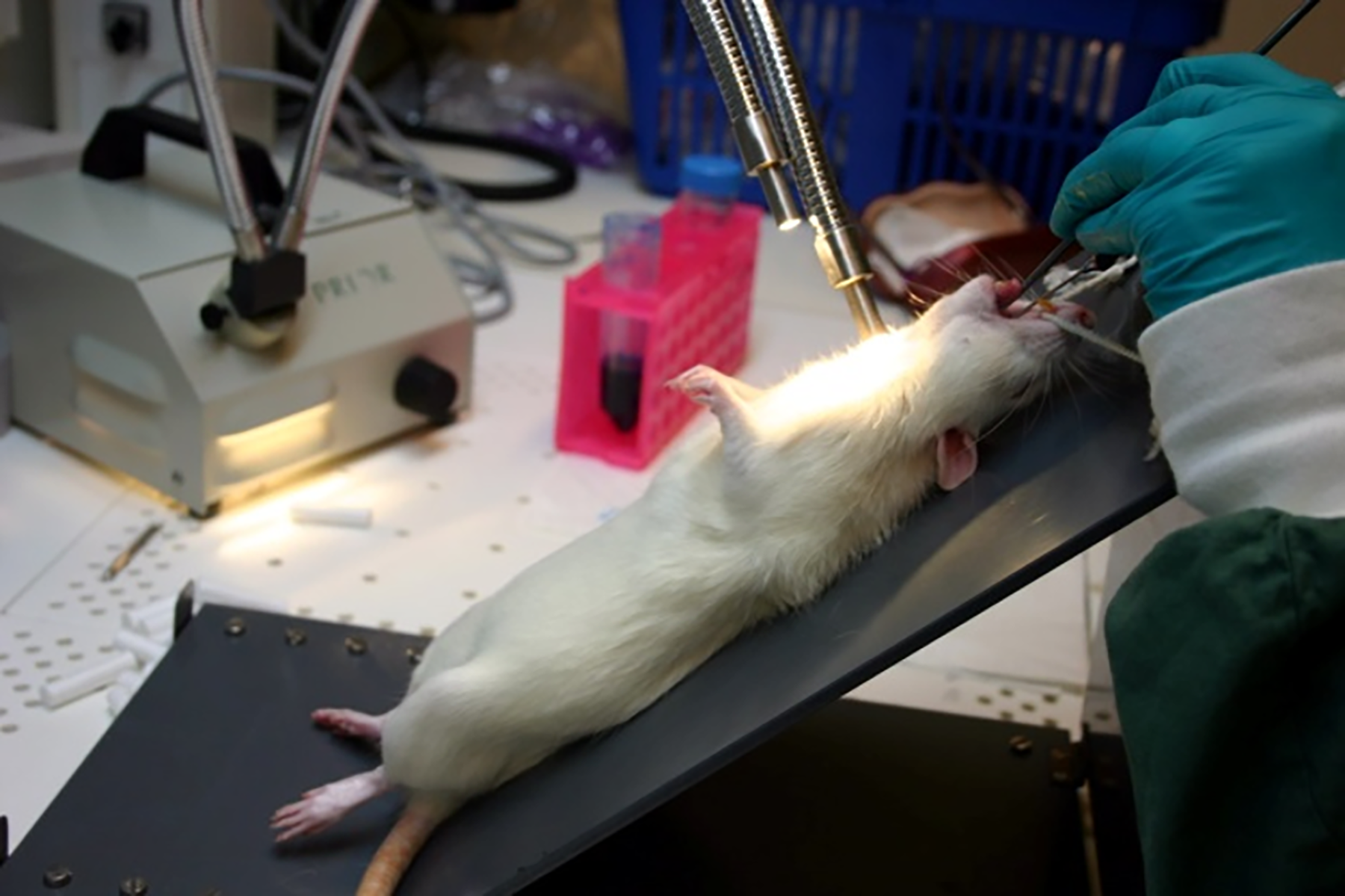
Intratracheal instillation dosing of an anesthetized rat.
There are also comparative studies reporting different inflammatory responses of the dosed material.12–14 The inhaled delivery induced smaller polymorphonuclear neutrophil levels from the granular biopersistent particles 12 as well as a more uniformly distributed inflammatory response at a lower dose of the lipopolysaccharide endotoxin13–14 compared with the intratracheal instillation.
The reported major different lung distribution of the anti-ricin antibody 4 resulted in an obvious difference in the efficacy of the antibody when the mice were challenged with ricin. The dosed antibody prevented death in all animals; however, the histological examination showed that ricin induced tissue lesions in the peripheral parts of the lung for all the intratracheal instilled mice, but the inhaled antibody covered the entire lung and therefore prevented peripheral lesions.
It has been reported that the important variables for the instillation procedure are the dose volume, the introduction of air into the syringe, and the method of intubation. 15 It was also concluded that a high instillation speed produced a better distribution in the lung than a low speed. A typical dose volume is 1–2 mL/kg bodyweight, and a smaller dose volume has been shown to distribute the drug less evenly in the lung. 8 This is supported by other researchers who concluded that a dose volume of 0.4 mL/kg pigment solution without air in the syringe resulted in an unsatisfactory lung distribution, that is, the pigment was only present around the main trachea and large bronchi of the lower lobes of the lung. 16 However, increasing the dose volume fivefold to 2 mL/kg, followed by an equal volume of air pushed the pigment further into the rat lung.
In our comparative studies, the same or similar lung doses of three different drugs designed for local delivery to the lung were administered to rodents by both intratracheal instillation and inhaled delivery. All three drugs were well retained in the lung, but due to two completely different mechanisms.
Two of the drugs are dual phosphatidylinositol-3 kinase or phosphoinositide 3-kinase (PI3K) γδ inhibitors developed for the treatment of asthma,17–18 and both have strong crystal lattices and high melting points, showing a limited capacity to dissociate from the solid form, which consequently results in extremely poor water-solubility, and they could be referred to as ‘brick dust’ molecules. 19 These two drugs were therefore dosed to the lungs as drug nanocrystals suspended in water and retained in the lung via the slow dissolution of the drug nanocrystals. One of them, AZD8154, has also been dosed inhaled using the drug nanocrystal suspension formulation to dogs 20 as well as to healthy human volunteers in a Phase 1 clinical study investigating the safety, tolerability, and pharmacokinetics of this drug. 21 AZD8154 was designed to have very low solubility and was therefore expected to have a long retention in the lung. 21 In addition to prolonged lung retention, it was shown to have low systemic exposure compared with anticipated local lung concentrations, suggesting that these parameters of the inhaled drug were well optimised. 21 Furthermore, AZD8154 demonstrated high pulmonary bioavailability, suggesting that the absorption was via the target organ. 21 Soluble and cell-permeable PI3Kδ inhibitors for long-acting inhaled administration had first been pursued 22 before a shift was made to a different chemical series with alternative long lung retention properties, that is, extensive residence in the lung was obtained through very low water-solubility. 20
The third drug is water-soluble enough to be able to be dosed as an aqueous solution, and it was thus not retained in the lung by slow dissolution but due to being a zwitterion with an acidic pKa below 4 and a basic pKa slightly below 7, which means that it is ionized at neutral pH, which reduces its permeability to the systemic circulation. Polarity is known to significantly reduce the permeation of small, hydrophilic molecules. 19
The aim of our studies was to compare the two different routes of administration regarding the exposure in lung and plasma as a function of time, as well as the tolerability of one of the drugs when administering the same high lung dose once daily for three days.
Materials and Methods
Chemicals
The three drugs, AZD8154, DRUG B, and DRUG C were manufactured by AstraZeneca and obtained from its drug library. AZD8154 and DRUG B are both dual PI3Kγδ inhibitors developed for the treatment of asthma,17–18 and their chemical structures correspond to compounds 58 and 36, respectively, in a previous publication. 17 The molecular weights of the drugs are 551.7 (AZD8154), 609.8 (DRUG B), and 654.7 (DRUG C) g/mol. A human serum albumin colloid (NANOCOLL, 0.5 mg human albumin particles ≤80 nm, GE Healthcare) was radiolabeled at Sahlgrenska University hospital with sodium pertechnetate (99mTc) a few hours before the colloid was used for in vivo dosing due to the short 6 hour half-life of 99mTc. All other chemicals were of analytical grade.
AZD8154 and DRUG B both form very stable drug crystals with high melting points, well above 200°C. The solubility in water of the crystalline form of both drugs is therefore extremely low and has been reported to be 0.05 µM for AZD8154. 17 The measured solubility in water for crystalline DRUG B at pH 7.4 was 1 µM. This means that the solubility in water will limit the systemic absorption of both drugs. AZD8154 has no dissociation constant (pKa) in the pH range 3–11, and is therefore not ionizable, which means that it was in the neutral form in the formulation. Drug B has a measured pKa of slightly above 8, which means that it was also largely in the neutral form in the formulation. These two drugs have measured partition coefficients in octanol/water (log P) between 3 and 4 and distribution coefficients at pH 7.4 (log D) in the same range as for the log P. The permeability of AZD8154 in Caco-2 monolayer permeability assays has been reported to be moderate and the reflux was little. 17
DRUG C had a low propensity to crystallize and was therefore synthesized as an amorphous material. The glass transition temperature of this material was determined to be above 100°C with differential scanning calorimetry analysis. The drug has a measured acidic pKa of approximately 3.5 and a measured basic pKa of slightly below 7, which means that it is a zwitterion and in an ionized form at physiological pH. The solubility in water of DRUG C is at least 40 g/L in its protonated form. The water-solubility of DRUG C is high enough not to limit the systemic absorption. The measured log P was approximately 4 and the measured log D was approximately 2 for this drug. The permeability of DRUG C was measured to be slightly below 0.2·10−6 cm/s in Caco-2 monolayer permeability assays, which would be considered a low permeability for a drug aimed for oral administration. 23
Formulations
Aqueous drug nanocrystal suspensions of AZD8154 and DRUG B containing DSPE-MPEG2000 as a stabilizer and glucose as a tonicity modifier were used for both administration routes. The suspensions were planetary bead-milled to a volume median diameter of approximately 0.1 µm using a Fritsch Pulverisette 7 classical line and 0.6–0.8 mm milling beads (Glen Creston), according to a previously published procedure.20,24 It was confirmed using X-ray diffraction analysis that both AZD8154 and DRUG B were crystalline, also after the bead milling process.
Aqueous solutions of DRUG C containing glucose as a tonicity modifier was used for both administration routes. The pH of the manufactured solutions was adjusted to approximately 6.
Inhaled delivery and intratracheal instillation
The drug aerosols were administered by a flow-past nose-only inhalation exposure system that enabled dosing of at least four rodents simultaneously. Each rodent was restrained in an acrylic tube with an adjustable backstop. The snout was located at the front end of the restraining tube. The restraining tube was connected to a dosing chamber in which the aerosol was generated. A jet nebulizer (Cirrus 2, Intersurgical) was used as the aerosol generator of the aqueous nanosuspensions of AZD8154 and DRUG B. A vibrating mesh nebulizer (Aerogen SOLO, Aerogen) was used as the aerosol generator of the aqueous solution of DRUG C. The inhaled dosing time was 10 minutes for the rats and 30 minutes for the mice. The nebulizer was filled with an overage of formulation, that is, more than would be required to nebulize the targeted dose. The droplet size distribution from the two different nebulizers was characterized as previously described. 20 The nebulized droplet sizes were measured by laser diffraction using a Malvern Spraytec instrument. The lung deposited dose achieved with the inhalation dosing system with the jet nebulizer was evaluated for two of the dose levels in rats by spiking the drug nanosuspension with a radiolabeled human serum albumin colloid. The radioactivity of a vial with spiked nanosuspension or a sampled dissected lung was measured using a dose calibrator (VDC 404, Veenstra) by putting the whole vial or the whole lung inside the ionization chamber and noting the instrument’s activity readout.
The intratracheal instillations were performed under isoflurane anesthesia as previously described for AZD8154, 18 and more thoroughly described for lipopolysaccharide 14 as well as generally. 15 Each individual rodent was briefly and superficially anesthetized with isoflurane, and when adequate anesthesia was observed placed heads up in a supine position on an inclined platform to give free access to the oral cavity. A metered amount of the aqueous drug formulation was injected into the trachea by a syringe equipped with a blunt cannula that was inserted through the mouth with its tip resting midway between the larynx and the first carina. The bolus injection was done instantly, and the rodent was then maintained in an upright position for a sufficient time to prevent the formulation from leaving the lung. The dosing volume for rats was 0.5 mL/kg followed by an equal volume of air or 1 mL/kg without air, and for mouse was 1.5 or 2 mL/kg followed by an equal volume of air.
All rodents were dosed only once for the pharmacokinetic evaluations of AZD8154 and DRUG B. A high 1 mg/kg lung dose of DRUG C was repeatedly dosed to the mouse once daily for three days for a tolerability evaluation.
Animals
The in-vivo studies were performed at an Association for Assessment and Accreditation of Laboratory Animal Care International (AAALAC)-accredited animal facility at AstraZeneca, Mölndal, Sweden, under approval of the Animal Ethics Committee of Gothenburg.
Male Han Wistar or Brown Norway rat and female CD-1 or BALB/c mouse were used in the studies.
The method of euthanasia of the rodents was by transcardial perfusion, that is, exsanguination under deep isoflurane gas anesthesia.
Bioanalysis
The lung and plasma levels of AZD8154 were assessed as previously described, 18 and DRUG B and DRUG C were assessed the same way. At termination, the animals were bled through the orbital venous plexus under deep isoflurane gas anesthesia and the blood was collected into K2 EDTA-treated test tubes and placed on water ice. The whole blood was centrifuged in refrigerated conditions, and plasma was collected and stored for future assessment at or below −20°C. The lungs were dissected out, weighed, immediately snap frozen, and stored for future assessment at −80°C. The lungs were homogenized in a Ringer solution using a Precellys bead-beating technology. Lung homogenates and plasma samples underwent protein precipitation using acetonitrile containing an internal standard and was followed by vortex mixing and centrifugation. The supernatants were diluted to match the initial mobile phase. The lung and plasma concentrations were determined by liquid chromatography with tandem mass spectrometric detection.
Results and Discussions
The aqueous drug nanocrystal suspensions of AZD8154 and DRUG B were because of the suspended particles chosen to be aerosolized with a jet nebulizer, whereas the aqueous solution of DRUG C was aerosolized with a vibrating mesh nebulizer. The two different nebulizers generated similar droplet size distributions (Fig. 2).

Measured droplet size distributions from the Cirrus2 jet nebulizer and the SOLO vibrating mesh nebulizer used for the inhaled delivery.
Previous experience before the comparative study was that a similar lung deposited dose was achieved with both nebulizers when dosing water-soluble drugs to the rodents, and thus that the same performance of the exposure system was achieved.
ACD8154 was dosed to the rodents as outlined in Table 1. The inhaled lung deposited dose was evaluated for the two dose levels of 25 and 35 µg/kg in rats by spiking the nanosuspension with a radiolabeled human serum albumin colloid. To spike the drug formulation with a radioactive tracer is a frequently used approach for nuclear imaging of inhaled drugs. 25 It is known that the albumin colloid used is small enough to deposit throughout the lungs but also large enough not to be adsorbed systemically from the lung.26,27 The average ratio between the amount of colloid and AZD8154 in the lung immediately after dose correlated very well, that is, within ±10%, with the ratio between the concentration of the colloid and AZD8154 in the spiked nanosuspension, which means that only a small fraction of the lung deposited dose of the drug had been absorbed systemically from the lung during the 10-minute dosing time. The amount of colloid in the lungs 24 hours after the dose was only reduced by 20% to 30% compared with the level immediately after the dose, which supports that the colloid was not adsorbed systemically and only cleared from the lung via mucociliary clearance. 27 The profile for the fraction of the inhaled lung dose over time (Figs. 3 and 4) shows that the targeted lung doses were achieved, and that the drug was well retained in the lung due to its poor solubility. About 90% of the targeted lung dose was no longer present in the lung 24 hours after the dose, and almost 10% was still retained. The result for the 85 µg/kg dose was very similar compared to the two spiked doses, which shows that the spiking had no significant impact. The same drug nanocrystal formulation of AZD8154 has also been dosed via inhaled delivery to both dogs 20 and humans. 21 The same absorption-dependent pharmacokinetics was observed in these studies.

Amount of AZD8154 in rat lung as a fraction of the targeted lung dose, immediately after dose (IAD),3 hours after dose (3hAD), and 24 hours after dose (24hAD) of the instilled lung dose of 55 µg/kg and the inhaled lung doses of 25, 35, and 85 µg/kg. The circles represent the individual results for each administration and time point. The boxes describe the statistics, i.e., the central pink mark indicates the median, and the bottom and top edges of the box indicate the 25th and 75th percentiles. The whiskers extend to the most extreme data points not considered outliers.

Amount of AZD8154 in mouse lung as a fraction of the targeted lung dose, immediately after dose (IAD) and 24 hours after dose (24hAD) of the instilled lung dose of 110 µg/kg, and the inhaled lung dose of 140 µg/kg. The circles represent the individual results for each administration and time point. The boxes describe the statistics, i.e., the central pink mark indicates the median, and the bottom and top edges of the box indicate the 25th and 75th percentiles. The whiskers extend to the most extreme data points not considered outliers.
Intratracheal Instilled and Inhaled Doses of AZD8154 in Rats and Mice

However, the fraction of the instilled lung dose in the lung immediately after the dose was for both species only about half compared with the inhaled delivery (Figs. 3 and 4). The fraction of the lung dose found in the lung 24 hours after dose was also tenfold higher, that is, almost 10% compared with only almost 1%, for the inhaled delivery compared with the intratracheal instillation in both mouse and rat (Figs. 3 and 4).
Extremely low water-solubility is known to result in low oral 19 as well as low nasal bioavailability, as shown for low solubility steroids such as fluticasone propionate, 28 fluticasone furoate, 29 and mometasone furoate. 30 The nasal absorption is limited by the poor water-solubility because the nasal mucosal contact time is limited 29 and most of the deposited drug is therefore cleared by the nasal cilia and swallowed before dissolution occurs. 31 The swallowed fraction of a nasal deposited dose will also be poorly absorbed orally due to an extremely poor water solubility, and the overall contribution to the systemic exposure will therefore be low. Low oral bioavailability in both rat and dog, that is, approximately 3%, has been confirmed for AZD8154 after peroral dosing of drug nanocrystals suspended in water.
The plasma profiles for the inhaled delivery of AZD8154 (Figs. 5 and 6) contain a peak more than 2 hours after dose for both species, which supports that the absorption from the lung to the systemic circulation continued well after the drug had been inhaled. There was, for two of the three inhaled doses in rats, a high initial plasma concentration immediately after the dose, followed by a drop in the plasma profile. The drop was most obvious for the 35 µg/kg inhaled dose, where the sampling was focused only on the initial 3 hours after the dose (Fig 5). Figure 7 shows a previously reported plasma profile for a similar inhaled lung dose of AZD8154 in dog, 20 and there is a clear similarity between the profiles for the two different species. One conclusion from the dog study was that the intranasal filtration in the nose was low, and that the systemic exposure originated primarily from lung-deposited drug. It is expected that the intranasal filtration is much higher for the rat compared with the dog, but the similar plasma exposure over 24 hours for both routes of administration (Table 1), and the fact that the intratracheal instillation completely bypasses the nose, support that the systemic exposure originated primarily from lung deposited drug also for the rat.
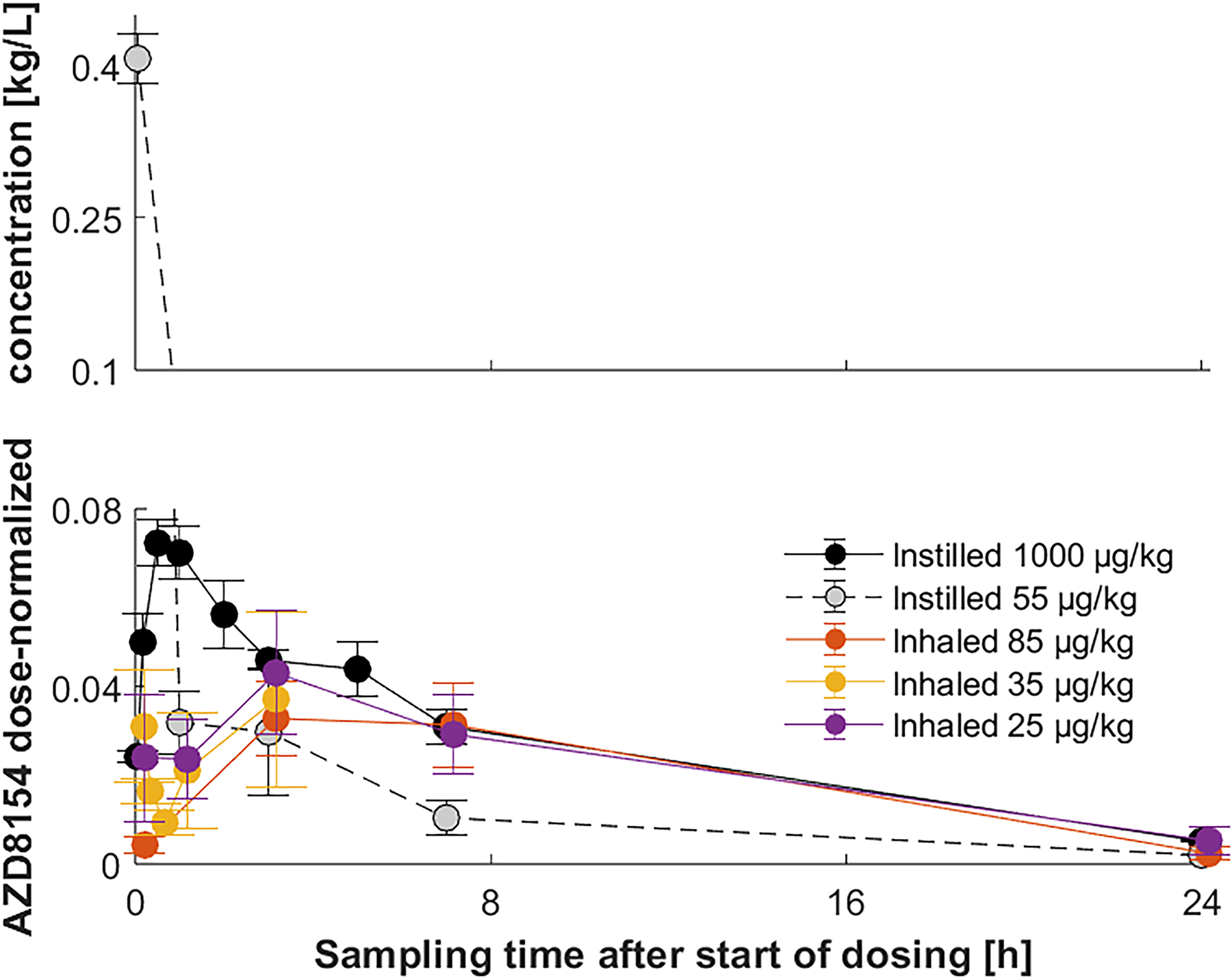
Dose-normalized rat plasma concentration of AZD8154 as a function of sampling time after initiation of dosing of the instilled lung doses of 55 and 1000 µg/kg and the inhaled lung doses of 25, 35 and 85 µg/kg. The vertical error bar at each data point is the standard error of the mean, i.e., the sample standard deviation divided by the square root of the sample size.
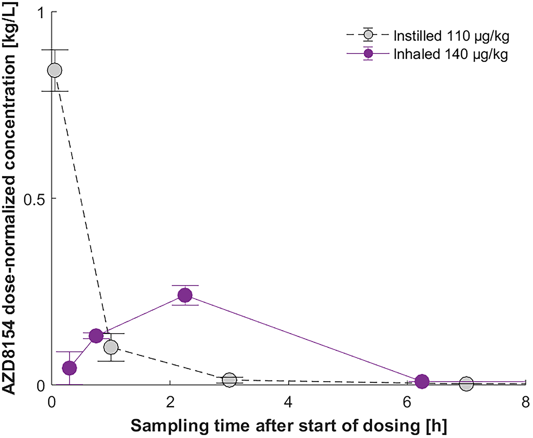
Dose-normalized mouse plasma concentration of AZD8154 as a function of sampling time after initiation of dosing of the instilled lung dose of 110 µg/kg and the inhaled lung dose of 140 µg/kg. The vertical error bar at each data point is the standard error of the mean, i.e., the sample standard deviation divided by the square root of the sample size.
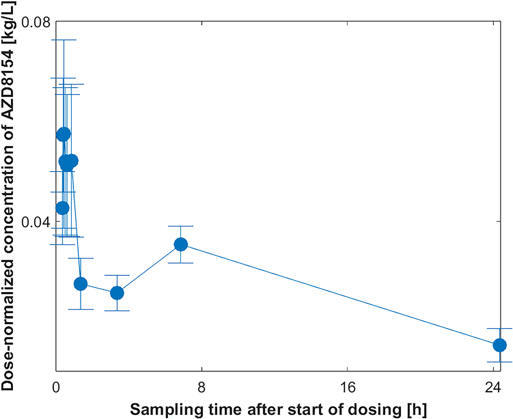
Dose-normalized dog plasma concentration of AZD8154 as a function of sampling time after initiation of dosing of an inhaled lung dose of 50 µg/kg, from previously reported results. 20 Three female beagles were all dosed at five separate occasions, so the total number of plasma profiles were 15 (3 × 5), i.e., n = 15 results at each time point. The vertical error bar at each data point is the standard error of the mean, i.e., the sample standard deviation divided by the square root of the sample size.
The initial plasma peak directly after the dose for both rat and dog indicates that there was an initial short burst of drug that was quickly dissolved and absorbed into the systemic circulation. The high fraction of the inhaled lung dose found in the rat lung initially means that this initial plasma burst must have originated from only a small fraction of the lung dose. The burst could have been caused by the smallest and fastest dissolving drug nanocrystals that had the greatest possibility to fit into the smallest aerosol droplets and therefore also could deposit in the most peripheral parts of the lung. A more peripheral deposition is expected to lead to faster absorption, due to the thin barrier to absorption in that region. 32 The plasma concentration distinctly dropped after the initial burst because less drug was absorbed from the lung, but it then increased again, leading to a second plasma peak just a few hours after the dose. The second plasma peak could be due to the dissolution of a major fraction of the lung dose, consisting of slightly larger and slower-dissolving drug nanocrystals in all parts of the lung except the most peripheral parts. The plasma concentration then slowly decreased again, because a major part of the lung deposited dose had been absorbed, and the absorption rate therefore decreased. This is supported by the fact that less than half of the lung dose was in the rat lung three hours after dose. It is plausible that the slower dissolution rate of the largest drug particles in the lung could have been a contributing factor to the slow decrease in the absorption rate after the second plasma peak. It should be noted that AZD8154 demonstrated an acceptable safety profile in healthy volunteers, 21 with no reports of serious adverse events or clinically significant drug-associated safety concerns. It is therefore extremely unlikely that any biological effects could have been the root cause for the second plasma peak a few hours after dose. The second plasma peak was therefore caused by the good drug retention in the lung from the designed low solubility, resulting in slow dissolution.
The plasma profiles for the instilled doses of AZD8154 were clearly different compared with the inhaled doses (Figs. 5 and 6). The instilled plasma profiles for the dose in the mouse and low dose in rats were similar in that the concentration was at its maximum immediately after dose and then dropped dramatically. The initial concentrations were in both cases more than a tenfold higher compared with the inhaled doses. This result was completely opposite to the expected result. Intratracheal instillation results in a non-uniform and central lung distribution1–7 compared with the uniform distribution in the entire lung for inhaled delivery.9–11 The instilled dose also reaches the lung instantly, whereas the inhaled dosing time in our experiments was 10 minutes for the rat and 30 minutes for the mouse (Table 1). It was therefore anticipated that the instilled drug should have been dissolved to a lesser extent due to the much shorter available time to dissolve in combination with a slower dissolution rate due to a greater concentration of the drug nanocrystals in a smaller area of the lung. The absorption from the lung was also expected to have been slower for the instilled dose due to the thicker membranes in the more central regions of the lung, where the drug will be mostly deposited. To find the opposite, that is, that the instilled drug had been absorbed faster compared with the inhaled drug was therefore very surprising and contradictory. A plausible explanation is that undissolved drug nanocrystals leaked from the lung into the systemic circulation already during the instillation procedure due to its invasive nature.
The plasma profiles for the instilled high rat dose (1000 µg/kg) and low rat dose (55 µg/kg) deviated. However, there was an early plasma peak also for the high dose, and the plasma concentration up to 3 hours after the dose was also much higher compared with the inhaled doses. The difference between the two instilled dose levels in rat was almost 20 times, but the installation procedure was also not the same (Table 1). No air was used in the syringe for the high dose, but for the low dose, an additional equal volume of air was used to securely push the formulation out of the syringe, which was the same procedure as for the instilled mouse dose. It is interesting that the different procedures correlated with the deviating plasma profiles, and it can be speculated that the additional volume of air may be the root cause.
Drug B is also very poorly water-soluble, and it was therefore also formulated as drug nanocrystals suspended in water. It was dosed to rats as outlined in Table 2. A similar difference in exposure as for AZD8154 between the two routes of administration of a similar lung dose was also observed for DRUG B. The initial rat plasma concentration immediately after dose was tenfold higher for the intratracheal instillation (Fig. 8) while the initial fraction of the lung dose in the lung was obviously lower (Fig. 9) compared with the inhaled delivery. The fact that at least about 80% of the inhaled lung dose was in the lung four hours after the dose shows that DRUG B was also very well retained in the rat lung (Fig. 9).
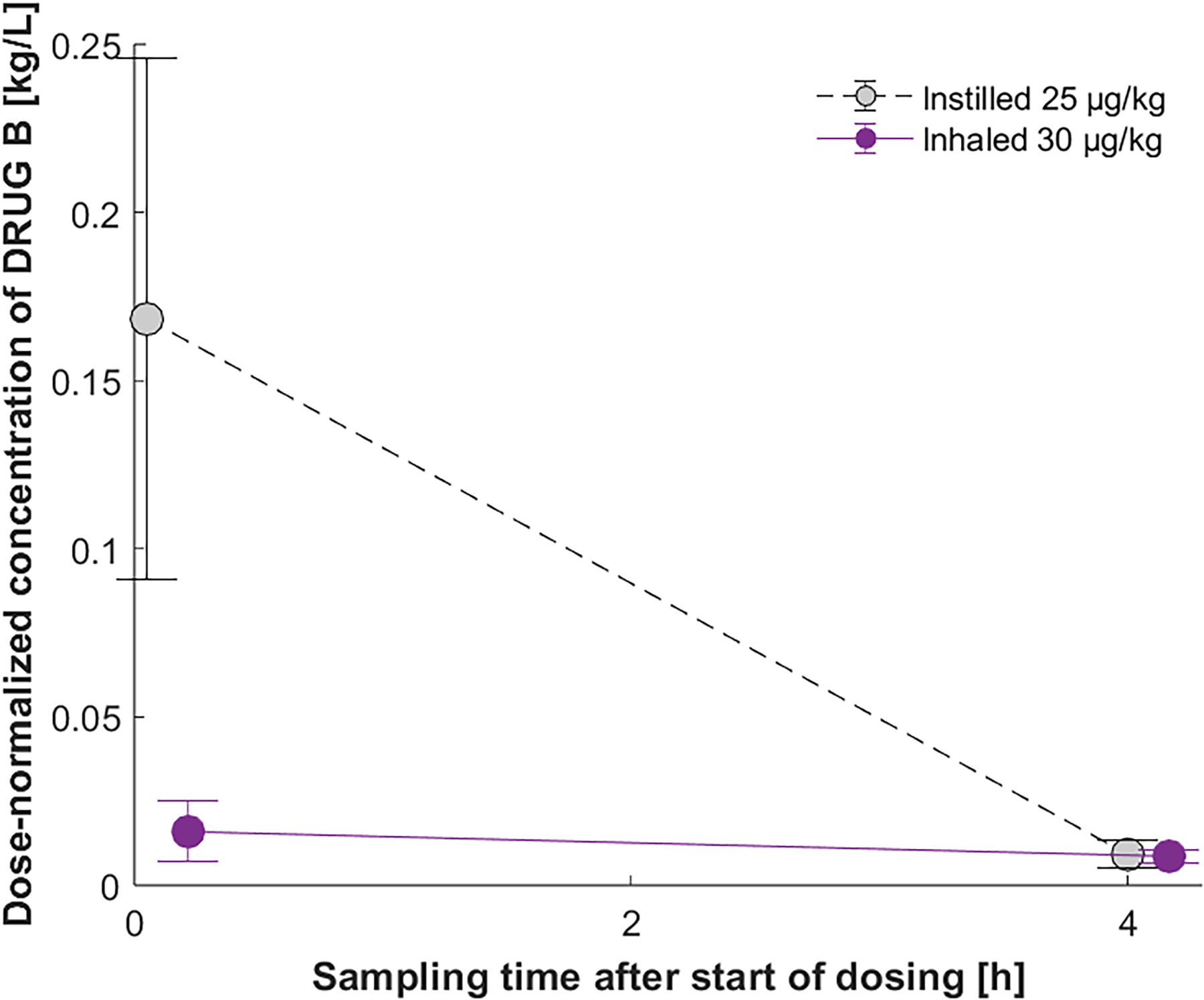
Dose-normalized rat plasma concentration of DRUG B as a function of sampling time after initiation of dosing of the instilled lung dose of 25 µg/kg and the inhaled lung dose of 30 µg/kg. The vertical error bar at each data point is the standard error of the mean, i.e., the sample standard deviation divided by the square root of the sample size.
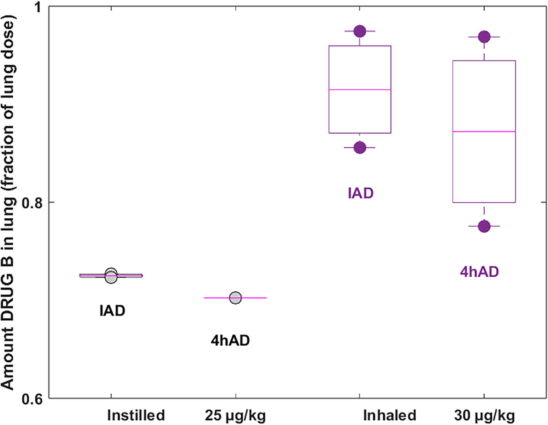
Amount of DRUG B in rat lung as a fraction of the targeted lung dose, immediately after dose (IAD) and 4 hours after dose (4hAD) of the instilled lung dose of 25 µg/kg and the inhaled lung dose of 30 µg/kg. The circles represent the individual results for each administration and time point. The boxes describe the statistics, i.e., the central pink mark indicates the median, and the bottom and top edges of the box indicate the 25th and 75th percentiles. The whiskers extend to the most extreme data points not considered outliers.
Intratracheal Instilled and Inhaled Doses of DRUG B in Rats (Wistar, Males)

A once daily instilled 1 mg/kg lung dose of the water-soluble DRUG C in seven female CD-1 mice using a dose volume of 1.5 mL/kg that was followed by an equal volume of air, resulted in observations such as hunched body posture, eyes half shut, decreased motor activity, and piloerection. The observations were made for all animals already after the first dose, and they were worsened after the second dose on day 2. The mice were pre-terminated for welfare reasons after the second dose, and the originally planned third dose was abandoned.
The same inhaled 1 mg/kg lung dose of DRUG C in 12 female CD-1 mice did not induce any treatment-related clinical observations. It was thus obvious that the inhaled dose was, in contrast to the instilled dose, tolerated by the mice.
The difference in tolerability correlated with a similar difference in exposure between the two routes of administration as for the two poorly water-soluble drugs. At the first sampling time, half an hour after the dose, the initial plasma concentration of the instilled DRUG C was fourfold higher, while the initial fraction of the lung dose in the lung was tenfold lower compared with the inhaled delivery. The fraction of the inhaled lung dose of DRUG C in the lung over time showed that the targeted lung dose was achieved and that the drug was well retained in the lung, that is, the fraction decreased from 60% to 50% between the initial and the second sampling time, 2 hours later. Drug C was, however, not retained due to poor solubility, but due to it being a zwitterion with an acidic pKa below 4 and therefore ionized at neutral pH, which reduces the permeability to the systemic circulation.
It has been reported8,16 that an instilled dose volume should be at least 1–2 mL/kg and preferably with an additional equal volume of air, to improve the poor lung distribution and push the dose further into the lung. It is plausible that the invasive instillation procedure will induce enough stress in the lung to cause a leakage of drug into the systemic circulation. It is uncertain if such a leakage would occur in or close to the trachea or further down in the lung. The trachea will be exposed to the highest pressure, but the tissue further down in the lung is thinner as well as more transfused and may therefore be more prone to leak drug into the systemic circulation.
Conclusions
All three drugs in our studies have very good lung retention properties, two of them are extremely poorly water-soluble and will therefore dissolve slowly in the lung, and the third is a water-soluble zwitterion with an acidic pKa below 4 and therefore ionized at physiological pH, which reduces its permeability from the lung to the systemic circulation.
The instilled dose was administered instantly to the lung and will be distributed centrally in the lung in a non-uniform way. The inhaled dose was administered during a dosing time of 10 or 30 minutes and will be distributed uniformly in the entire lung. It was therefore expected that the instilled drug would be absorbed slower and that a smaller fraction of the lung dose would have reached the systemic circulation immediately after the dose compared with the inhaled drug. However, the contrary was instead observed, that is, immediately after the dose, the plasma concentration was much higher, while the fraction of the lung dose in the lung was obviously lower for the instilled compared with the same inhaled dose of all three drugs.
There was also an obvious observed difference in the tolerability of the same lung dose of the water-soluble drug, and the inhaled dose was, in contrast to the same instilled dose, tolerated by the mice.
A plausible explanation for the observed difference in the exposure is that the drugs leaked from the lung into the systemic circulation already during the instillation procedure due to its invasive nature. This insight is important because intratracheal instillation administration is widely used during the early phase of pulmonary drug development as an alternative to inhaled delivery, and the observed difference in exposure and tolerability has, to the best of our knowledge, never been reported before.
Footnotes
Acknowledgments
The authors thank Britt-Marie Fihn, Astrid Collin, Mio R. Blomqvist, and Veronica Kühn for their contributions to the in vivo studies. The authors are grateful to Eva Lamm Bergström for her insightful support.
Author Disclosure Statement
No competing financial interests exist.
Funding Information
This research received no specific grant from any funding agency in the public, commercial, or not-for-profit sectors.
Author’ Contributions
M.B.: Conceptualization, methodology, investigation, formal analysis, writing—original draft. E.H.: Investigation, writing—review & editing. R.H.: Investigation, formal analysis, writing—review & editing.