
Abstract
Introduction:
A major roadblock to the investigation of emerging “omic” technologies is the availability of clinically derived tumor tissue. This problem is compounded by tissue being processed in labs using formalin-fixation, paraffin-embedding. A novel approach that circumvents these barriers was developed and tested. This approach represents an opportunity for biobanks to generate hard-to-obtain specimens from clinical tumor specimens for emerging “omic” research studies.
Objectives:
This study demonstrates a specimen processing method capable of creating new samples dedicated for multi-omic studies from clinical tissues, all without detracting from the current formalin-fixed, paraffin-embedded process.
Methods:
Using this new method, aliquots for the study of exosomes and metabolites can be generated from primary bladder cancers excised via transurethral resections. Once procured, the nature of tumor-derived exosomes can be examined using an exosome protein microRNA one-stop biosensor and metabolites via liquid-chromatography tandem mass spectrometry. Intact cells are also recovered and can be prepared for examination by either Thin-Prep cytology methods or the creation of cell blocks. The latter methods are used to confirm the phenotype of the cells present in these aliquots.
Results:
Populations of diagnostic tumor cells were confirmed to be recovered and morphologically consistent with the originating parent tissue. Isolation and characterization of exosomes from these dedicated samples confirmed the presence of tumor-specific signal molecules. The untargeted profiling of other dedicated aliquots found identifiable metabolites of multiple different classes that had been extracted from these tumor cells.
Conclusion:
The fight against cancer will involve understanding its complexities. Developing technologies to under-studied analytes of cancer will be integral to this process. The adoption of the described tumor specimen processing approach in primary bladder cancer in this study represents a novel means for biobanks to generate and collect dedicated aliquots for research into these analytes of increasing importance.
Introduction
The fight against cancer is constantly improving because of advances in knowledge and technology. Once dominated by the field of genomics, cancer is now known to involve multiple deregulated cellular pathways. 1 These reprogrammed pathways can potentially serve as “hallmarks” for cancer detection because they create altered cellular signals. These altered signals can now be detected by emerging technological platforms that are capable of identifying products that are generated or transmitted by cancer cells, either as altered metabolites or transported cellular elements (e.g., proteins, nucleic acids, etc.) in exosomes.2–11 These altered signals may eventually develop into clinical assays used for the early detection, or the identification of recurrent cancer, by interrogating for their presence in the peripheral blood or other liquid specimens such as urine.12,13 Traditionally, investigation of tumor biomarkers begins with the examination of tumor tissue or cells because they are the originating site for these biomarkers. Human tumor tissue is the preferred source for these research studies as they do not demonstrate the bias that may develop as cell cultures.14,15 However, clinical samples for research are sometimes difficult to obtain, and are processed through a method called formalin-fixation, paraffin embedding (FFPE) that is optimal for microscopic examination but creates problems for the examination of nucleic acids.16–18 Similar problems have been noted in the investigation of metabolites in FFPE tissue that include diminished recovery, differential recovery of specific classes and chemical modifications.19–22 Exosomes are predominantly studied from peripheral liquid sources like the blood, and not from tissue specimens.23,24 When they are studied from tissues, they are sourced infrequently from extracts of whole tissues, predominantly only for noncancer studies, and often require extensive labor and are pulverized, making them unusable for clinical diagnosis.25–28 The processing of tissue by FFPE is particularly problematic for exosomes in the interstitial fluid as it involves clearing the tissue of all fluid before infiltration with wax, which may lead to diminished levels of exosomes in tissue-based studies. Additionally, tissue should be the starting material to be studied, as cell lines, which are often used instead for exosome and metabolomic studies, may not be fully representative of tumor cells from the human body.29–31 A novel method needs to be developed capable of recovering both metabolites and exosomes from clinical tumor samples without compromising the specimen for traditional FFPE microscopy-based pathological diagnosis. Herein, we describe a simple and effective method capable of generating multiple analyte-specific daughter aliquots from a cancer specimen that does not compromise the tissue or interfere with standard FFPE processing protocols. The adoption of this approach represents the potential to generate a rich source of specimens for biobanks that can eventually be made available to the burgeoning exosome and metabolomic cancer research communities.
Materials and Methods
This project was approved by an Institutional Review Board (IRB) (protocol T-31-011, Lake Erie College of Osteopathic Medicine) and because it involved the collection of material that would otherwise be lost or discarded, included a waiver of consent. Primary bladder cancer specimens were chosen as the tissue source based on how they are acquired and their availability. In everyday clinical practice, once a bladder cancer is identified, the tumor is typically excised using a resectoscope, a specialized cystoscope equipped with a wire loop. This instrument allows for curettage of the tumor and portions of the underlying tissue in a procedure known as a Trans Urethral Resection of Bladder Tumor (TURBT). This procedure fragments the tissue so it can be recovered through the narrow orifice that is the urethra. Included in these fragments of tumor are elements of the underlying tissue stroma, which is clinically needed to establish staging by evaluation of tumor involvement in either the lamina propria or detrusor muscle. The fragmented pieces of tissue from this curettage specimen are then sent to the pathology laboratory. At the hospital where the study specimens were acquired, these specimens arrive fresh and in a dedicated container, with no formalin and minimal amounts of fluid. Typical processing in the pathology laboratory includes placement of these fragments into a tissue cassette, which are then processed through a series of reagent exchanges prior to infiltration with wax for eventual sectioning. Because these cassettes often have sizeable pores meant to allow for the flow of differential concentrations of alcohol reagents to mix with the tissue fragments, and because the tissue is comprised of variably sized pieces, a biopsy bag is often used. Biopsy bags have a much finer pore size, often between 0.2 and 0.3 mm in size, are disposable and are meant to hold the tissue in the cassette and help prevent fragments from leaking out and possibly ending up or in the cassettes from other cases and contaminating them. This could potentially lead to difficulties in interpretation and in making the correct clinical diagnosis.32,33 However, individual cells and small groups of cells are still capable of leaking out of these biopsy bags and potentially ending up in the cassettes of other clinical cases. The approach described takes advantage of this feature, the ability to recover these single cells and small groups of cells that would otherwise contaminate other tissue cases. In doing so, new specimens consisting of these tumor cells are created. These new specimens can now be the originating starting point for a number of different purposes. In this study, we sought to determine if metabolites and exosomes could be directly sourced from them.
Specimen processing
Tumors were processed as they were received with an average of 1 hour between receipt to the start of processing. Curetted urothelial cell carcinomas taken out as TURBT specimens and sent unfixed to the pathology laboratory were de-identified and processed as shown in Figure 1 and depicted in Figure 2. The fragments of tissue from the specimen cup were transferred to a number of biopsy bags that would adequately fit the received specimen, then placed into a tissue cassette(s), and then closed with the accompanying cover. These cassettes were then transferred into a separate specimen cup with enough 1 × Phosphate Buffered Saline (PBS) to be able to bathe the cassettes in fluid, followed by gentle mixing for 1 minute and followed by sitting at room temperature for 4 minutes. Because each collected tumor case was different, the amount of tissue placed into cassettes was dependent on the size of the clinical specimen. Five cassettes could fit into a 90 mL capacity specimen cup for a washing. The volume used to bathe the tissue present in these cassettes averaged to be about 50 mL. The purpose of this gentle bath was to collect exosomes, which are known to be continuously secreted by living cells as part of their cell-to-cell communication. The cassettes with the tissue in them were transferred to a collection basin and routed for additional formalin fixation and the process of FFPE. The fluid in the specimen cup was transferred to 1.5 mL microcentrifuge tubes and then spun down using a tabletop minicentrifuge for 30 seconds at a speed of 7000 rpm/2680×g (Fig. 3). The supernatant was aspirated out, frozen and kept at −20°C and transported to investigators for the evaluation of exosomes (described below). To confirm that these cells were indeed the same as the originating tumor tissue, one of the newly generated pellets was processed for visual examination as a cytological specimen. Afterwards, the pellet was resuspended in PreservCyt® Solution (Hologic, Marlborough, MA) and thin-layered slides for cytological examination were prepared using a Thin Prep 2000 Processor (Hologic, Marlborough, MA). For additional visual confirmation, a second pellet was FFPE as a cell-block using the sodium alginate-based method. 34 The cells from both these preparations were compared with the tissue where they had originated from that had undergone FFPE processing (Fig. 3).
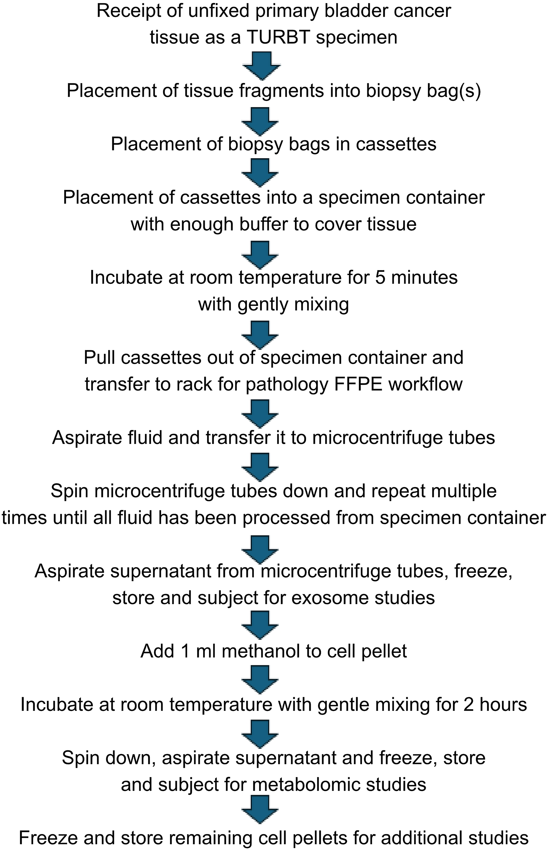
Workflow schematic for the generation of new, analyte-specific specimens from primary bladder tumor tissue. FFPE, formalin-fixed, paraffin-embedded; TURBT, transurethral resection of bladder tumor.
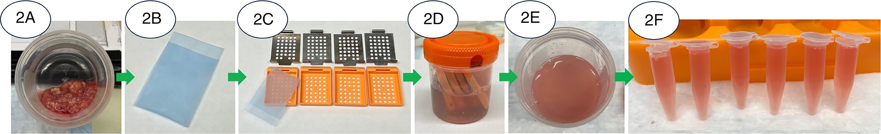
Application of the workflow for the generation of new, analyte-specific specimens from primary bladder tumor tissue.
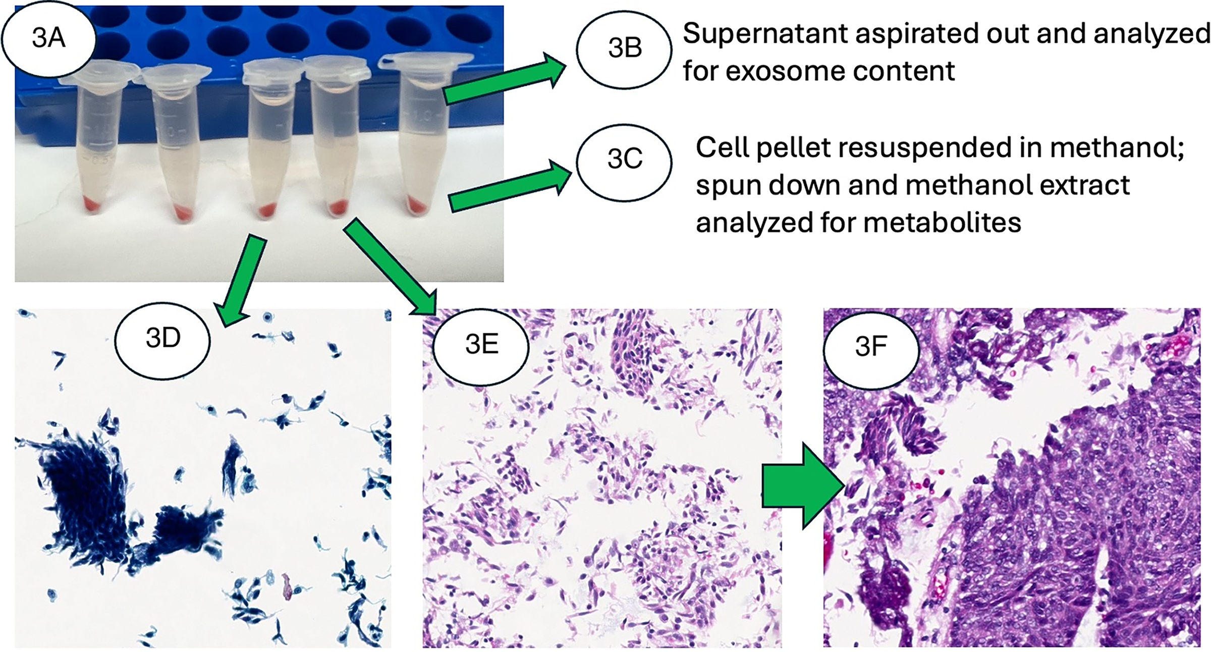
Further processing of the new aliquots.
Exosome examination
The recovered supernatants were examined for the presence of exosomes. This was done first by analyzing for size and concentration using a nanoparticle tracking analysis (NTA) system (NanoSight, LM10, Malvern Instruments Ltd.), a method used to characterize submicroscopic particles in liquid media. Briefly, the samples were diluted in PBS until 50–100 nanoparticles were detectable within the field of view of the NTA system. All measurements were performed using identical instrument settings: a detection threshold set to 6, a screen gain to 8, and a camera level to 14.
Exosomal CD63 and microRNA-21 (miR-21) levels were measured using a newly developed exosome protein microRNA one-stop (Exo-PROS) assay. 35 CD63 is a protein frequently found on exosomes, while miR-21 is a small, non-coding RNA molecule found to be overexpressed in cancer cells. This Exo-PROS assay employs surface plasmon resonance as the sensing mechanism to enable sensitive detection of exosomal biomarkers including both proteins and microRNAs. Briefly, the surface of the Exo-PROS biochip was functionalized with anti-CD63 antibodies and background signal levels were established. Samples were then applied onto the biochip to allow for the capture of exosomes. Unbounded exosomes were washed off, the intensity of reflected laser light recorded and the expression level of exosomal CD63 calculated. Next, molecular beacons (MBs), which are oligonucleotide hybridization probes specific for miR-21, were added and incubated with the captured exosomes. Levels of miR-21 were quantified in the captured exosomes, and their expression levels calculated.
Metabolomic studies
One or more of the pellets from each case was dedicated for metabolomic studies. Metabolites were extracted from the pellets using a 2-hour incubation with 80% methanol as previously described. 36 After this 2-hour incubation, the cells were pelleted down using a minicentrifuge for 30 seconds at 7000 rpm, and the supernatant extract was collected, frozen at −20°C, and transported to a laboratory for metabolomic analysis. An untargeted, or discovery-based metabolomic profiling approach was undertaken so as to be able to analyze the breadth of detectable metabolites in the samples. Briefly, solid phase extraction was performed on a Waters Oasis HLB cartridge with the optimized method consisting of conditioning with methanol (MeOH), followed by equilibration with formic acid (FA), washing with 5:95 MeOH:water, and elution with MeOH. The samples were analyzed with an optimized gradient liquid chromatography tandem mass spectrometry (LC-MS/MS) workflow. An Ultimate 3000 ultraperformance liquid chromatography (UPLC) was coupled to a ThermoFisher Q-Exactive Orbitrap to conduct the analysis. A ThermoScientific Acclaim Polar Advantage II C18 column (3 µ, 120 Å, 2.1 × 100 mm) was used for the separation. The separation was performed at a flow rate of 0.2 mL/min, injection volume of 1 μL, m/z range of 150–1000, run in data dependent Mass Spectrometry (ddMS) discovery mode and collision energy of 27 V. The gradient was t = 0.0 minutes at 5% B, ramped to 65% B in 3.0 minutes, ramped to 85% B in 6.0 minutes, ramped to 100% B in 20.0 minutes, held at 100% B in 25.0 minutes, ramped down to 5% B in 25.5 minutes, and held at 5% B in 30.5 minutes. Data was analyzed using a Thermo Xcalibur 4.2.47 and Compound Discoverer 3.3 SP3.
Results
A total of six bladder tumor specimens were collected and processed by this method, yielding enough material for exosome and metabolomic studies, and the generation of cytological and cell block preparations for the visual corroboration that the recovered tumor cells in these pellets were morphologically the same as present in the parent tissue that was FFPE (Fig. 3). The initially clear fluid that was added to these specimens became cloudy after the short incubation period in the specimen container (Fig. 2). The short centrifugation step after dispensing this cloudy fluid yielded a clear supernatant and a tan pellet. There was enough of this to create anywhere between 4 and 10 new specimens in microcentrifuge tubes, depending on the starting amount of tissue received in the specimen container from the operating room. These 4–10 new specimens consisted of new aliquots consisting of exosomes, and depending on the operator’s preference, an additional 4–10 for metabolomic studies or of cell pellets. They did not contain any tissue fragments needed for microscopic examination. The cells in the pellets were found to consist of intact single and small groups of tumor cells. These tumor cells consisted of both low- and high-grade urothelial cell carcinomas. The tumor cells in the cytology specimens that came from the recovered cell pellets corresponded to the same grade observed in the parent tissue that was FFPE. Grading of tumor was based on current criteria for urothelial cell carcinoma. Lamina propria and detrusor muscle fragments were present in all of the parent FFPE-processed specimens and not in the cell pellet specimens. Only single cells and small groups of cells were present in the Thin-Prep cytology slides or the cell block preparations.
NTA detected exosomes in all washing samples and successfully measured their size and concentration. The exosomes had an average diameter of approximately 130 nm and a number concentration ranging from 1 to 3.5 × 10¹¹ exosomes/mL (Table 1).
Size and Concentration of Exosomes in Washing Samples From Six Bladder Tumor Tissues, Measured by Nanoparticle Tracking Analysis

HG, high grade; LG, low grade.
The Exo-PROS assay was used to capture exosomes from the supernatant samples and to quantify exosomal Cluster Designation 63 (CD63) and miR-21 expression levels. Because CD63 is a universal exosomal surface marker, the Exo-PROS biochip was functionalized with anti-CD63 antibodies to facilitate exosome capture. The expression of miR-21 in the captured exosomes was then measured using MBs as the sensing probes. miR-21 was selected as the biomarker due to its strong association with cancer, as demonstrated in recent meta-analyses, and its specific relevance to bladder cancer.37–41 The Exo-PROS assay successfully detected both exosomal CD63 and miR-21 in the supernatant samples. Interestingly, expression of exosomal miR-21, normalized to CD63 to account for variations in exosome concentration, correlated with both bladder cancer grade and tumor size (Fig. 4). The samples from higher grade and larger tumors showed higher exosomal miR-21 levels than those from the low grade and smaller tumors, findings that warrant further examination in larger specimen cohorts. These findings confirm the presence of exosomes in tumor washing samples and highlight the potential utility of exosome-derived biomarkers in informing personalized patient care.
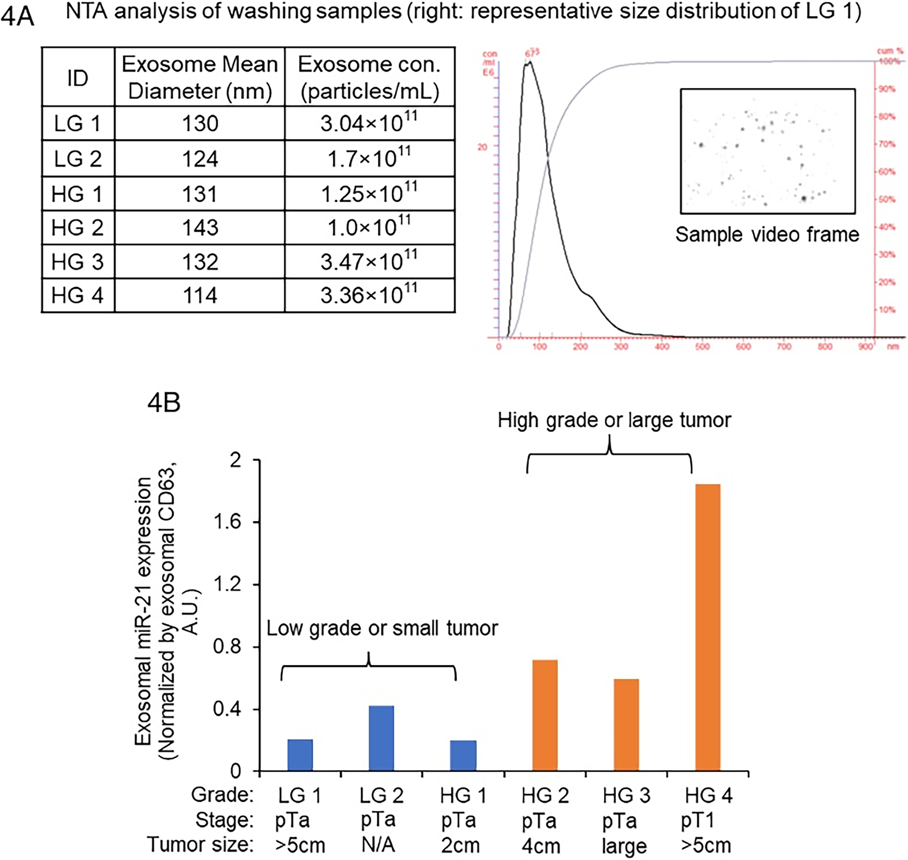
Characterization of exosomes in washing samples from bladder cancer patients.
From the methanol extracts, detected features, or the signals extracted from the raw data that corresponded to potential metabolites, ranged from 72 to 99 for each specimen. Some of the identified compounds included fatty amides, endocannabinoids, glycerolipids, straight-chain aliphatic dicarboxylic acids, and drugs (Table 2). The findings from these extractions favored smaller molecules and polar molecules. The most abundant classes were lipids and fatty amides. Some common entities identified included the fatty acid oleamide and hexadecanamide. Three features were found across all cases. Two specimens had signals and different adducts not detected in the other bladder methanol extracts. For 4 of the specimens, there were 23 unique features and different adducts detected and not observed in any of the other bladder biopsies. Changes due to degradation could not be confirmed or denied based on the single acquisition point of the specimens, and storage conditions prior to examination. Overt modification could not be commented on without examination of positive and negative control material, which was not present for this study. These findings support the feasibility of this approach in creating analyte-specific specimens for metabolomic profiling from the recovered tumor cells from biopsy bags.
List of Metabolites Identified in Common From the Methanol Extracts
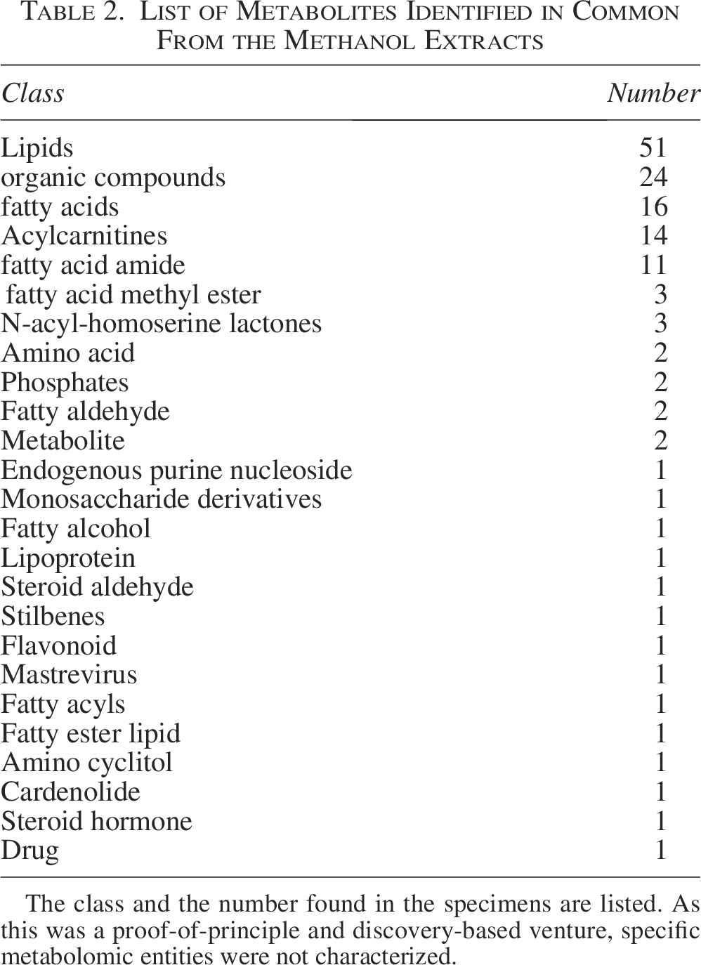
The class and the number found in the specimens are listed. As this was a proof-of-principle and discovery-based venture, specific metabolomic entities were not characterized.
Discussion
Cancer results in the generation of multiple alternative pathways instigated by tumor cells to develop a growth advantage. These alterations begin with changes in the cell’s nucleic acids, but are carried out by the signaling machinery of the cell. Currently, the emphasis in diagnostic testing resides in the examination of tissue and cells and their nucleic acids. This leaves out a number of other analytes that are currently not tested but can serve to potentially improve cancer management through their detection. In low-grade bladder cancer, in which cytological examination of urine specimens is the gold standard, the sensitivity of cancer detection has been reported to be as low as 16%. 42 Thus, screening for bladder cancer represents a field rife for improvement. Fortunately, bladder cancer also represents an ideal source to create adjunctive specimens for additional studies because of the nature of the current surgical procedure known as the TURBT. In this project, we used the observation that despite efforts to keep bladder cancer specimens from “contaminating” other clinical tissue cases through the use of biopsy bags, stray tumor cells still can be found where they are not wanted. This served as the foundation for not only reducing these stray cells and contaminating other clinical cases, but also using them to generate additional aliquots of material for analytes not often studied directly from tissue sources. A sizeable amount of contaminating cells can be recovered from biopsy bags, enough to not only create aliquots for exosomes and metabolomic studies, but to confirm by cytology that they are the same cells as the originating parent tissue, with additional material available for even more unrealized studies.
While tissue biopsies remain the clinical standard for tumor characterization, they often fall short in capturing the full extent of tumor heterogeneity. In this context, metabolites from, and exosomes secreted by, tumor cells may offer a more comprehensive molecular snapshot of the tumor landscape. Detecting these analytes in body fluids have been gaining interest as a new frontier in cancer research, but is made difficult due to the dilutional effect of analytes coming from all the cells in the body and mixing together to be present in these fluids. This may potentially reduce their diagnostic specificity. In contrast, metabolites and exosomes generated in the aliquots using this method create highly concentrated specimens of these analytes because the fluid used to develop them bathed, or was in direct proximity to the tumor cells with little to no contribution from other types of cells. This highly concentrated specimen is ideal for discovery-based studies, but what makes it even more valuable is that it is coming from material that is predominantly, if not all, tumor. In other words, there is no dilutional effect from other tissues in the body as in peripheral blood or urine samples. Additionally, this method does not engender the difficulties associated with tissue that is formalin fixed and paraffin embedded. Finally, the gatekeepers of the tissue specimen, pathologists, should not pose as barriers to biobanks to create these additional aliquots for research purposes, as the tissue fragments remain intact and are not compromised for diagnostic purposes. Only fully representative tumor cells, albeit as single cells and small groups of cells, serve as the basis for the creation of aliquots for biobanking and research purposes. The results in this study demonstrate the presence of exosomes and metabolites in these washing samples and suggest that exosome-derived and metabolite-based biomarkers are present and may provide patient-specific information to support personalized care.
For biobanks, after the collection of these new aliquots, methods to store, preserve, and ensure quality include consistent rinsing protocols (e.g., using a fixed volume of PBS per unit tissue weight, minimizing processing delays, and maintaining samples at cold temperature prior to processing) and would be important considerations to reduce variability. Additionally, standardizing quality control steps, such as monitoring protein concentration, exosome number concentration and exosomal markers (e.g., positive markers CD9, CD63, CD81, TSG101, Alix; and negative markers Calnexin, GM130), in pilot aliquots, may further improve reproducibility across different samples and time points. As for metabolites, the reader is referred to two recent, thorough publications that delve in much greater depth on quality control for metabolomics.43,44 To reduce the effects of cold-ischemia, instead of starting the collection and processing protocol as was described in this study “when they were received,” biobanks interested in initiating this method may opt to collect the TURBT specimen directly from the operating room, thus greatly reducing this variable.
A limitation of this pilot study was the absence of an examination and comparison of the exosomes and metabolites from the urine of the patients from which these TURBT specimens were derived. These types of studies will help to further narrow down which biomarkers from these different analytes may serve as candidate biomarkers to improve the sensitivity of cancer detection. A second limitation is that this study was the first of its kind and hence was not tested on other types of cancer and types of specimens. A possible third consideration, the absence of a comparison with the current gold standard, tissue that has been FFPE, should not be regarded as an experimental constraint because of the limitations already noted in specimens that have been processed as FFPE material.19–26
Looking forward, these same samples may also provide the basis for new methods in the management of cancer patients, as in the longitudinal monitoring needed in their care. Bladder cancer is currently regarded as the most expensive tumor to manage because of the significant time needed for the monitoring of tumor recurrence and or progression. As an example, once key biomarkers are identified in tumor samples processed through this method, their levels could be tracked noninvasively in follow-up urine samples through metabolomic or exosome studies to improve sensitivity in assessing therapeutic response over time or the emergence of tumor progression. The generation of multiple aliquots consisting of different analytes opens up the possibility for an expanded investigation of the hallmarks of cancer, as in this case for bladder cancer. Additional studies to assess whether this approach can be applicable to other tumors and other types of specimens are currently being undertaken.
Conclusions
The method described in this article represents a simple method to recover diagnostic tumor cells that have been found to be problematic in the clinical pathology laboratory as part of current processing protocols for primary bladder tumors, and turn them into useful sources for the generation of aliquots for dedicated analyte studies. Larger tissue fragments, consisting of lamina propria and detrusor muscle, are not part of these new specimens, and are retained for microscopic examination as part of the parent tissue and needs for tumor staging. Exosomes, which are continuously secreted by viable cells, are recoverable from the cells that make these new aliquots, and exhibit contents reflective of oncogenesis. Metabolites, extracted from these same cells using methanol, yield a number of classes of analytes that can be further characterized using tandem mass spectrometry. This simple and easy processing method represents a means for biobanks to generate and provide the specimens needed for the investigation and discovery of potentially new biomarkers in the form of exosomes or metabolites that are often difficult to obtain from clinical tumor tissues. Because there is no need for complex laboratory instrumentation, this collection protocol can be practiced in almost any laboratory, leading to the creation of the large numbers of samples often needed for biomarker studies to reach statistical significance. 45
Authors’ Contributions
W.M.: Conceptualization, data curation, formal analysis, methodology, resources, visualization, and writing. Y.W.: Conceptualization, data curation, formal analysis, methodology, resources, supervision, and writing. J.H.: Data curation, formal analysis, and investigation. T.W.: Conceptualization, data curation, formal analysis, methodology, resources, supervision, and writing. A.I.: Data curation, formal analysis, investigation, and writing.
Footnotes
Author Disclosure Statement
None.
Funding Information
None.