
Abstract
Properties of the chitosan films can be improved by incorporating clay minerals. So, solvent-cast films of the β-chitosan containing stevensite-rich or kaolinitic-illitic clays (up to 50 mass %) were characterized for their structural and mechanical properties. The effects of molecular weight (MW) and deacetylation degree (DD) of chitosan and the clay/chitosan mass ratio on the inhibition growth of Escherichia coli and Staphylococcus aureus were studied using the response surface methodology (RSM). The films consisted of exfoliated/intercalated or flocculated composites, and the electrostatic bonds formed between the functional moieties of the chitosan and the clay particles active sites essentially influenced their mechanical strength. The results of the study using RSM showed that the optimal value of MW required for the inhibition of the bacteria varied according to the film used, and high antibacterial activity necessitated high DD (89–97%).

Keywords
Introduction
Chitosan is a deacetylated derivative of the chitin, which is a constituent of shellfish and crustaceans exoskeletons, among others. It is a copolymer composed of β-(1-4)-2-amino-D-glucose and β-(1-4)-2-acetamido-D-glucose units. Three polymorphic forms (α, β, and γ) of chitosan are identified. β-chitosan is more soluble and more reactive than α-chitosan, 1 which is the most abundant form. Chitosan is suitable for many practical uses2–6 because of its non-toxicity, biodegradability, film-forming character, and high density of positive charges associated with the protonated amino-groups. The chitosan exhibited a good antibacterial activity. Referring to some authors, the antibacterial activity of β-chitosan was higher than that of α-chitosan. 1
The inhibition processes depend on chitosan characteristics, mainly its deacetylation degree (DD) and molecular weight (MW), as well as on bacteria strain.7–9 It can be summarized that the higher the DD, the better the inhibition. This fact is linked to the abundance of protonated amino-groups, and to their ability to interact with the specific functional groups of cells membrane macromolecules. 10 The studies dealing with the influence of MW on the inhibition of bacteria growth, such as Escherichia coli, showed different results. 10 Disparate effects of MW on chitosan antibacterial properties were cited elsewhere. 11 According to Raoka et al., 12 the effect of MW on the antibacterial activity of chitosan is greater than that of DD.
Clay minerals, especially those of the smectite group, can be delaminated. So, nanosized particles with high aspect ratio are obtained. Compared to kaolinite and illite, smectite minerals have high cation exchange capacity (CEC) and surface area. 13 Owing to these characteristics and to their anionic character, these clay minerals are suitable materials for medical and cosmetic purposes. 14
To improve mechanical and barrier properties of chitosan films for drug carrying and food packaging uses, the chitosan was mixed with limited amounts (up to about 5 wt%) of montmorillonite.15–17 The addition of the montmorillonite (di-octahedral clay mineral of the smectite group) to chitosan resulted in the formation of nanocomposites with efficient bactericidal effects.16,18,19 So far, no attention has been paid to the study of antibacterial activity of β-chitosan films containing stevensite or kaolinitic-illitic clays. Stevensite is a tri-octahedral swelling clay mineral, which belongs to the smectite group such as montmorillonite. So, chitosan association with limited amount of stevensite should result in intercalated and/or exfoliated nanocomposites formation. The lack of studies related to bacterial inhibition by chitosan-stevensite films could be due to stevensite scarcity. Kaolinite and illite are non-expandable natural clay minerals, 13 and chitosan intercalation in the interlayer spaces of their pristine structures could not occur. Thereby, chitosan association with kaolinite or illite should lead to tactoids formation. In such a condition, kaolinite as well as illite behaves like conventional fillers. To obtain chitosan-kaolinite nanocomposites films, some authors prepared exfoliated kaolinite by mechano-chemical treatment. 20
The use of β-chitosan-montmorillonite films as bactericidal materials has been ignored in spite of the potential reactivity of β-chitosan, which consists of parallel chains with weak intermolecular bonds. 21 For the same amount of montmorillonite (up to 5 mass%), β-chitosan-based films showed higher tensile strength and Young’s modulus than those of α chitosan-based films, and both films manifested similar water solubility. 22
The influences of chitosan characteristic (DD and MW) and clay/chitosan ratio changes on the inhibition of pathogenic bacteria growth have not been investigated, probably because of the numerous experiences required. This drawback could be overcome by using the response surface methodology. 23
In this study, the structure of solvent-casted films composed of β-chitosan and stevensite-rich clay (RH) or kaolinitic-illitic clay (BN2) was examined. Moreover, the changes of the physical/mechanical properties of films according to clay additions were followed. In addition, the growth inhibition of the most prevalent bacteria (Escherichia coli and Staphylococcus aureus) according to chitosan DD, chitosan MW, and clay/chitosan ratio was assessed using the RSM.
Materials and methods
β-chitosan
The β-chitosan was prepared from the chitin, extracted from the pens of local squid. For this purpose, finely ground squid pens were demineralized with HCl solution (0.55 N) and deproteinized with heated NaOH solution (80°C, 0.33 N). The pens powder/solution ratio was 1:10 (w/v). To prepare chitosan, chitin was deacetylated for 24 h using NaOH solution (40 mass%) and standard reflux setup.
24
The X-ray diffraction pattern of the prepared chitosan is shown in Figure 1. Taking into consideration the latter trace, and using the Scherrer’s equation and the crystallinity index determination method given in Focher et al.,
25
the chitosan crystallinity index and apparent crystal size were found to be 77 and 2.1 nm, respectively. Referring to the thermal curve shown in Figure 2, the chitosan was thermally stable up to about 266°C. X-ray diffraction trace of the prepared chitosan. Thermal curves of the prepared chitosan, BN2- chitosan (a) and RH-chitosan (b) films. (clay/chitosan mass ratio: 0.25/1).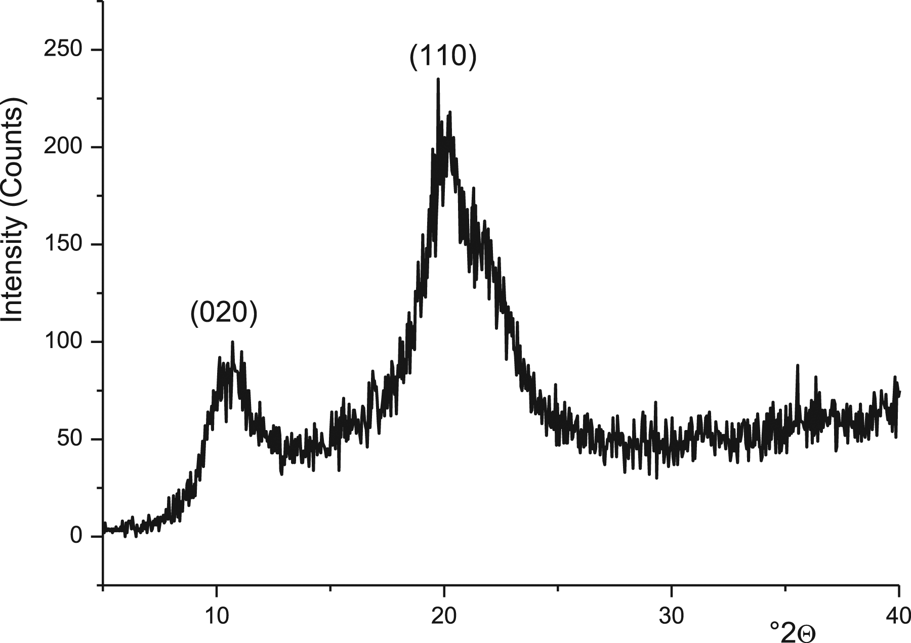

The molecular weight (MW) of the chitosan was calculated using the Mark–Houwink equation.
26

η is the intrinsic viscosity; K and a are determined constants. The deacetylation degree (DD) of the chitosan was determined by potentiometric titration following the experimental procedure in Tolaimate et al.,
24
and using the following equation
27
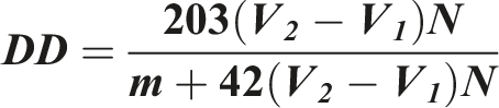
V1 and V2 are the volumes of NaOH solution at the first and the second neutralization points. N is the strength of the alkali solution used. m is the weight of the chitosan sample.
Clays
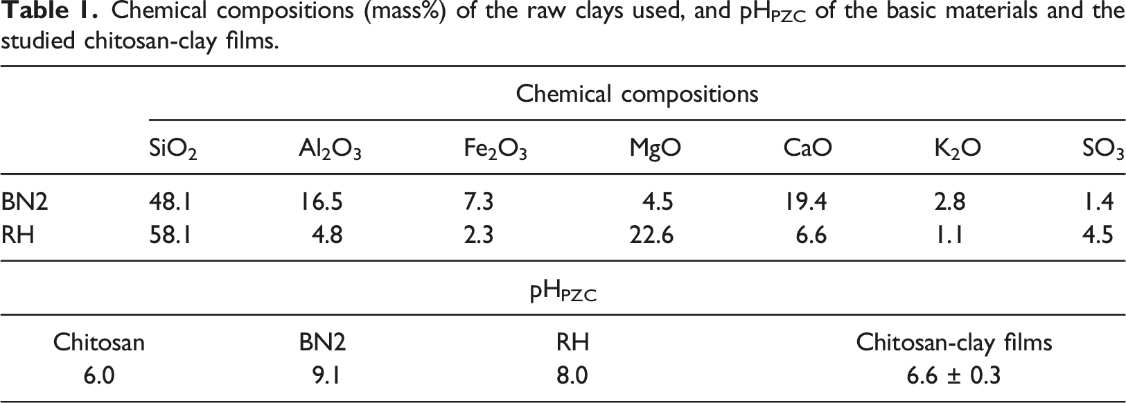
Chemical compositions (mass%) of the raw clays used, and pHPZC of the basic materials and the studied chitosan-clay films.
Chitosan-clay films
Chitosan-clay films (clay/chitosan mass ratio ≤100%) were prepared with the solvent casting technique. 30 For this purpose, sample (≤1 g) of the sodium-saturated clay was dispersed in 5 mL of acetic acid (1% v/v) solution, and stirred for 24 h. On the other hand, a portion of chitosan (1 g) was introduced in 100 mL of an acetic acid solution (1%v/v), and stirred for 2 h. The limpid solution of chitosan was mixed with the clay dispersion, and the mixture was homogenized for 24 h. 25 mL of the mixture was poured in a Petri dish and kept at 30°C for 48 h, so to evaporate water. The film formed was retrieved and soaked with a solution of NaOH (0.5 M) for 12 h, rinsed with distilled water, and dried at 25°C. The thickness of the films was of about 10 µm.
Physical/mechanical properties of the film
The mass loss of films was determined by weighing disks (∼3 cm diameter) before and after immersion in 50 mL of distilled water stirred at 350 rpm for 24 h. The tensile strength and the elongation at break of films were measured on strips according to the ASTM D882, 31 using an Instron universal machine operating with a crosshead speed of 50 mm/min. The measurements were done in triplicate.
Analysis techniques
Fourier-transform infrared (FT-IR) analyses were carried out on thin disks composed of 1 mg sample and 99 mg KBr. The spectra were recorded with a Perkin Elmer 1725 spectrophotometer functioning in the range of 4000–400 cm−1 at a resolution of 4 cm−1. The X-ray diffraction (XRD) analyses were performed on powder samples with a Philips X’Pert MPD diffractometer operating with a copper anode (λKα = 1.5418 Å). The step size (°2θ) and the scan step time were set at 0.013 and 1 s, respectively.
Percentage inhibition of bacteria growth
To determine the percentage inhibition (PI) of bacteria growth, a bacteria suspension (considered as a blank suspension) was adjusted to 105–106 CFU/mL using sterile saline water (0.9% w/v). A film sample (9 cm2) was introduced in a tube containing 5 mL of nutrient broth, and autoclaved at 120°C for 20 min. Then, the tube was inoculated with 0.5 mL of the bacteria suspension, and incubated at 37°C for 24 h. The optical density (OD) of the incubated and the blank suspensions were measured by using an UV-visible spectrophotometer operating at 620 nm.
32
These data were used to calculate PI
Effects of the chitosan characteristics and the clay/chitosan ratio on the growth inhibition of the bacteria

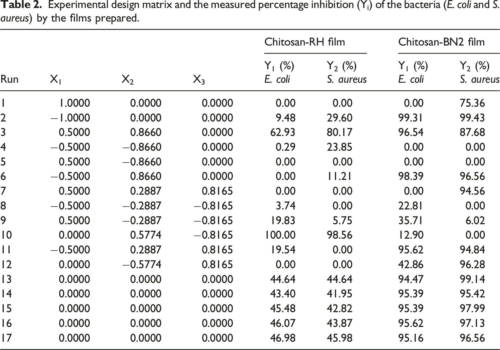
Experimental design matrix and the measured percentage inhibition (Yi) of the bacteria (E. coli and S. aureus) by the films prepared.
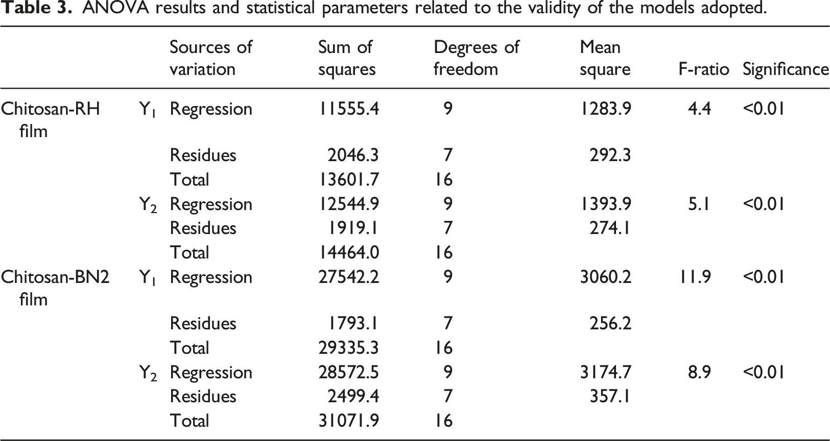
The validity of the polynomial models was assessed on the basis of the analysis of variance (ANOVA) and checked by the probability plot of residuals.
Results and discussion
Structural characterization of the chitosan-clay films
As can be deduced from the typical XRD patterns shown in Figure 3(a), the association of β-chitosan with BN2 clay did not have impact on the position of the basal reflexions of illite (°2θ = 8.78) and kaolinite (°2θ = 12.48). So, the interlayer space of both clay minerals remained unchanged, and consequently intercalated composites did not occur in this case. So, tactoid structure took place.
36
Considering that the points of zero charge of chitosan and chitosan-clay films (Table 1) were somewhat similar, the surface charge densities of these materials were comparable. So, BN2 particles were layered with chitosan. XRD patterns of the clays (a, a’), the sodium-loaded clay fractions (b, b’) and the chitosan-clay films (c, c’). (a) and (b) are associated to BN2 and RH, respectively. I: illite, K: kaolinite, Q: quartz, C: calcite, D: dolomite, St: stevensite, G: gypsum.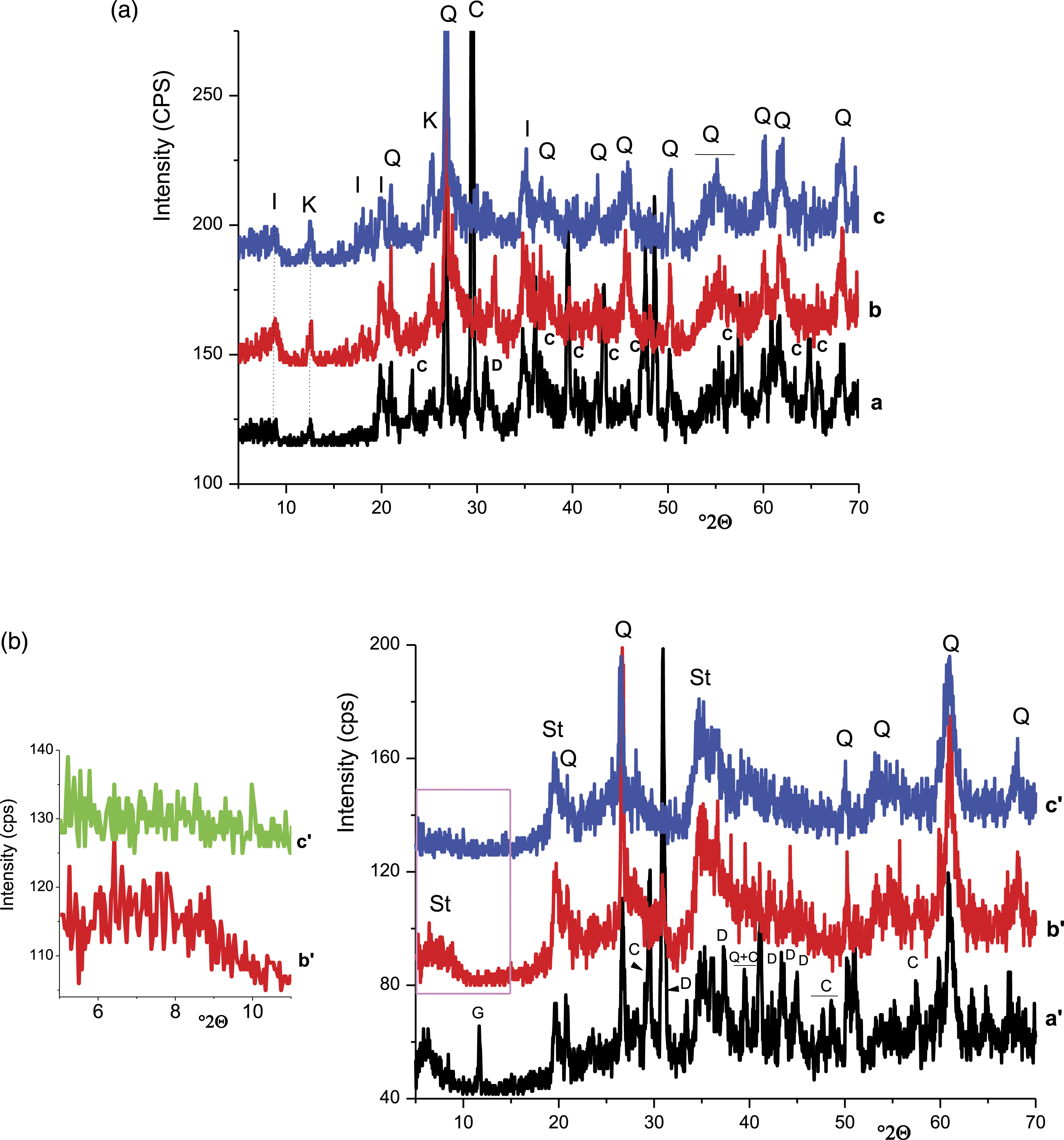
Referring to Figure 3(b), the basal X-ray reflexion of the stevensite (°2θ = 6.02) vanished as a result of the association of RH clay with β-chitosan. This fact was associated to stevensite delamination caused by excessive expansion of the interlayer due to the excessive insertion of chitosan chains. Such a phenomenon was also observed in the case of mixing montmorillonite (smectite clay mineral) with chitosan. 37 Stevensite particles, as was the case with BN2 particles, were coated with chitosan because the pHPZC of stevensite-containing films and chitosan were close (Table 1). However, in this case, exfoliated/intercalated structure was formed.
Considering the FT-IR analyses shown in Figure 4(a), the spectrum of the film (b) consisted of an additional band at 1566 cm−1, which is associated to the protonated amino-groups of chitosan. In addition, the band at 1417 cm−1, which is attributed to the deformation of the CH2 in the CH2OH group of the chitosan, intensified. The bands related to the clay minerals bonds were not modified. These results allowed the deduction that the functional groups (-NH3+ and CH2OH) of the chitosan were involved in its retention by clay particles surfaces, which are negatively charged. FT-IR spectra of the clay fractions (a, a’), the chitosan-clay films (b, b’), and the β-chitosan (c, c’). (a) and (b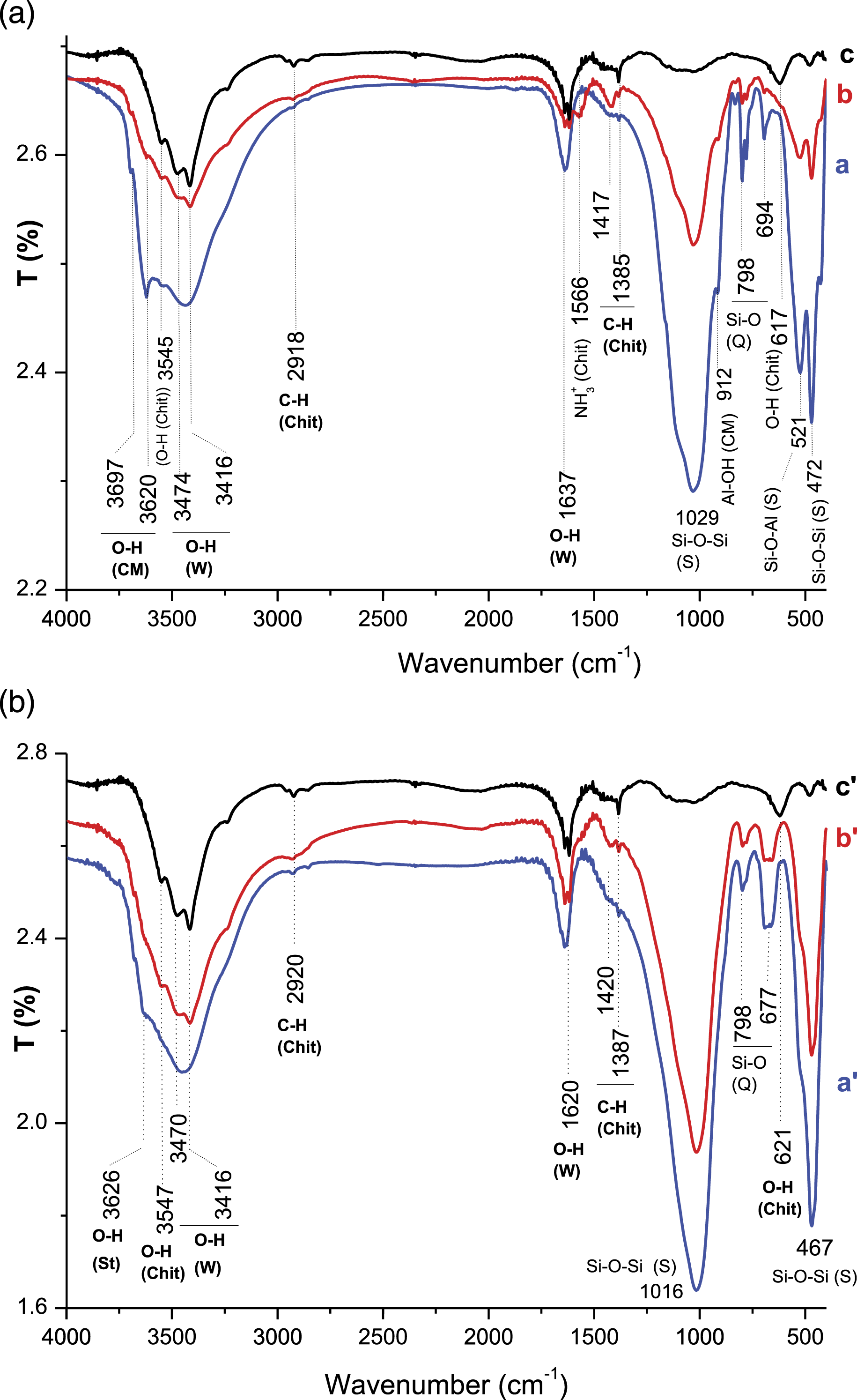
The comparative examination of FT-IR spectra given in Figure 4(b) showed that the band at 1420 cm−1 associated to the CH2 in the CH2OH group of the chitosan was well distinguished on the spectrum of the film (b), whereas the remaining bands were not affected. So, it was believed that the primary hydroxyl group of chitosan developed hydrogen bonds with stevensite particles, known as anionic clay mineral. These bonds played a key role in the adsorption of the chitosan on the clay particles.
SEM examinations of chitosan-BN2 films showed clay aggregates such as seen in Figure 5(a) (micrograph a) together with embedded particles (Figure 5(a), micrograph b). In addition, chitosan-rich zones were identified (Zone 1). These observations allowed the deduction that chitosan-BN2 films consisted of segregated domains and the inter-aggregates spaces were chitosan-rich zones. The SEM observations realized on chitosan-RH films revealed scarce clay aggregates (Figure 5(b), micrograph b), and chitosan-coated clay particles such as shown in Figure 5(b) (micrograph b). The scarcity of clay aggregates was associated to the clay exfoliation and the formation of exfoliated/intercalated composites, as previously mentioned. SEM micrographs of chitosan-BN2 (a) and chitosan-RH (b) films, and EDS spectra of different areas. Zone 1 (a) and zones 2 (b) are chitosan-rich domains. Zone 2 (a) and zone 1 (b) are clay-rich areas.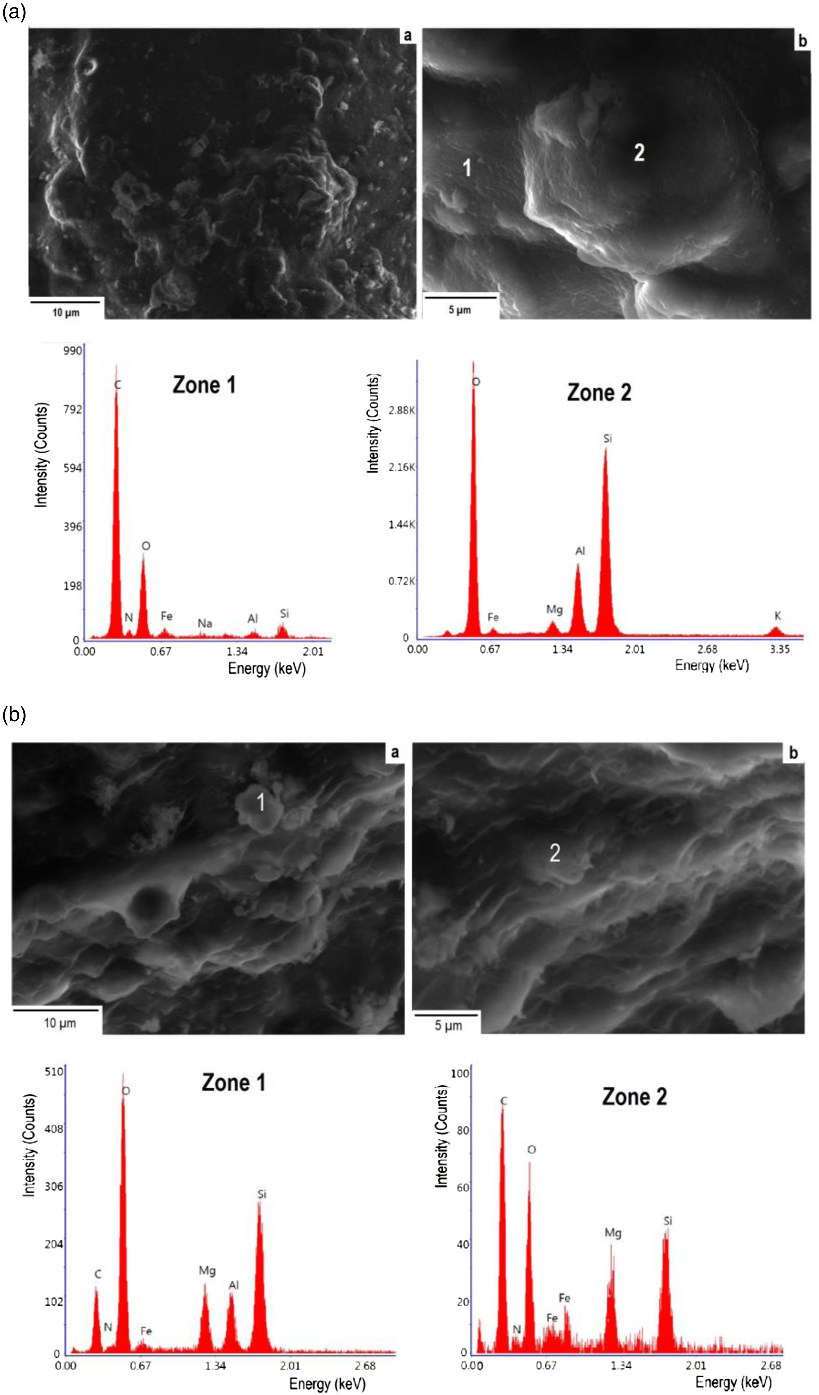
Physical/mechanical properties and thermal stability of the films
Films mass loss
The mass loss of films containing up to about 0.15 g of RH/g of chitosan was somewhat constant (3.5 ± 0.7 mass%) (Figure 6, curve a). However, it increased almost linearly with the increase of clay content, and the increment was of 10.3xMc (Mc: mass of the clay added). Considering chitosan and clay characteristics (DD = 90%, CEC (cation exchange capacity) = 0.8 meq/g), the estimated amount of glucosamine unit per one gram of β-chitosan was of 5.56x10⁻³ mole, and the quantity of clay active sites was of 8x10⁻⁴ mol/g. Hence, the portion of chitosan bonded to RH particles (0.15 g) was not significant (∼2%), and the clay particles were thick-coated with chitosan. In the clay-rich films, a portion of clay particles was scarcely coated with chitosan. So, in such a condition, the clay particles were easily lost. Variation of the mass loss of films versus the clay content. (a) chitosan-RH film and (b) chitosan-BN2 film.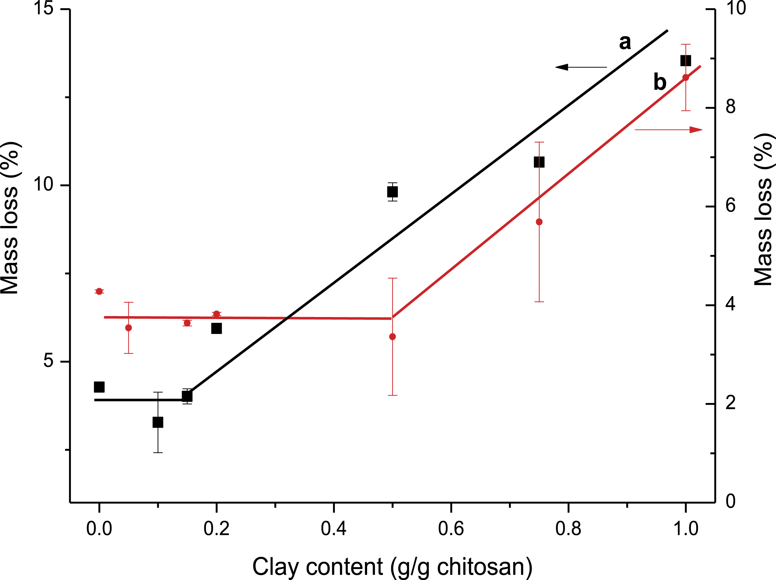
The mass loss of chitosan-BN2 films, which consisted of up to 0.5 g BN2 per g chitosan, was almost constant (Figure 6, curve b). However, it increased linearly in the case of clay-rich films. In this case, the mass loss per gram of clay was estimated to be 10.5.
Taking into consideration the DD of chitosan and the CEC of BN2 clay (0.3 meq/g), together with the fact that the hydroxyls of chitosan were implicated in the association of film constituents, the fraction of chitosan involved in the binding process was estimated to be 10% for the film containing 0.5 g BN2. Thus, a high amount of chitosan was stacked over the clay particles, and flocculated composite formed.
In view of the above results, the abundance of flocculated composites in chitosan-clay films contributed to the reduction of clay particles loss.
Thermal stability of the films
The thermal curves of chitosan-BN2 and chitosan-RH films displayed endothermic peaks at 89 and 92°C, and exothermic effects at 277 and 280°C, respectively (Figure 2). The endotherms were associated to the loss of physisorbed water. The exotherms were assigned to the chitosan decomposition. The relative thermal stability shown by clay-containing films could be associated to the clay characteristics (nanosized structure and high aspect ratio) and to their barrier effect. 38
Mechanical properties
Small additions of RH (<∼0.14 g/g chitosan, i.e., 12 mass%) to β-chitosan improved the Young’s modulus as well as the tensile strength of films (Figure 7(a)), but they led to the reduction of the elongation at break and the tensile energy absorption (Figure 7(b)). Excessive additions (>20 wt %) resulted in the decline of the films stiffness. Variation of the mechanical properties of the chitosan-RH films as a function of the clay content. (a): Young’s modulus and tensile strength. (b): Elongation at beak and tensile energy absorption.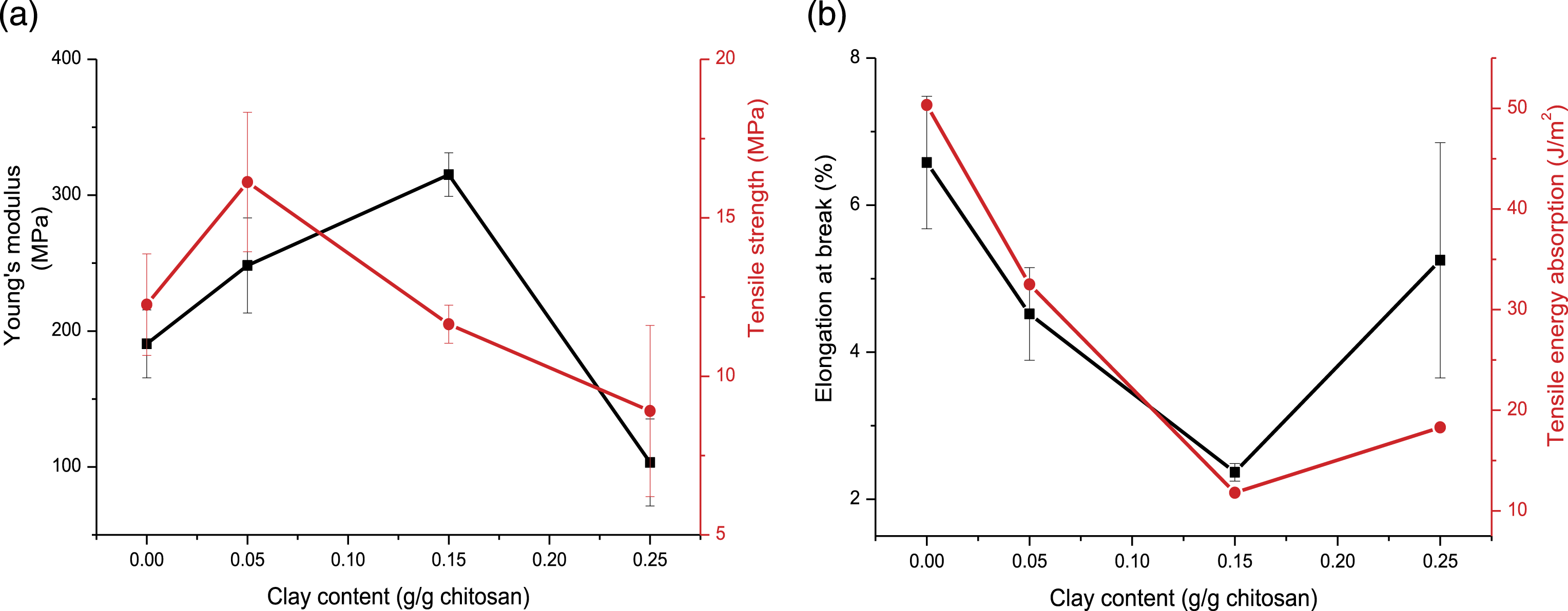
The tensile strength of the chitosan-BN2 films containing up to about 0.25 g clay/g chitosan increased with respect to that of the chitosan film (Figure 8(a)), whereas the Young’s modulus (Figure 8(a)), the elongation at break, and the tensile energy absorption (Figure 8(b)) fluctuated. For clay-rich films, the mechanical properties declined for the reasons given below. Evolution of the mechanical properties of the chitosan-BN2 films versus the clay content. (a): Young’s modulus and tensile strength. (b): Elongation at beak and tensile energy absorption.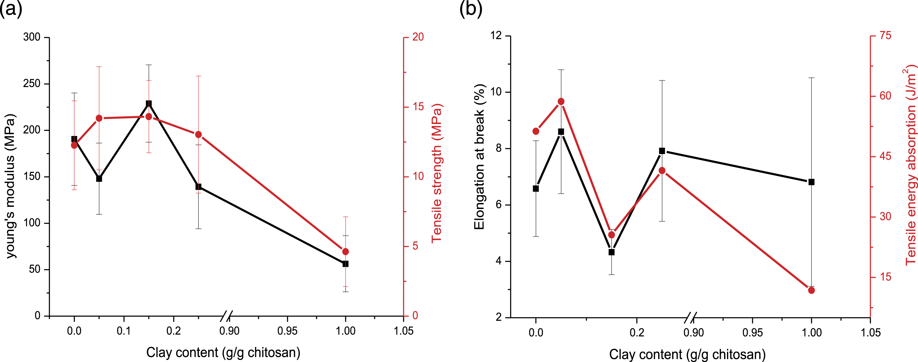
In overall, the mechanical properties of polymer-based composites are affected by the inherent characteristics of the components used, the amount and the distribution of fillers, the interfacial bonding as well as by the processing methods. 39 Considering chitosan-RH films, the improvement of films mechanical resistance was essentially due to the formation of hydrogen bonds between the chitosan and the delaminated clay particles. In fact, the formation of tough interfaces clay/chitosan facilitated the load transfer across the film, and consequently the stress was homogeneously distributed. 40 However, as the volume fraction of RH exceeded 1.6% (v/v), the clay addition had a detrimental effect on film mechanical resistance. It was believed that because of the swelling character of RH clay, the clay-rich films were the object of microcraks due to the drying shrinkage. The formation of such defects together with the uneven distribution of the clay particles could be responsible for the decline of mechanical strength.
For BN2-containing films, the presence of the aforementioned electrostatic forces seemed to play a main role in the improvement of mechanical resistance of films with clay fraction <3.2% (v/v). Further clay additions resulted in a drop of mechanical strength, possibly because of the flocculation and/or the heterogeneous distribution of clay particles.
Kinetics of the growth inhibition of the bacteria
Referring to the kinetics curves given in Figure 9(a), PI of the E. coli growth by the films approximated 90–100% after 24 h. The kinetics of the inhibition of the bacteria growth followed the pseudo-first order equation Kinetics curves associated to the inhibition growth of E. coli (a) and S. aureus (b) by the chitosan-RH (a) and the chitosan-BN2 (b) films.
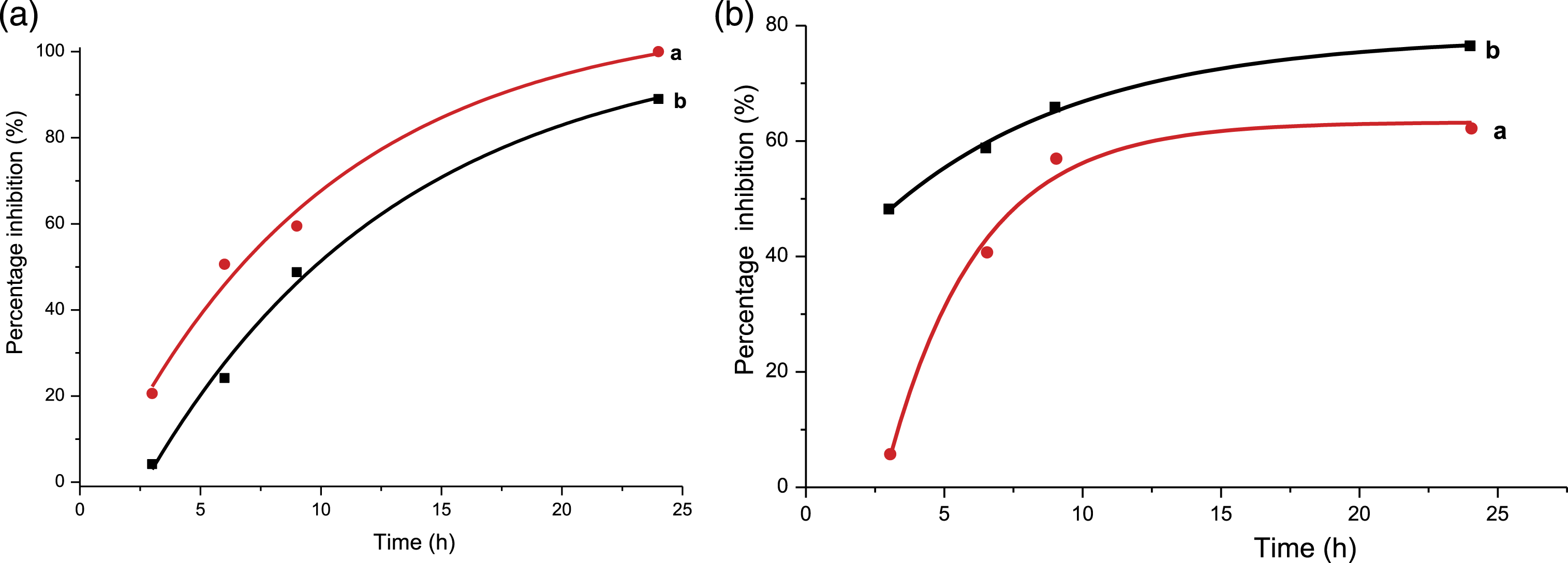
(PI)t and (PI)e are the instantaneous and the equilibrium percentages inhibition of the E. coli growth, respectively, k and t are the rate constant and time. The estimated values of k were of 3.12x10⁻⁵ s−1 and 3.45x10⁻⁵ s−1 for RH- and BN2- containing films, respectively. Based on the linear evolution of
Taking into consideration the curves shown in Figure 9(b), the kinetics of the inhibition of the S. aureus growth by the RH-chitosan film was relatively slow, but for both films, the change of the PI versus time fitted better the following linear form of the pseudo-second kinetics equation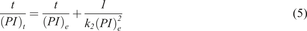
In view of the above results, the kinetics of inhibition process was mainly dependent on the interaction between microorganisms and film constituents, mainly chitosan. Indeed, according to some authors, 10 the lipoteichoic acid, which is a constituent of the cell wall of the gram-positive bacteria, such as the S. aureus, can interact with chitosan, and results in the disturbance of the cell membrane functions. The built up of chitosan around the cell wall might impede the entrance of nutriments and the release of residue by the cell. Therefore, cell metabolism was drastically altered. On the other hand, the PI of the E. coli was controlled by diffusion, seemingly owing to the loss of the selective permeability of the cell membrane. This fact was attributed to the interaction between the anionic groups of bacterial membrane lipopolysaccharide and the protonated amino-groups of chitosan. 10
Evaluation of the effects of the factors studied on the bacteria inhibition
ANOVA results and statistical parameters related to the validity of the models adopted.
The validity of these models was also assessed by the probability plot of residuals.
35
The almost linear distribution of the residuals, shown in Figure 10, supported the adequacy of these polynomial models. Probability plots of residuals related to the models used.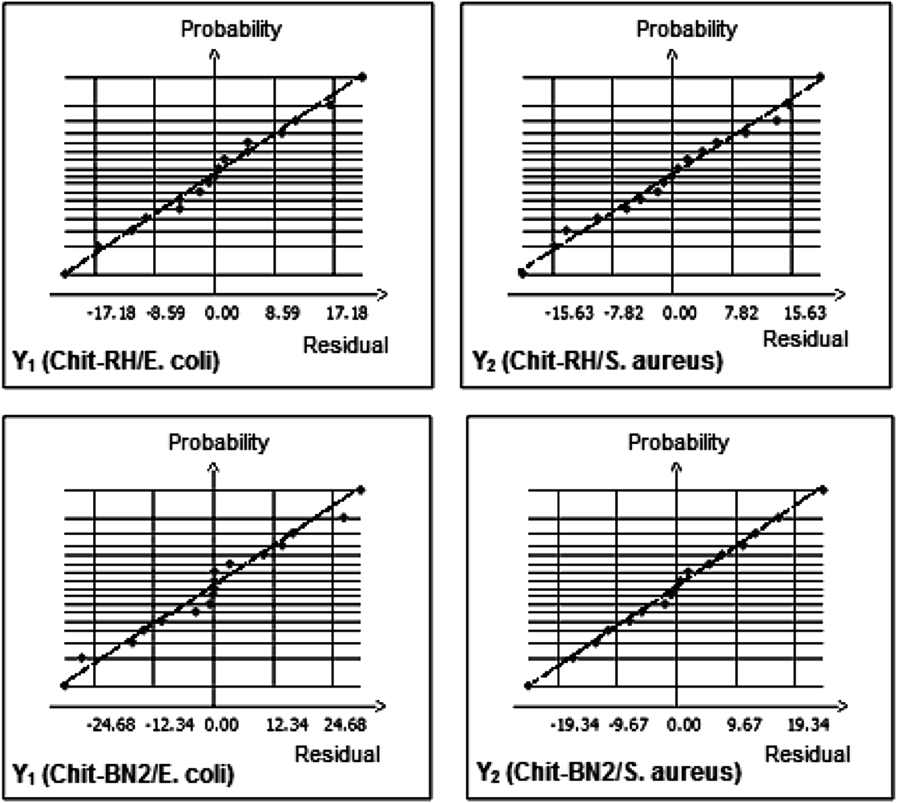
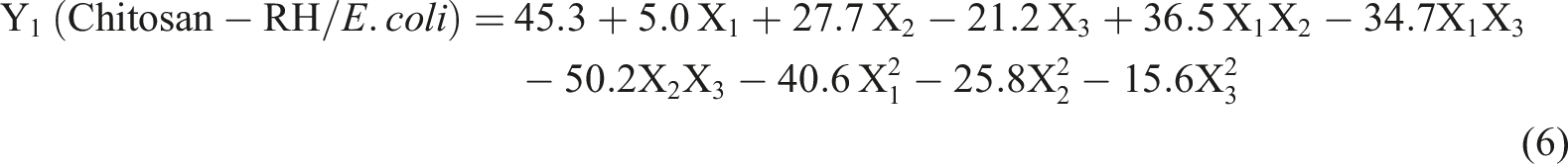
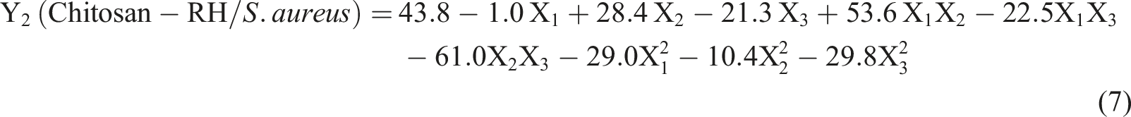
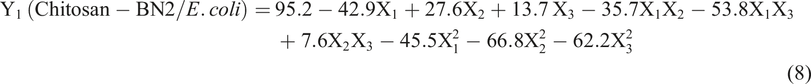
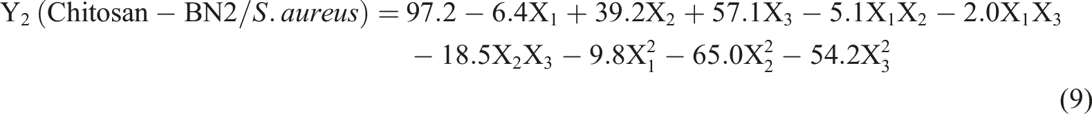
The comparison of the algebraic values of the linear terms b2X2 (equations (6)–(9)) pointed out that the increase of DD, which is correlated to the increase of the chitosan amino-groups amount, improved the inhibition growth of both strains of bacteria. This result was in conformity with the above discussion that is the amino-groups were the main functional moieties involved in the inhibition of the bacteria growth. Considering the linear terms of the equations (6) and (7), the weights of the studied factors effects on PI followed the order: DD > MW > R. Moreover, PI using chitosan-RH films decreased with the increase of MW. In contrast, it increased with the use of chitosan-BN2 films (equations (8) and (9)). These results supported the disparate effects of MW on the bactericidal activity. 10 Recalling the above equations, the weight of the effect of R on PI was relatively low, except in the case of the equation (8) where the increase of R resulted in the decline of PI.
Taking into consideration the bijXiXj terms of the equations (6), (7), and (9), the most important influencing interaction was that occurring between DD and MW. This interaction was categorized as an antagonistic one because the simultaneous rise of DD and the decrease of MW or vice-versa should result in the increase of PI. A typical response surface representing the antagonistic effect of the interaction between DD and MW is shown in Figure 11(a). In the case of the equation (8), R and MW interacted antagonistically and their interaction had the prominent effect on PI. The response surface showing the variation of PI against R and MW is given in Figure 11(b). Variations of the percentage of inhibition of E. coli (Y1 (chitosan-RH), Y1 (chitosan-BN2)) against the factors studied. (a): clay/chitosan mass ratio (R) = 0.5; (b): deacetylation degree (DD) = 85%.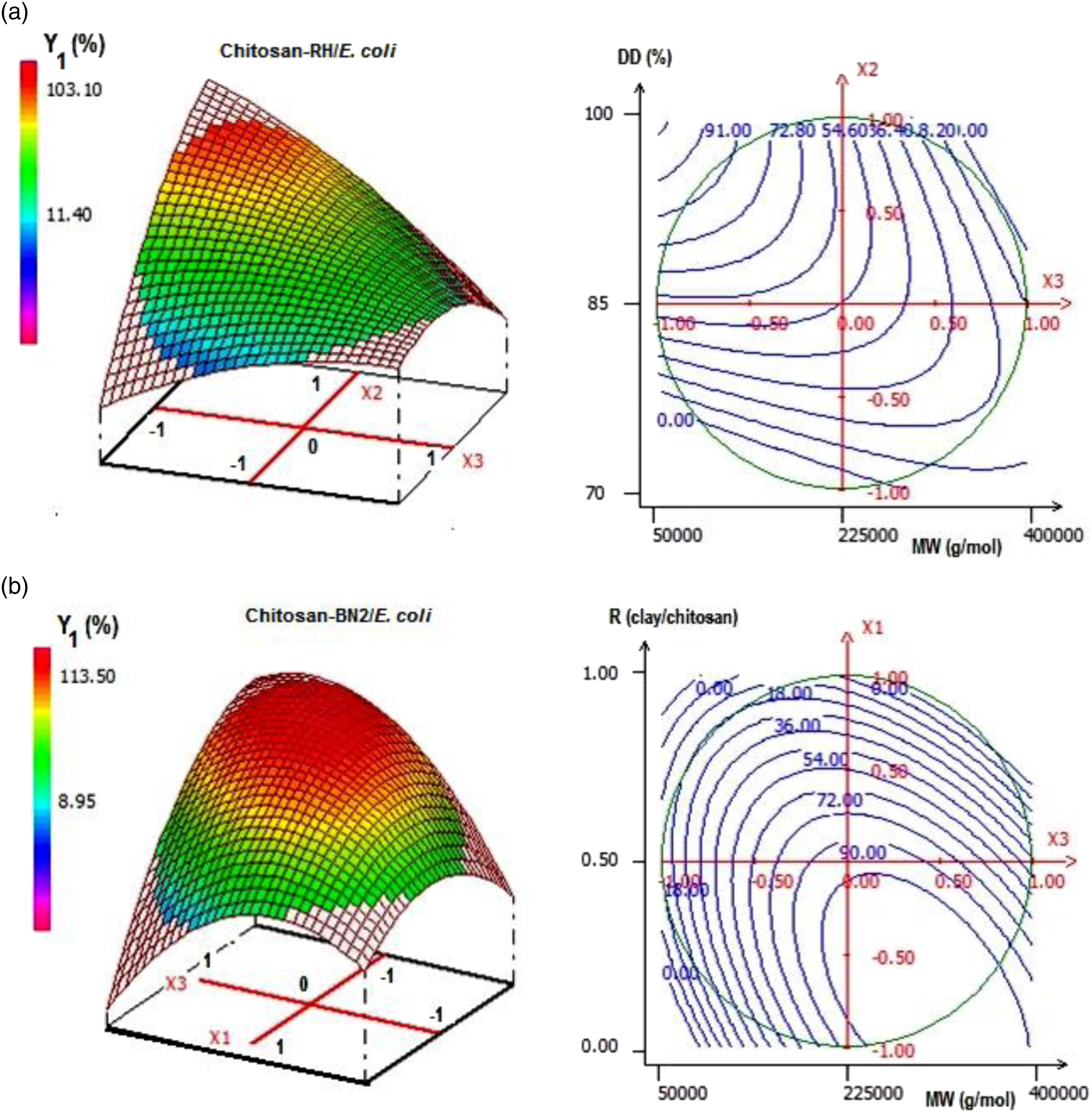
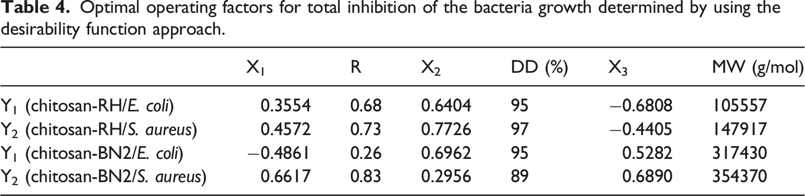
Optimal operating factors for total inhibition of the bacteria growth determined by using the desirability function approach.
Considering the PI = f (clay/chitosan) curves plotted on the basis of the equations (6)–(9) (Figure 12), the antibacterial effect of the chitosan film was less important as compared to that of the chitosan-RH films, which consisted of exfoliated/intercalated composites. In contrast, it was almost equivalent or somewhat better than that of the chitosan-BN2 films, which were composed of tactoids. Variation of the percentage inhibition of the bacteria growth by the prepared films versus clay/chitosan mass ratio. DD = 85%, MW = 225000 g/mol.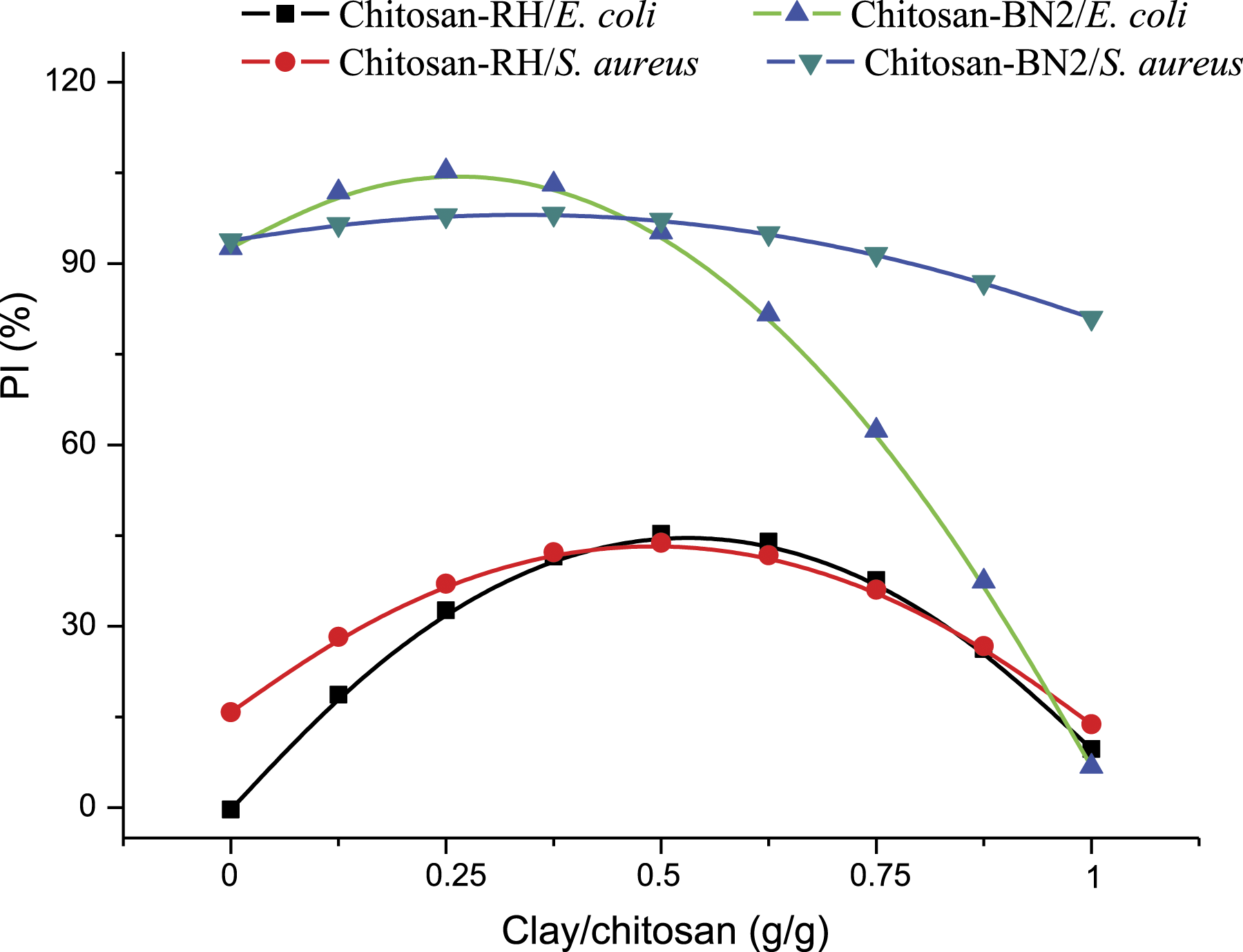
Conclusions
Stevensite-containing chitosan films consisted of exfoliated/intercalated nanocomposites, and the association between the films components were presumably achieved through hydrogen bonding. Limited additions of RH (up to 0.14 g/g chitosan) improved films tensile strength and Young’s modulus, but they had a negative impact on their elongation at break.
Chitosan films containing kaolinitic-illitic clay were essentially composed of flocculated composites. The amino-groups together with the hydroxyls of chitosan were involved in its association with the clay particles. The addition of BN2 clay (up to 0.25 g BN2/g chitosan) improved the mechanical strength of films.
The results of the kinetics of the bacteria growth inhibition by the studied films allowed the deduction that the inhibition process depended on the bacterial strain. In the case of E. coli, the cell membrane seemed to be damaged due to its interaction with the protonated amino-groups of chitosan, and the vital substances flowed out. For S. aureus, the nutrients seemed to be impeded to get into the cells because of the stack of chitosan over their membranes.
The polynomial models obtained by using the RSM allowed the prediction of the effects of the factors studied and of their mutual interactions on PI. The rise of MW resulted in the decrease of the PI of E. coli, but it increased that of the S. aureus. The increase of DD yielded to the rise of PI, and the optimal value should be in the range of 89–97%. Based on the RSM results, the antibacterial activity of the chitosan film was a good as that of the tactoid-rich films. However, it was less significant as compared to that of the films composed of exfoliated/interacted clay.
Footnotes
Declaration of conflicting interests
The author(s) declared no potential conflicts of interest with respect to the research, authorship, and/or publication of this article.
Funding
The author(s) disclosed receipt of the following financial support for the research, authorship, and/or publication of this article: This work was supported by the CNRST (grant number PPR/26/2015).