




Abstract
Radiotherapy is an effective cancer treatment, particularly for pelvic tumours. The number of patients with pelvic cancer being diagnosed and successfully treated is growing. Radiotherapy to the pelvis causes lasting side-effects collectively referred to as pelvic radiation disease (PRD), including bowel, bladder, sexual dysfunction, vaginal and cervical stenoses, and menopause. There is growing interest in management of menopause in cancer survivors, with the primary focus on the oncologic risk of hormone replacement therapy (HRT). Research examining if the modality with which the cancer was treated causes menopause-specific side effects is rare; however, malabsorption syndromes and anatomical changes in the pelvis post-radiotherapy may complicate effective delivery and monitoring of HRT. Consideration of these changes may significantly benefit patients in this young and growing cohort; thus, there is an urgent need to raise awareness of PRD among all clinicians, including those providing menopause care.
This article discusses the management of menopause in women who have been treated with pelvic radiotherapy for cancers in the pelvis, specifically focusing on the anatomical and physiological changes caused by radiotherapy, and how these changes can impact on the effective delivery and monitoring of HRT. It will provide a basic introduction to pelvic radiation disease (PRD) and discuss cases seen at a specialist PRD clinic highlighting these important issues.
Background
I work as a GP, a Menopause Specialist and clinical lead of the pelvic radiation late effects service at the Beatson west of Scotland cancer centre in Glasgow. This service provides support for any patient, male or female, who has had curative radiotherapy for cancers in the pelvis experiencing long-term side effects of treatment. This role has given me a unique insight into the ‘lifechanging’ consequences of cancer treatment – something I had previously failed to fully appreciate despite working in general practice for over 30 years caring for numerous cancer survivors. Although the primary focus of the service is on management of gastrointestinal side effects of pelvic radiation, our aim is to provide a holistic approach to caring for patients with pelvic radiation disease (PRD). Many of the patients we see are young women also experiencing treatment-induced menopause, so possessing menopause management skills and a desire to provide holistic care the service has accidentally ‘morphed’ into an undercover menopause clinic. In this role, I have become aware of physiological and anatomical changes in the irradiated pelvis that have the potential to impact on and complicate effective delivery and monitoring of hormone replacement therapy (HRT).
Definitions
Radiotherapy is an effective cancer treatment, particularly for pelvic tumours. It is often combined with surgery, chemotherapy, hormone, and/or newer immunological therapies. While advances in radiotherapy techniques have reduced the risk of some side effects, radiotherapy to the pelvis continues to cause lasting side effects including bowel, bladder, sexual dysfunction vaginal and cervical stenoses, and treatment-induced menopause – collectively referred to as pelvic radiation disease (PRD).
Symptoms arise as a result of damage to healthy tissues caught in the radiation field. Particularly susceptible are bowel, bladder, and reproductive organs, referred to as organs at risk (OAR). PRD differs from the acute side effects of radiotherapy which usually settle once treatment is completed, for some people acute symptoms become chronic (Box 1).
• One or more ongoing symptoms of variable complexity that may affect people who have previously had radiotherapy to the pelvic region to treat their cancer. • Symptoms starting or continuing 3 months or more after the end of radiotherapy. Sometimes they start many years or decades after radiotherapy.Box 1: Definitions
Symptoms are not limited to the pelvis: PRD is a chronic, multi-symptom, multi-organ condition that often affects a person’s psychological wellbeing through its effects on the gastrointestinal and genitourinary tracts, endocrine, sexual health, skeletal health, skin, vascular, and lymphatic systems. 1
Gastrointestinal symptoms of pelvic radiation disease
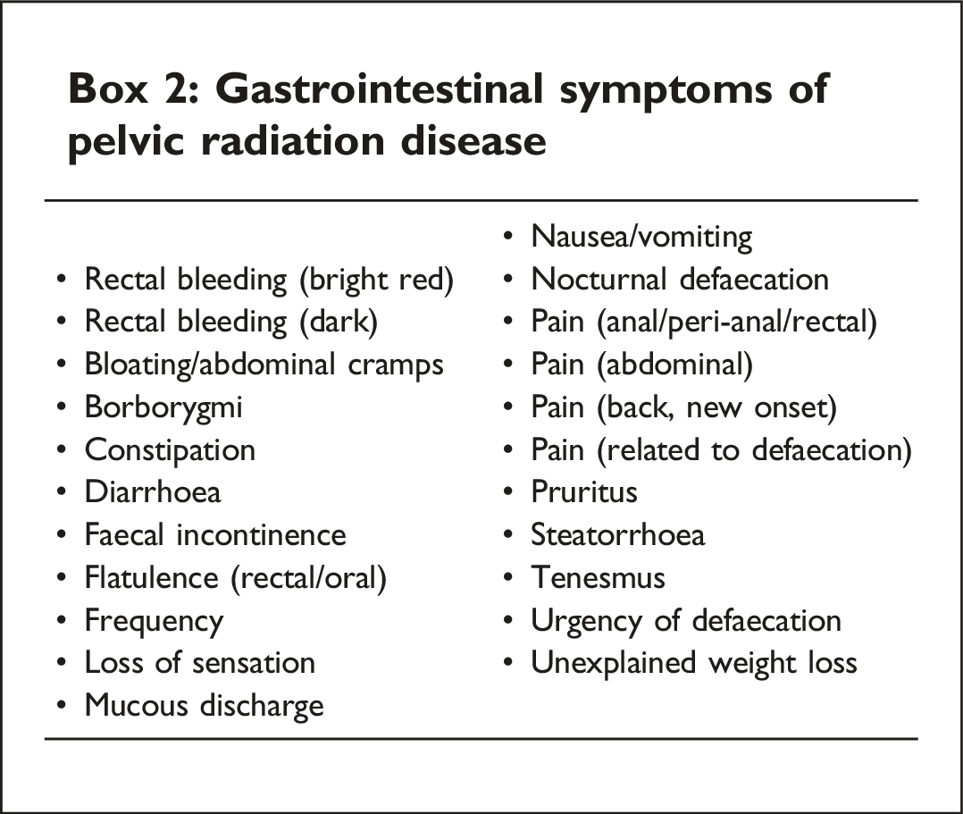
Almost any gastrointestinal (GI) symptom can be caused by radiotherapy (Box 2).
Box 2: Gastrointestinal symptoms of pelvic radiation disease
There are 3 common conditions we regularly diagnose and manage in our service – as detailed in Box 3. They all cause malabsorption, with symptoms of abdominal pain diarrhoea and stool frequency and have the potential to impact on absorption of orally delivered medication, such as HRT.
Radiation damage to terminal ileum affects the normal reabsorption of bile salts. Unabsorbed bile salts increase colonic mucosal permeability which then increases fluid and mucus secretion causing colonic contraction resulting in watery diarrhoea, pain, and bloating.
Radiotherapy causes changes in the normal bacterial microbiome by reducing gastric acid production and altering gut motility. This results in the presence of excessive bacteria in the small intestine or SIBO. This can cause bloating, diarrhoea, and abdominal pain. Complications of SIBO range from minimal vitamin deficiencies to severe malabsorption.
Irradiation of the pancreas can affect its function, reducing the production of enzymes and impacting on the break down and absorption of nutrients. This disrupts normal digestion resulting in symptoms of abdominal pain and diarrhoea and malabsorption.Box 3: Malabsorption syndromes commonly diagnosed post-pelvic radiotherapy
We conducted a service review examining clinic data between December 2018 and December 2022: this confirmed a high incidence of malabsorption syndromes in patients post-pelvic radiotherapy especially in the cohort of female patients likely to require HRT (Box 4). Patients referred to our specialist PRD service, over 4 years, were identified retrospectively (n = 156). Fifty (32%) of our patients were females in the premenopausal age group (defined as < 50 years of age) at risk of treatment-induced menopause. These women had been treated for the following tumours. • Cervical 42 (84%), • endometrial 2 (4%), • vulval 2 (4%), • vaginal 1 (2%), • anal 2 (4%), and • rectal 1 (2%). Cervical cancer was the commonest cancer and most of the tumours were suitable for HRT if required. In this group of patients, we diagnosed the following: • Bile acid malabsorption (BAM) affected 60%. • Small intestinal bacterial overgrowth (SIBO) affected 44%. • Pancreatic exocrine insufficiency – (PEI) affected 28%.Box 4: Summary of service review data (Beatson PRD LE service)
Anatomical changes post-pelvic radiotherapy
In addition to physiological changes resulting from pelvic radiotherapy, anatomical changes such as vaginal, cervical stenosis, and vaginal adhesions are common and have the potential to cause problems such as dyspareunia and sexual dysfunction. Studies report that as many as 88% of women treated with pelvic radiotherapy for cervical cancer and other gynaecological malignancies develop vaginal stenosis as a consequence of therapy.2,3 We know women experience sexual dysfunction after radiotherapy for colorectal cancer, but the percentage of women who develop vaginal stenosis after this treatment is not well reported. It has been suggested this is because these women are followed up by colorectal surgeons rather than gynaecologists, so there is possibly less focus on genital and sexual health. 2 Following cervical cancer treatment, stenosis is thought to be associated with brachytherapy and the upper vagina is most often affected.4,5 In rectal cancer, the lower portion of the vagina commonly receives the high dose of radiation. The lower vagina, introitus, and vulva are therefore affected, causing narrowing of the introitus but relatively normal vaginal length. All these changes result in dyspareunia and sexual dysfunction. If sex is painful women avoid it, so if these problems are not addressed early on, they only get worse. Less use of the vagina leads to further shortening and narrowing.
Strategies for prevention and management
Topical estrogen therapy
As an immediate response to radiotherapy, there is loss of most of the epithelium in the vagina receiving the maximal surface dose. This loss lasts for 3–6 months. When re-epithelialisation occurs after radiation the surface is thin with an incomplete layer of basal cells.2,6 Using topical estrogen therapy immediately after completing treatment can promote epithelial regeneration, and its effects may be even greater for patients more than 3 months after radiation.
A controlled double-blinded study of 93 women compared those treated with Dienestrol 0.01% immediately after completing radiotherapy for cervical cancer, with a control group using a placebo vaginal cream. Women who received active estrogen cream had significantly less dyspareunia and were twice as likely to have normal vaginal calibre as those who received placebo. 7
Vaginal dilators
Vaginal dilators can help prevent stenosis and treat established stenosis. Attention should be paid to choosing the best dilator size, shape, and composition suitable for the woman as well as providing correct instruction on how to use. Dilators should be introduced soon after completing radiotherapy and used for 15–20 min at a time 2–3 times a week with a lifelong commitment to use. 8 Although many women do not resume regular vaginal intercourse after pelvic radiotherapy – keeping the vagina open for future examination is important, patients need to be educated on this matter and understand the purpose of continued dilator use. It’s not just about sex! From my experiences in the PRD late effects service, very few women report or remember receiving adequate advice and tuition on use of dilators immediately post-treatment or were prescribed local estrogen therapy.
In reality onco-gynae clinical nurse specialists have discussed, documented, and provided dilators and lubricants to most patients, but when women are reminded of this, they state that ‘sex was the last thing on my mind’ or they forgot about or tried the dilators but gave up due to pain and didn’t see the point in persevering with their use. Clearly, there is a need to be more proactive in preventing these problems occurring in the first place and ensure we educate and enquire about compliance with dilator use at follow-up appointments.
How do these anatomical and physiological changes impact on HRT delivery and monitoring?
When managing menopause after cancer and considering prescribing HRT currently the primary focus is on the oncologic risk of HRT.
This often requires discussion with the oncologist and a careful risk vs benefit discussion with the patient to enable a shared decision-making process.
If HRT is to be initiated, then important practical prescribing choices remain which should involve consideration of any long-term consequences or side effects of the cancer treatment that have the ability to impact on delivery of HRT. These include changes in gastrointestinal physiology and pelvic anatomy resulting from pelvic radiotherapy.
Which regime? Which route? Monitoring side effects?
Each will now be considered with respect to the special circumstances post-pelvic radiotherapy.
Regime
Endometrial and ovarian activity after pelvic radiotherapy
The ovaries are highly sensitive to radiotherapy, and they are not included as organs at risk in the planning stages of treatment. Unless fertility-sparing procedures are employed, pelvic radiotherapy will destroy ovarian function resulting in treatment-induced menopause in those pre-menopausal at the time of treatment. Although patients are generally treated with radiation doses that are thought to ablate the endometrium, several studies have demonstrated that normal endometrium can tolerate a high dose of radiation and has the ability to recover from radiation injury and preserve its proliferative activity. 9 This means if HRT is being considered for these women, a continuous combined regime should be prescribed. 10
Route
There are currently no specific guidelines for HRT prescribing in patients with GI/malabsorption issues resulting from PRD. However, there is guidance from recognised bodies on prescribing in GI disorders. It would seem reasonable to extrapolate these to this patient group and thus avoid the oral route (Box 5).
‘The transdermal route is advised if GI disorder affecting oral absorption of HRT’.
‘Oral contraception may be less reliable in women with IBD who have malabsorption due to severe small bowel disease or resection, or who have vomiting or severe diarrhoea for more than 24 hours’.
‘Many drugs will be incompletely absorbed by patients with a short bowel and may be needed in much higher amounts than usual or may need to be given intravenously’.Box 5
A continuous combined transdermal patch would therefore seem to be the most appropriate first choice for this group. However, we know ‘one size doesn’t fit all’ when prescribing HRT and some women will require ‘tailoring’ of their regime. Driven by recent publicity, there is currently a preference among patients for transdermal estradiol and oral micronised progesterone, particularly among cancer survivors. This is suitable for women who don’t have severe malabsorption issues as long as prescribers adhere to licensed dosing regimens. Some prescribers might consider delivering the progesterone component vaginally to avoid issues with oral malabsorption; however, there is a risk of reduced endometrial protection if the progesterone is being absorbed through ischaemic, fibrotic irradiated vaginal tissues and vaginal adhesions and cervical stenosis might further reduce the local effect on the endometrium. These issues are important and need consideration. It is vital these women get adequate progestogenic opposition and avoid any endometrial activity causing bleeding, as bleeding is extremely problematic in these patients.
Monitoring and managing side effects of HRT
Most minor and transient side effects can be managed in the same way as they would be in any other patients, either with dose adjustments or reassurance they will settle. However, bleeding issues are much more problematic for this group.
Bleeding problems
There are 2 types of bleeding problems experienced in this group: visible unscheduled bleeding and concealed unscheduled bleeding. Visible unscheduled vaginal bleeding causes anxiety; it often mimics tumour presentation and can then spark a ‘fear of recurrence’. It can be more challenging to investigate due to vaginal stenosis, adhesions, or other anatomical changes caused by radiotherapy. Speculum examination, transvaginal ultrasound scan, and Pipelle sampling may not be possible. Examination under anaesthetic might be necessary with risk of perforating fragile irradiated tissue. Additional MRI or CT scans may be required to exclude tumour recurrence. Concealed unscheduled bleeding is an even bigger problem and can result in haematocolpos, haematometra, and haematosalpinx. It usually presents with pelvic pain, which is a common feature in PRD and has many possible causes (Box 6). This can result in patients presenting to several different specialities. In the absence of ‘visible bleeding’, gynaecology services are often not the initial ‘port of call’. Bone pain – radiation-related insufficiency fractures (RRIF)/osteonecrosis. Radiation-induced lumbar plexopathy (RILP). Pelvic bleeding – haematometra/haematocolpos. Gastro-intestinal pain. Bladder pain. Skin pain. Vascular pain. Tumour recurrence/secondary tumour.Box 6: Causes of pelvic pain post-pelvic radiotherapy
Our service is currently aware of at least 8 cases of concealed unscheduled bleeding developed after initiation of HRT. All were unsuspected, misdiagnosed, and mismanaged for some time. The diagnoses ranged from pelvic inflammatory disease to appendicitis. Once correctly diagnosed concealed bleeding can present complex management problems. If haematometra or haematocolpos are suspected, it is vital to get histology to exclude tumour recurrence. This requires Dilatation and Curettage which can be very hazardous or impossible in irradiated tissue, with a risk of perforation. Since histology of the endometrium is mandatory, if D&C fails a hysterectomy might be required, but it is a very high risk procedure in this patient group. 11 As vaginal and cervical stenoses are common post-pelvic RXT, this issue must be on our radar when monitoring this patient group. Ongoing management with HRT, once diagnosed, can also be challenging as our case studies demonstrate.
Case study 1
A 42-year-old woman was diagnosed with locally advanced cervical cancer and treated with primary chemoradiation, receiving external beam radiotherapy and brachytherapy. She experienced a treatment-induced menopause and was commenced on HRT by her GP shortly after completing therapy. She was initially prescribed transdermal estradiol patch (50 mcg twice weekly) and oral micronized progesterone (100 mg nightly). The estradiol patches were gradually increased to 100 mcg to gain adequate symptom control; progesterone dose was unchanged at 100 mg nightly. Over the next 2 years, she developed troublesome GI symptoms – erratic bowel habit, constipation, diarrhoea, and urgency. However, her main concern was severe pelvic pain and lower abdominal distention. She felt permanently ‘Pregnant’. There were no reports of any vaginal or rectal bleeding. Now at the age of 44 years, she was referred to PRD LE service. Unfortunately, prior to her first appointment her pain became so severe she attended Accident and Emergency. After a pelvic USS, she was told she had ‘raging Pelvic Inflammatory disease’, with a pelvis ‘full of pus’. She was commenced on antibiotics and discharged home with gynaecological outpatient follow-up. She was shocked and distressed by this diagnosis and the way it was delivered – having been unable to have sexual intercourse for some time due to pain and vaginal shortening – she was mortified having to explain the diagnosis of PID to her partner. Her symptoms did not improve with antibiotics and when reviewed by the gynaecologist a repeat USS scan was requested and the report stated: ‘The endometrial cavity is expanded by an echogenic mass/material measuring 4.7 cm which appears surrounded by a small volume of free fluid. While appearances are thought most likely to represent clot, other pathology should be considered’.
Subsequent examination under anaesthetic, dilatation, and curettage and analysis of uterine contents confirmed primarily blood – in keeping with a diagnosis of haematometra.
Her pain was relieved almost immediately post-procedure but returned a few days after recommencing her previous HRT regime despite increasing progesterone dose to 200 mg. She was advised to switch to a transdermal continuous combined patch hoping a synthetic progestogen delivered transdermally would provide stronger progestogenic opposition and prevent endometrial activity. Unfortunately, after only a week her pain returned and became intolerable, her lower abdomen was swollen and tender, and she had no vaginal bleeding. At this point, an urgent MRI scan was performed it reported:
“Post-treatment appearances of the cervix. No evidence for recurrent disease. The endometrium however is distended measuring 15 mm, with fluid appearances are suggestive of haematometria presumably secondary to cervical stenosis. In the right adnexa, there is a dilated tubular area and this could either represent some endometriosis or an haematosalpinx.”
Conclusion
No definite evidence of recurrence. Haematometra and likely right haematosalpinx? Cervical stenosis
On recommencing her transdermal HRT, she had developed a recurrence of haematometra secondary to cervical stenosis and further drainage was required. She discontinued her HRT perioperatively. Although this relieved her pelvic pain, she developed severe menopausal symptoms. She was initially recommenced on half a combined transdermal patch. Within 3 days, she reported a return of severe pelvic pain and had to discontinue application. The pain subsided 24 hours later, but menopausal symptoms returned. As her endometrium appeared to be exquisitely sensitive to any estrogenic stimulation, non-hormonal therapies were considered. Transdermal oxybutynin was commenced, providing effective management of her hot flushes but had no impact on her mood and cognitive function. Addition of an SSRI has improved her mood and reduced anxiety and provided an additional reduction in her menopausal symptoms. Currently, she is relieved that her symptoms are under control, but clearly this is not a long-term solution. At 44 years old, she requires estrogen for long-term prevention of chronic disease. This case illustrates the complexity of diagnosis and management of unscheduled bleeding post-pelvic radiotherapy.
Case study 2
A 33-year-old woman diagnosed with 2B cervical carcinoma was treated with chemo-radiotherapy (EBRT and brachytherapy). Prior to her treatment, she had regular periods and was not on any exogenous hormones. She experienced a treatment-induced menopause and reports onset of severe menopausal symptoms shortly after completing treatment, and her main issues were severe sweating and anxiety. Systemic HRT was commenced 8 weeks after her final treatment – transdermal continuous combined patches (Evorel Conti) which provided immediate symptom relief. Unfortunately, after 6 weeks on HRT she started to develop intermittent pelvic pain which became gradually worse, at times so severe it felt like ‘labour pains’; she never experienced any vaginal bleeding. The pain became more severe and persistent and resulted in a multiple A + E attendances, prescriptions for stronger analgesia, and several courses of antibiotics for presumed PID. Between July and December 2023 she had 4 pelvic CT scans, 1 pelvic MRI, and 3 pelvic USS. The USS scans reported distended endocervical canal, clot, and hydro/pyo salpinx and recommended direct visualisation. MRI reported haematometra, haematosalpinx, and fluid in the pelvis probably due to retrograde menstruation. During one admission, a repeat USS within days showed the haematometra increasing in size. Her case was discussed at a regional MDT meeting; hysterectomy was considered to be high risk as her rectum was adherent to the posterior wall of the uterus. She was advised to discontinue HRT and booked for elective EUA and division of adhesions, and this was done as an emergency due to a further acute episode of severe pain. Following the procedure, she reported instant resolution of her symptoms and has not experienced any further pelvic pain but she has been advised to remain off her HRT and is now experiencing severe menopausal symptoms and was referred to our pelvic radiation late effects service for further management.
She is now only 34 and concerned about the long-term consequences of untreated premature menopause and her quality of life and relationship are suffering due her symptoms. She appreciates the potential complications of surgery/hysterectomy following pelvic radiotherapy, she is upset, and frustrated about the number of times she had to attend A + E with severe symptoms which were misdiagnosed as urinary tract infection or PID. We have discussed alternative management with non-hormonal therapies and diet and lifestyle interventions to maximise her bone and cardiovascular health, but she is desperate to recommence HRT. She has commenced a trial of transdermal oxybutynin patches, with the aim of reducing her sweats and hot flushes and reducing her bladder frequency and urgency and commenced local estrogen (estriol ultra-low dose muco-adhesive gel) to alleviate her severe UGA – she has been advised to discontinue this if she experiences any hint of pelvic pain.
Once again this doesn’t feel like a satisfactory long-term solution, even if her symptoms do improve, but currently there are no guidelines on managing these patients who are frequently young and present significant practical problems balancing management of premature menopause against causing further pain and bleeding. It is vital clinicians who are involved in oncology, gynaecology, and menopause care, document, and discuss these cases to enable prompt diagnosis but more importantly consider if any change in treatment plans could prevent these complications. When considering the tariff of cancer treatment we must ensure quality of life issues and the cost to the NHS of managing the complications of treatment are included in the equation.
Discussion
What really worries me is our service is only seeing the tip of the iceberg. The lack of awareness of, and limited access to, specialist PRD clinics means many patients probably go unrecognised. We all encounter these patients in menopause clinics and primary care. They come bearing a cancer label, however, how many have malabsorption label or red flag warning ‘beware irradiated pelvis’.
These women frequently look clinically well. However, their clothing may be concealing clues to underlying malabsorption risks – perhaps a PICC line for parenteral nutrition or an abdomen covered in surgical scars, a colostomy, ileostomy bag, or both. Many of these women have vaginas so shortened and stenosed they barely permit an estrogen pessary – let alone allow sex! If seen in virtual clinics or not examined these clinical signs might be missed and dyspareunia assumed to be due to urogenital atrophy rather than vaginal stenosis, adhesions, or radiation-induced skin reactions. As part of a routine menopause symptom assessment, we always enquire about frequency of flushing – but we don’t mean the toilet – some of our PRD patients are opening their bowels more than 20 times a day. We always ask about bladder frequency and urgency – but don’t ask about bowels. In general, there is a lack of awareness of pelvic radiation disease among medical professionals and the public and it is vital this is addressed especially among clinicians managing menopause in patients treated with pelvic radiation.
Conclusion
Much attention has been focused on management of menopause in cancer survivors, primarily looking at tumour hormone sensitivity determining if it is safe to prescribe HRT. Little attention has been paid to how the treatment modality and its side effects can impact on effective delivery of HRT. There is no consideration of these issues in current guidelines on practical prescribing and monitoring HRT. Clinicians and future research must begin to consider the consequences of the cancer treatment and how they impact on menopause management. Going forward, we must develop preventative measures to limit risk of treatment-induced menopause or side effects that limit safe delivery and monitoring of HRT.
• Collect and share data – to fill the ‘void’ on managing menopause after pelvic radiotherapy and other systemic anti-cancer therapies (SACT). • Quantify the incidence of treatment-induced complications impacting on menopause management and having a negative impact on QOL, long-term health, and delivery of HRT. • Improve liaison between menopause services, oncologists and onco-gynaecologists, and colorectal surgeons managing these complex patients. • Encourage oncologists and onco-gynaecologists to review current management pathways taking a broader view of treatment outcomes and not only consider cancer survival rates but also factor in avoidance of potential complications managing TIM, for example, ‘prophylactic’ TAH and BSO prior to RXT. • Amend menopause symptom questionnaires/APPS – to include significant GI symptoms and ‘flag’ potential issues with PRD. • Raise awareness of PRD and cancer survivorship issues. • Post-COVID Era – Patients still need to be examined. • Establish working groups at national levels to develop guidelines and consensus statements. • Engage Royal College Radiology – investigate possibility of ovarian sparing/dose constraints to ovaries at treatment planning stages. • Prevention better than cure – more proactive advice/tuition re-vaginal dilator use and early introduction of local estrogen to prevent vaginal stenosis and adhesions. • PRDA – prda.org.uk. PRDA Best Practice Pathway 2022. MacMillan Cancer Support: https://www.macmillan.org.uk.Future development/suggestions
Useful resources
Footnotes
Acknowledgements
Dr K Cavanagh, Dr A Peters, C Smeaton (CNS – PRD Late Effects), S Millar (Therapy Radiographer),and G Giebner (Dietician), Beatson West of Scotland Cancer Centre, Glasgow, Scotland. UK. Professor AJ Chalmers, School of Cancer Sciences, University of Glasgow, Scotland. UK. Beatson Cancer Charity. Macmillan Cancer Support.
Declaration of conflicting interests
The author(s) declared no potential conflicts of interest with respect to the research, authorship, and/or publication of this article.
Funding
The author(s) disclosed receipt of the following financial support for the research, authorship, and/or publication of this article: The PRD late effects service is partly funded by Beatson Cancer Charity, Macmillan Cancer Support, and NHS Greater Glasgow and Clyde.
Ethical approval
I gained local Caldicott guardian approval, and formal ethical approval was not required.
Guarantor
SD Sally Darnborough.
