
Abstract
Aptamers are single-stranded synthetic oligonucleotides that bind noncovalently to targets with high affinity and selectivity. They are generated through an in vitro selection process known as Systematic Evolution of Ligands by Exponential Enrichment. Aptamers show significant promise in cancer diagnostics and therapeutics due to their specificity, versatility, and tunable biochemical properties. Aptamers can be used to target cellular biomarkers, which are detectable molecules that can indicate specific physiological states of cells, organs, and organisms, making them valuable indicators for diseases. Clinical trials on therapeutic aptamers are providing further insight into their potential use. Leukemia is a hematological malignancy characterized by the uncontrolled proliferation of abnormal blood-forming cells, primarily affecting the bone marrow and peripheral blood. This review will highlight the applications of aptamers in relation to the diagnosis and treatment of leukemia. Novel research on leukemia-related aptamers will be presented along with future directions for aptamer-based diagnostic and therapeutic methods.
Leukemia is a type of cancer that starts in the blood stem cells and affects the blood and bone marrow. It is a severe and often fatal disease 1 that, as the disease progresses, the bone marrow and other blood-forming tissues produce more immature or abnormal white blood cells. Leukemia can be caused by external factors, inherited genes, or by chance, 2 and it has also been linked to environmental factors such as benzene toxicity 3 ionizing radiation and alkylating agents. 4 Leukemia malignancies can be classified into different subtypes, including acute myeloid leukemia (AML), acute lymphoblastic leukemia (ALL), chronic myeloid leukemia (CML), and chronic lymphocytic leukemia (CLL). These subtypes have distinct characteristics and originate from different stages in the development of blood cells. 5
Accurate classification of leukemia requires an integrated diagnostic approach combining hematological assessment with immunophenotyping and molecular analysis.6,7 The rate of diagnosed leukemia in the Canadian population is increasing annually by 3.9%. 8 A population-based study of 184 countries found that Australia, New Zealand, North America, and Western Europe have the highest prevalence of leukemia among both males and females, with a higher incidence observed in males. 9 The main form of leukemia in children is ALL, but cases of ALL occur in adults and infants as well.2,10 In adults, higher CLL, AML, and CML rates are observed.9,11–13
Biomarkers for Leukemia
Biomarkers are biological molecules found within the body that mark the presence of a certain cell type. In clinical practice, biomarkers can serve many roles—for example, helping define disease biology, supporting diagnosis and monitoring, guiding prognosis, predicting treatment response, and assessing therapeutic parameters such as pharmacokinetics, pharmacodynamics, and safety.
They are unique to the cell type and serve as indicators of normal or abnormal cells. 14 Examples of biological biomarkers include proteins, DNA, RNA, 15 and microRNAs. 16 Identification of candidate cancer biomarkers typically involves studying the biology of the tumor and surrounding environment or examining how a pharmaceutical agent is being metabolized. 17 Disease-specific biomarkers aid in quick diagnosis and improve patient outcomes by providing a target for rapid diagnostic tests. 18 Biomarkers can be useful in various clinical settings, including risk assessment, screening for early cancer detection, diagnosis, staging, prognosis, and monitoring of anticancer therapies. 14
Current research in this field attempts to identify specific biomarkers that can distinguish between different types of leukemias. This would enable more rapid and more precise diagnosis and treatment. Researchers are also trying to classify leukemia patients as low, intermediate, or high risk using molecular and cytogenetic biomarkers.19,20. Table 1 lists some commonly agreed upon biomarkers in leukemia. For a comprehensive review of leukemia biomarkers, the reader is directed to the following references.21–40
Commonly Agreed Upon Biomarkers for Leukemia

Ang-1, angiotensin 1; Ang-2, angiotensin II; BCR-ABL, breakpoint cluster region–Abelson murine leukemia viral oncogene homolog 1 test; CDKN2A, cyclin-dependent kinase inhibitor 2A; CEBPA, CCAAT/enhancer-binding protein alpha; CRS, cytokine release syndrome; DNMT3A, CD38, ADP-ribosyl cyclase/cyclic ADP-ribose hydrolase 1; FIBA, fibrinogen alpha chain; FLT3, tyrosine kinase 3; GATA2, rndothelial transcription factor GATA-2; GM-CSF, granulocyte-macrophage colony-stimulating factor; IFNγ, interferon gamma; IKZF1, IKAROS family zinc finger 1; IL6, interleukin 6; IL8, interleukin 8; MCP1, monocyte chemoattractant protein-1; MIP1α, macrophage inflammatory protein 1 alpha; MIP1β, macrophage inflammatory protein 1 β; NPM1, nucleophosmin 1; PCNA, proliferating cell nuclear antigen; PF4, platelet factor 4 variant; sIL2Rα, soluble interleukin 2 receptor-alpha; sgp130, signal transducing β-receptor glycoprotein 130; (sIL6R), soluble interleukin-6 receptor; S100A8, calgranulin A; T-ALL, T-cell acute lymphoblastic leukemia; Tie-2, TEK tyrosine kinase; UBA1, ubiquitin-like modifier-activating enzyme 1; VEGF-C, vascular endothelial growth factor C; cytosine-5-methyltransferase 3A DNA; Zap-70, tyrosine-protein kinase ZAP-70.
Current Leukemia Detection and Treatment
When diagnosing and classifying leukemias, multiple different techniques are often used simultaneously. 6 The disease is typically detected through abnormalities observed in blood tests or during routine clinical evaluation. Since individuals with leukemia have abnormal blood cell counts, a complete blood count, which measures the number and quality of white blood cells, red blood cells, and platelets, is commonly used as a preliminary indicator of the disease. Diagnosis can further be supported with techniques such as cytomorphology and histomorphology combined with cytochemistry and flow cytometry. 41 In addition, chromosomal abnormalities can be assessed using fluorescence in situ hybridization (FISH) and quantitative polymerase chain reaction (qPCR). Cox et al. compared FISH with PCR-based methods for the identification of the breakpoint cluster region–Abelson murine leukemia (BCR–ABL) viral oncogene homolog 1 test translocation and reported that FISH using BCR/ABL probes is a sensitive approach for detecting BCR–ABL fusion-positive cells, which can complement PCR-based testing; thus, FISH may be used either as a standalone assay for detecting the translocation or to corroborate PCR results. 41 Another study also compared the diagnostic and clinical utility of FISH and qPCR for detecting different leukemia types at different stages. A strong concordance was observed between qPCR and FISH for detection of BCR–ABL in patients with CML and ALL, particularly at higher transcript levels. However, qPCR was found to be more precise for measuring low levels of BCR–ABL transcripts and thus rendering FISH not suitable for monitoring minimal residual disease (MRD). 42 Immunophenotyping is another way of diagnosing and classifying leukemia. One technique used to assess immunophenotype is flow cytometry. 7 Flow cytometry helps to identify the unique features of leukemia cells, which assists in prognosis and determining the response to treatment using MRD. When an adequate number of events is acquired, typically several hundred thousand total cells per sample, standardized flow cytometric MRD analysis can reach sensitivity comparable to PCR-based methods. 43 Flow cytometers are becoming smaller and less expensive and, therefore, create more opportunities for clinical laboratories to use this technique in the routine diagnosis and management of disease. 7 Flow cytometry allows for highly sensitive detection and quantification of intracellular antigens in malignant and normal hematopoietic cells. 44 It is also a method that has shown to have one of the highest detection sensitivities for early monocytic commitment of bone marrow cluster of differentiation (CD) hematopoietic precursors as well as of monocytic aberrations and maturation blockades, which are common in myeloid disorders. 45 A study demonstrated that flow cytometry was an effective, reliable, rapid, and economical analytical technique. 46 This was demonstrated using the FLT-3 protein present on leukemia cell surfaces. This method provided good quantification of FLT‐3 biomarker levels, which is critical for physician decisions in diagnosis and treatment of acute leukemias. Although flow cytometry has proven to be a very sensitive and valuable method in the diagnosis and monitoring of leukemia, it is highly dependent on the interpretation of results. 47 Therefore, knowledge of diseased cell populations is as important as recognizing normal and reactive patterns. Thus, due to the complexity of recognizing individual disease states and the requirement for skilled technical/medical interpretation, flow cytometry most likely has not reached its full potential.
Other assays for diagnosing and managing leukemia include the Xpert BCR–ABL monitor assay. This assay was developed to measure BCR–ABL transcript levels in blood samples from CML patients. Philadelphia (Ph) chromosome, which contains the BCR–ABL fusion gene, is a genetic marker for CML and ALL. The Xpert BCR–ABL monitor assay provides a standardized alternative to laboratory-developed assays and can be operated by nonspecialized personnel. 48
Traditional treatments for leukemia and other cancers have included both chemotherapy and radiation therapy. Chemotherapy is the main method of treatment for AML, while CML is untreatable by this method. 49 A lack of specificity in these treatments causes unwanted toxicity to healthy tissues.49–51 Treatment has proven to be effective in some cases of leukemia; however, there is a high rate of relapse among those who enter remission. Methotrexate, doxorubicin, and daunorubicin are commonly used chemotherapeutics in leukemia treatment.10,49,50,52,53 Modern research in this field aims to increase the specificity and lower the non-specific toxicity of therapeutic treatment.
Aptamers
Background
Aptamers are chemically synthesized, single-stranded oligonucleotides (DNA, RNA or XNA) that fold into three-dimensional shapes, enabling high-affinity and specific noncovalent binding to target molecules.54–59 Some of these targets include metals, small organic molecules, nanoparticles, chemical linkers, peptides, proteins, fluorophores, toxins, drugs, viruses, bacteria, and whole cells (see Fig. 1). 59 – 68
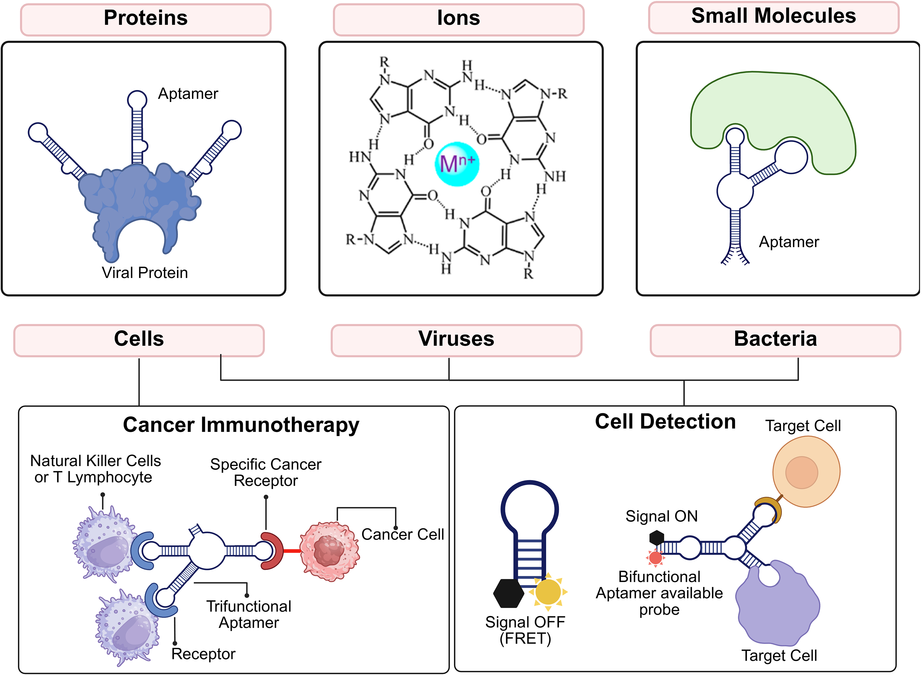
A schematic illustrating the diverse targets of oligonucleotide aptamers (proteins, 60 ions, 61 small molecules, 62 cells, 63 viruses, 64 and bacteria, 65 accompanied by relevant examples of binding interactions, highlighting some of their potential applications. Of note, the ion panel shows the interaction of one g-quadruplex region of an aptamer sequence that is stabilized by a metal cation. In addition, in the cell detection panel, the yellow symbol indicates a fluorophore (donor), and the black symbol indicates a quencher/FRET acceptor. “Signal OFF (FRET)” reflects donor quenching when fluorophore and quencher are proximal; “Signal ON” reflects decreased FRET and increased fluorescence upon target binding that separates the pair. Image recreated from original using BioRender under the Creative Commons Attribution License from. 66 The ion-binding aptamer panel was adapted from Zhou et al. Chemical Reviews, 2017; Figure 2E. 61
Aptamers have applications in a variety of fields including biological imaging,69,70 food safety, and medicine.71,72 In recent years, they have been studied as a tool for the diagnosis52,66,67,69,73–75 and therapeutic treatment of a variety of cancers.10,49,76,77 Aptamers are ideal vectors for cancer therapies, as they can bind with specific biomarkers associated with cancer cells and have the means to induce cytotoxic effects without affecting healthy cells in the process. 77 With respect to therapeutics, aptamers have been studied in a variety of contexts, including as a delivery method for chemotherapeutic drugs or as a standalone therapeutic through preclinical studies and clinical trials.13,49,52,76–79 Pegaptanib (Macugen) is a selective RNA aptamer that targets VEGF165 and was the first aptamer drug to receive regulatory approval. It also became the first anti-VEGF therapy approved for neovascular age-related macular degeneration. Clinical studies showed that it significantly slowed vision loss compared with sham injections and maintained an acceptable safety profile over as long as 3 years of treatment.80–82
Antibodies and aptamers operate in a similar fashion in that they share the same targets and have high affinity/specificity for those targets.14,54–56,67,83 Despite the similarity in mechanism of action, aptamers have been shown to have several advantages over antibodies, including low production cost,10,49,51,68 ease of production and modification,5,10,49,52,67 better tissue penetration due to their small size (8–25 kDa compared with ∼150 kDa for antibodies),49,66,68,76,84–86 and little to no immunogenicity or toxicity. 87 Of note, aptamers that have been successful through the clinical trial process tend to be RNA aptamers that are heavily modified and therefore have increased costs of production associated with them. The requirement for modifications is a practical bottleneck of bench to bedside translation for aptamer research. Because aptamers are oligonucleotides, they do not generate an immune response like antibodies (or modified oligonucleotides) and therefore may not elicit the unwanted side effects associated with an activated immune response.5,10,51,52,67,68,73,76,88 In addition, aptamer selection may be performed rapidly in vitro, whereas antibodies are typically produced in vivo, which is a much longer process. Antibody production often requires the use of animals or cells, which limits target classes for antibodies to those that are nontoxic, unlike aptamers that can essentially be selected to any target that can form hydrogen bonds.67,73,76,89 Not only is in vitro chemical synthesis a faster process, but it also allows for better reproducibility51,67 and the synthesis may be automated, which increases efficiency of the process. 73 In vitro selection also provides more flexibility in the experimental conditions, allowing the aptamers to function under nonphysiological conditions. 90 Moreover, aptamers are also more stable at higher temperatures. When the temperature is raised, they may denature but will return to their native state when the temperature is lowered, allowing a return of function. 90 Conversely, antibodies will completely lose function when denatured, which is irreversible.10,52,66,68,79
Despite the advantages of aptamers, there are some associated challenges with their use. Unmodified aptamers are hard to translate into the clinic mainly because they do not last long enough in the body and are cleared too quickly. Because they are nucleic acids, aptamers are susceptible to nuclease degradation and clearance, which results in a short half-life in vivo 79 and can eliminate activity. Their small size, while helping tissue penetration, also means they are rapidly filtered out by the kidneys, sometimes within minutes after intravenous dosing, so there is often not enough time to achieve a meaningful therapeutic effect. Aptamers also have a relatively simple chemistry since they are made from only four nucleotides, which can limit how well they interact with certain targets and increases the need for engineered formats or chemical modifications. Although modifications can improve stability and extend circulation, aptamers typically still have much shorter half-lives than monoclonal antibodies, so stability remains the main hurdle.49,54–56,79 One notable modification is the replacement of the 2′-hydroxyl of the ribose sugars with a fluoro, O-methyl, or amino group.76,79 Another known modification for this issue is polyethylene glycol (PEG)ylation, which has been used in different studies.49,68,71,72,91 The PEG modification has also been seen to increase the molecular weight of the aptamer in question to counteract rapid renal secretion.68,79 Although this may be helpful for the concern of renal secretion, increasing the molecular weight may cause problems with target binding 79 and may initiate an immune response. 92 PEGylation is a well-established drug delivery strategy that is widely used to improve pharmacokinetics and pharmacodynamics across many types of therapeutics, including aptamers. More than 30 PEGylated drugs are already in clinical use, and many others have advanced into clinical trials. 93 However, PEGylation is not risk-free: preexisting anti-PEG antibodies have been linked to acute hypersensitivity reactions after first exposure to pegnivacogin, a PEGylated RNA aptamer, highlighting a clinically important immunological concern that needs to be considered when using PEG in therapeutic design. 94 Finally, even though clinical tumor resistance to aptamer therapies has not been clearly documented, it should still be considered when designing and evaluating new aptamer-based treatments. 95
As an alternative to chemical modification, Spiegelmers may be used to prevent nuclease degradation
Selection
Systematic Evolution of Ligands by Exponential Enrichment (SELEX) is a method through which oligonucleotide aptamers can be selected from DNA or RNA libraries for specific targets. Figure 2 illustrates the SELEX process. This evolutionary process consists of four main steps: incubation, partitioning, recovery, and amplification via PCR. During the incubation step, a strategically designed partially randomized oligonucleotide library is incubated with the target of interest, such as proteins, whole cells, or other targets. 50 For a comprehensive discussion of SELEX considerations, see DeRosa et al. 96 During the partition stage, bound oligonucleotide sequences are separated from the unbound sequences through a variety of heterogenous methods such as filtration, affinity chromatography, and magnetic bead-based separation, as well as homogeneous partitioning techniques such as capillary electrophoresis.43,97,98 Bound sequences are then eluted from the target and subsequently amplified by PCR. DNA sequences may be directly amplified, while RNA sequences must be reverse transcribed to generate cDNA before amplification. 50 PCR products then undergo subsequent rounds of SELEX to enhance the affinity of the sequences for the target. 50 There are a few different methods that can be used to improve selectivity. This includes the use of competitors, increasing washing times to remove oligonucleotides that do not bind as well and decreasing the amount of the target (molecules, proteins, cells, etc.). 76 Traditionally, oligonucleotides would then be cloned and sequenced. However, next-generation sequencing can now be used to bypass the cloning step. 99 In general, DNA aptamer selections are becoming more and more common, but RNA selections are also possible. In the case of RNA selections, an extra step is involved where the PCR product postamplification must be reverse transcribed before moving to the newest selection round.
With this general procedure in mind, different SELEX methods vary slightly. For example, when selecting Spiegelmers, the incubation stage cannot utilize a native target, but rather one of the selection against the mirror image of the target. 98 Other changes have led to new SELEX methods such as protein-based SELEX, cell SELEX, and in vivo SELEX, to name a few. 50 Protein-based SELEX is a common method that uses proteins as the aptamer target, mainly purified recombinant protein biomarkers. 50 However, this method is limited because it may not be used when the identity of the protein biomarker of interest is unknown, insoluble, or has a complex structure. 50 It has also been seen that aptamers selected through this method sometimes cannot recognize their protein targets in vivo because the proteins adopt a different conformation in vivo than in vitro, or the protein may be posttranslationally modified, unlike the recombinant protein that was used as the target. 5
The cell SELEX method (Fig. 3A) utilizes live cells that express the biomarker of interest.50,66,91 The most common target of cell-SELEX is proteins on or embedded in the surface of the cell. However, it is possible that aptamers in cell-SELEX could also bind to other targets such as lipids or sugars. The advantage of cell-SELEX is that the target is in its natural state; for example, proteins will be in their native conformational state on the cell surface. 66 Because proteins do not need to be purified for this method, the protein target does not need to be known,50,66 and it can therefore be used for the identification of biomarkers in cancer cells. 91 Cell SELEX involves the general steps of SELEX as shown in Figure 2; commonly, a counterselection step that follows the core SELEX process is added prior to the incubation of the library with target. This counterselection step helps ensure aptamers will only bind with cells containing the target of interest.50,66,91 During this process, cells should be viable and nonmalignant to minimize nonspecific binding and selection of off-target aptamers; if not, nonspecific binding aptamers may become more prevalent. 50 Additional SELEX variations, including in vivo SELEX and related approaches, are illustrated in Figure 3B and C.

Schematic representation of the Systematic Evolution of Ligands by Exponential Enrichment process steps (left) and available partitioning methods (right). Image recreated from original using BioRender under the Creative Commons Attribution License from. 66

Panel A: Schematic diagram illustrating Cell-SELEX cycle. Panel B: Aptamers are easily chemically modified, and so they allow for the conjugation of molecules such as siRNAs (1), drugs (2), and nanoparticles (3) via common functional group addition to the aptamer terminals (amine, carboxylic acid, thiol, etc.). 100 Typically, the efficacy of therapeutic aptamers and their conjugates are assessed in cellular and/or rodent models (Panel C). Image recreated from original using BioRender under the Creative Commons Attribution License from. 101 In this figure, the linker is labeled in purple, while the nanoparticle is shown in yellow.
In vivo SELEX uses live animals to generate specific aptamers in vivo and does not require the counter-selection step, as cell-SELEX does. 50 This method has been seen in a study conducted by Mi et al. 102 whereby the group used in vivo selection to generate RNA aptamers selected to target colorectal cancer metastases; the aptamers were shown to accumulate selectively in liver tumors while sparing normal tissues. The molecular target was identified as p68 RNA helicase (DDX5), with binding confirmed at a dissociation constant (Kd) of 14 nM.
Leukemia SELEX examples
Earnest et al. 103 developed aptamers against AML cells associated with oncogene MLL-AF9, which is expressed in human blood stem/progenitor cells. Three candidate sequences were identified, from which one lead aptamer (KGE02) was selected based on binding affinity and specificity. Flow cytometry determined that the lead aptamer, modified with a 5′ fluorescein tag, had an approximate binding constant of 37.5 ± 2.5 nM to AML cells. To further confirm the lead sequence binding, fluorescence and confocal microscopy were performed to understand the localization of the cognate biomarker for KGE02. Fluorescein-tagged KGE02 was incubated with the cells and showed distinct localization in the membrane of the AML cells, which necessitates further analysis for drug-aptamer conjugates.
In another study, Amano et al. 104 demonstrated that RNA aptamers were developed to bind with the Runt domain (RD) of the AML1 protein, an essential transcription factor involved in the development of hematopoietic cells. Genetic variations of AML1 can lead to abnormal hemocytopoiesis and immunodeficiency, often detected in human leukemia. This study suggested that RNA aptamers, by specifically binding to the AML1 RD, could offer new insights into the treatment of AML1-related diseases.
Aptamer KH1C12 was compared with two other control cell lines (K562 and NB4) using flow cytometry and exhibited significant selectivity to the target AML cell line (HL-60) and was able to recognize the target cells within a complex sample of normal bone marrow. 75 They also identified another aptamer, KHG11, which can bind with significant selectivity to target AML cell lines such as HL-60 and human promyelocytic leukemia cells. 75
Using the Cell-SELEX approach, Shangguan et al.105,106 identified the DNA aptamer Sgc8c-7, which targets protein tyrosine kinase 7 (PTK7). PTK7 is highly expressed on the T-ALL cell line CCRF–CEM, suggesting that Sgc8c-7 could be useful for developing more precise and effective targeted approaches for ALL.
Aptamer-Based Leukemia Diagnostics
Aptamers for leukemia diagnosis
There are many emerging studies on applying aptamer technology to proof-of-concept systems for diagnosing leukemia. Yu et al. 107 have developed a simple and rapid electrochemical aptamer cytosensor for the direct detection of CML K562 cells based on a specific aptamer and a biotin-conjugated concanavalin A (bio-ConA) detection probe. Recoveries of 79.6%–93.3% were obtained when testing to detect K562 cells in human blood samples, which suggests that the biosensor could be a potential tool for CML K562 cell detection in biological sample. Although this method has strong potential as a leukemia diagnostic tool, reproducibility is still challenging. 108
Recently, a study used the thickness-shear mode acoustics method (TSM) and single-molecule force spectroscopy (SMFS) to study the interactions between DNA aptamers (sgc8c) specific to the PTK7, which is known to be a biomarker of leukemia lymphoblasts (MOLT-4). 74 In this study, the frequency changes concerning MOLT-4 cell lines are more significant in comparison with Jurkat cells and the U266 (control) cell lines. In this method, the sensing layer formed by thiolated Sgc8c aptamers is very sensitive to both low concentrations of MOLT-4 and Jurkat cells. Therefore, the TSM method allowed the development of a highly sensitive, label-free biosensor for the detection of leukemia cells. The SMFS method also proved the high selectivity of the Sgc8c aptamers to the PTK7 receptors. 74
Yang et al. 109 used the cell-SELEX approach and worked toward selecting DNA aptamers that target NB4 AML, a human promyelocytic leukemia cell line. The target protein of the K19 aptamer was identified as the sialic-acid-binding immunoglobulin-like lectins (Siglec-5). The aptamer was able to successfully detect small amounts of AML cells in human bone marrow samples. In this study, the researchers employed a combination of flow cytometry, fluorescence imaging, and mass spectrometry to evaluate aptamer binding and identify target proteins on AML cells. Flow cytometry was the primary quantitative tool used to assess aptamer binding affinity and specificity across leukemic cell lines and primary bone marrow samples by measuring fluorescence intensity after aptamer staining. Fluorescence microscopy provided visual confirmation of aptamer localization on the cell surface, supporting the flow cytometry data. To identify the molecular target of the lead aptamer (K19), mass spectrometry was conducted following affinity purification, which led to the discovery of Siglec-5 as the aptamer’s cognate receptor. These complementary techniques enabled both the functional characterization and molecular validation of the aptamer-based diagnostic strategy. 109
A method based on aptamer-modified fluorescent silica nanoparticles (FSNPs) has been developed to detect leukemia cells. Amine-labeled Sgc8 aptamer was conjugated to carboxyl-modified FSNPs, which resulted in a highly sensitive and specific aptamer-modified FSNP system. This was due to the amplified fluorescence signal caused by the high density of FITC fluorophore in the nanoparticles and specific target binding by the aptamer. The study suggested that the Sgc8-FSNPs system may be a good candidate for leukemia diagnosis. 110
Mallikaratchy et al. 111 identified the aptamer TD05, which binds to Ramos cells. They chemically modified TD05 to covalently cross-link with its target on Ramos cells to capture and enrich the target receptors using streptavidin-coated magnetic beads. This process was followed by mass spectrometry, which revealed that the target for the TD05 aptamer is membrane-bound immunoglobulin.
Simple colorimetric sensors/assays can eliminate the use of analytical instruments to obtain results and, therefore, are attractive as convenient rapid test methods. Bamrungsap et al. 112 developed aptamer-conjugated magnetic nanoparticles (ACMNPs) to sensitively detect leukemia cells. A PTK7-binding DNA aptamer (sgc8c) was used to recognize CCRF–CEM cells and confirmed specificity with clear sequence controls: sgc8c-ACMNPs gave a strong signal on CCRF–CEM cells, while a control aptamer conjugate (TDO5-ACMNPs) produced little to no signal, and the same control logic was applied using Ramos cells. The assay detects cells by measuring changes in spin–spin relaxation time (T2) without requiring wash steps, and it still worked in mixed samples containing both target and nontarget cells, using a random-sequence DNA–MNP conjugate as an additional negative control. Importantly, they also tested the method in more realistic biological samples such as serum, plasma, and whole blood, which supports its potential for clinical leukemia cell detection.
A recent study investigated the detection of leukemia cells using aptamer-conjugated, gold-coated magnetic nanoparticles on a nitrogen-doped graphene-modified electrode. A thiolated sgc8c aptamer was immobilized on gold-coated magnetic Fe3O4 nanoparticles (Apt-GMNPs). Ethidium bromide was intercalated into the stem of the aptamer hairpin, serving as a signal for quantifying leukemia cells. This approach demonstrated high sensitivity, selectivity, robustness, and simplicity for the early detection of leukemia. In addition, this aptasensor was successfully applied to detect leukemia cells in complex samples, such as human blood plasma, without significant interference. 113
Aptamer-Based Leukemia Therapeutics
In recent years, aptamers have been studied as potential alternatives to traditional cancer treatment methods such as chemotherapy, radiation therapy, and antibodies. Currently, there are five categories of aptamer-based cancer therapies being explored: biotherapy, cell-selective chemotherapy, gene therapy, targeted nanomedicine, and immunotherapy. 49 Each method works in a different way, but they all aim to cause cell death. These methods will be discussed, and the mechanism of action for each method can be seen in Figure 4.

Aptamer-based biotherapy
In the area of biotherapy, aptamers are used as standalone therapeutics to interfere with intracellular systems or signaling to induce apoptosis. 49 AS1411 and NOX-A12 can be considered the most notable examples, as they have both shown promising results in clinical trials for the treatment of leukemia. 49 AS1411 is a 26-nucleotide, guanine-rich DNA oligonucleotide that has often been described as a nucleolin-targeting aptamer. However, it differs from most aptamers in an important way: AS1411 was not discovered through SELEX but was instead designed to form a G-quadruplex structure. 114 Later studies, including work from the original group, showed that AS1411 enters cells mainly through macropinocytosis and that this uptake does not depend on nucleolin expression. 115 In addition, its uptake can be reduced by nonspecific competitor oligonucleotides, suggesting that internalization is largely nonspecific rather than driven by a specific receptor interaction. 63 Despite these mechanistic limitations, AS1411 advanced to phase II clinical testing in AML (NCT00512083), where it showed a good safety profile and modest clinical activity. Overall, AS1411 may not behave like a classical, target-specific aptamer, but it remains an important early oligonucleotide therapeutic and a clear reminder that rigorous target validation is essential in aptamer-based drug development.
It has been proposed that AS1411 can interact with nuclear factor-κB, which lowers the activity of BCL-2 mRNA and consequently inhibits cancer cell proliferation and induces cell death.52,78 The phase II clinical trial (NCT00512083) initiated in 2007 tested intravenous injection of AS1411 into 70 patients with AML. 79 Although reduced side effects were observed, therapeutic efficacy was evaluated independently through measures of tumor burden reduction and cell viability. 49 After the promising initial results, AS1411 was eventually used in a study for metastatic renal cell carcinoma.76,79 However, this study did not receive the same positive results as the AML study and, therefore, showed that biomarkers in AS1411-responsive tumors are required.116,117
NOX-A12 is a 45-nucleotide Spiegelmer that has been studied for the therapeutic treatment of CML, which is known to be untreatable with conventional chemotherapeutic methods. 49 Because NOX-A12 is a Spiegelmer, it has the advantage of being resistant to nuclease degradation. 13 NOX-A12 targets CXCL12, which binds with CXCR4 and CXCR7 in the cell, each of which possesses a role in cancer cell proliferation, metastasis, and motility.13,118 A preclinical study conducted by Hoellenriegel et al. 13 found that NOX-A12 also caused chemosensitization, allowing the cancer to be treated with chemotherapeutic drugs. 13 Currently, there are eight clinical trials that investigate this reagent, two of which are ongoing, one started in 2019, and the other in January of 2026 (https://clinicaltrials.gov/search?term=NOX-A12%20). Apoptosis was induced as a consequence of the aptamer-mediated disruption of CXCL12 signaling rather than by a conventional cytotoxic agent. 49 NOX-A12 has also undergone phase II clinical trials (NCT01486797) for CLL among other cancers. 13 The trial found that when in combination with bendamustine and rituximab, the treatment had success compared with traditional chemotherapy. 49
Cell-selective chemotherapeutic delivery
Several studies have been conducted involving the use of aptamers as a mode of delivery for chemotherapeutic drugs, siRNA, miRNA, and shRNA to target cancer cells. 119 Aptamers may be conjugated to cellular delivery systems such as liposomes to enable targeted transport of therapeutics to malignant cells.51,67 Conversely, therapeutic payloads can be attached to aptamers through covalent linkers or associated noncovalently (e.g., via intercalation), enabling aptamer-guided delivery of small molecules and nucleic acid cargos.57,67 Upon receptor binding, many aptamer–drug delivery constructs are internalized primarily through receptor-mediated endocytosis and subsequently traffic to endo/lysosomal compartments, enabling intracellular release of the payload. 67 When the therapeutic has been internalized, it then begins to induce its effects without damaging surrounding healthy tissue. Kotula et al. 77 used a nucleolin aptamer AS1411 as a delivery tool to help a chimeric aptamer enter leukemia cells. This chimera carried a β-arrestin 2–inhibitory aptamer (β-arr2A3) and was tested in leukemia cells from a mouse CML model as well as in human leukemia samples. In these systems, the β-arr2A3 construct interfered with β-arrestin-related signaling and was associated with reduced leukemia cell growth.
Another study carried out by Zhang et al. 120 showed the development of a polyvalent aptamer system for the delivery of Dox to leukemia cells through binding with PTK7. Experimentation found that the poly-aptamer approach showed greater binding affinity with the target compared with monovalent aptamer approaches as well as a greater ability to eliminate cancerous cells. Furthermore, Huang et al. 121 showed that conjugation of Dox to aptamer Sgc8c-7 resulted in highly efficient targeted delivery of Dox to CCRF–CEM cells, with minimum uptake by off-target cells; the results mentioned above highlight the benefits of using aptamers in clinical applications.
Zhao et al. 53 developed an aptamer targeting the CD117 receptor, which is highly expressed on AML cells. In follow-up experiments, they found that the aptamer preferentially bound membrane-associated IgM (mIgM) on the cell surface, suggesting that CD117 expression may be linked—directly or indirectly—to how mIgM is displayed on leukemic cells. Notably, the aptamer did not bind soluble IgM in plasma, which supports the idea that its interaction is specific to cell-surface targets rather than circulating IgM. CD117 is a transmembrane receptor that is highly expressed on leukemia cells in 95% of relapsed AML patients. The anti-CD117 aptamer sequence contained a G-rich core section and G-quadruplex functional structure, with a length of 79 bases. The result of this study showed the aptamer 1-F specifically binds to CD117 as the AML biomarker with high-affinity Kd (4.24 nM) and was able to specifically be internalized into CD117-expressing cells. This aptamer was later conjugated to methotrexate to form a therapeutic Apt-MTX. 53
Other therapeutic methods
Apart from biotherapy and cell-selective chemotherapy, other aptamer-mediated therapies studied include gene therapy, targeted nanomedicine, and immunotherapy. Gene therapy utilizes an aptamer conjugated to different types of RNA (siRNA, miRNA, etc.) to silence oncogenes and consequently cause cell death. 49 While there are limited studies in this area as it relates to leukemia, there are studies in relation to other cancers. 122
In targeted nanomedicine, aptamers are conjugated with nanoparticles, which are delivered to target cells to induce cell death. 49 As mentioned above, there are cases where nanoparticles have been conjugated with aptamers and other chemotherapeutic drugs for delivery to the cells. For example, Ho and colleagues 123 recently developed a new CRISPR-Cas9 delivery system. This system targets the critical gene interleukin-1 receptor accessory protein (IL1RAP) in human leukemia stem cells (LSCs). They achieved this by using mesenchymal stem cell membrane-coated nanofibril (MSCM-NF) scaffolds loaded with lipidoid-encapsulated Cas9/single guide RNA ribonucleoprotein and the chemokine CXCL12α.352. The MSCM-NF scaffolds effectively facilitated the targeted delivery of Cas9/IL1RAP sgRNA to LSCs, reducing the growth of LSCs and decreasing leukemic burden in a mouse model of AML.
Mei et al. 124 employed rolling circle amplification to generate self-assembled DNA nanoflowers (NFs) with a high drug loading capacity of 71.4% w/w. These nanomaterials were designed to overcome multidrug resistance (MDR) in cancer therapy by incorporating cell-targeting aptamers (e.g., KK1B10 for leukemia and Sgc8 for breast cancer) directly into the DNA template. The aptamers enabled selective recognition and internalization into cancer cells, including drug-resistant variants, thereby enhancing doxorubicin retention and minimizing off-target toxicity. NFs delivered doxorubicin into target chemosensitive and MDR cancer cells, preventing drug efflux and enhancing drug retention in MDR cells. Moreover, NF-Dox induced potent cytotoxicity in target cells while minimizing side effects in nontarget cells, effectively circumventing MDR and reducing side effects.
In addition, Douglas et al. 125 developed a novel DNA hexahedral barrel nanostructure to control a switch using the conformational change of a nucleic acid aptamer. A DNA aptamer-based lock secures a closed, hexahedral barrel that holds molecular cargo, such as proteins or small molecules. In this design, the barrel is kept closed by an aptamer sequence paired with a partially complementary strand. When the nanorobot encounters its specific antigen (or target), these antigens bind to the aptamer sequences on the locks. As the aptamers bind to the antigens, their conformational changes cause them to dissociate from their partially complementary strands. The dissociation of the aptamer–antigen complex results in the release of the locks, effectively “unlocking” the barrel. Once the locks are removed, the structural constraints that maintain the closed state are lifted, allowing the barrel to open and release its payload. Research has shown that this structural system enhances selectivity for target tumor cells and enables controlled antibody drug release, effectively suppressing the proliferation of leukemia cells.
In 2013, Wu and colleagues 126 developed a multifunctional aptamer nano assembly (AptNA) for precise cancer therapy targeting. The assembly involves loading the antisense nucleotide (AS) of MDR-1 onto a specially designed DNA nano assembly (Apt-NA) attached to a specific aptamer. This treatment has proven effective in eliminating drug-resistant myeloid leukemia cells. The aptamer, KK1B10, recognizes the drug-resistant cell line K562/D, and the AptNA is selectively taken up by the cell. MDR-1-AS is then released to combat the cell’s drug resistance through inhibition of P-glycoprotein expression. Experimental findings have confirmed the specific cytotoxic impact of AptNA on the targeted tumor cells.
Aptamer-based immune redirection has also been investigated in other contexts through dual targeting designs; however, the examples discussed here emphasize CAR-T engineering and RNAi-mediated gene silencing.49,122,127 Leukemia immune-engaging therapies have progressed more in antibody-based formats than in aptamer-based systems. Blinatumomab, a monoclonal antibody that engages T cells, is one example. Clinical trials have demonstrated its ability to treat ALL, showing that malignant cells can be targeted effectively by cytotoxic T cells (MT-103-211). 128 A comparison of aptamers used for diagnostic and therapeutic purposes is summarized in Table 2.
Comparison of Aptamers Used for Diagnostic and Therapeutic Purposes

ALL, acute lymphoblastic leukemia; AML, acute myeloid leukemia; CLL, chronic lymphocytic leukemia I N-heterocyclic carbene gold; CML, chronic myeloid leukemia; DOX, doxorubicin; MTX, methotrexate; NHC-Au, complex; NR, not reported.
Future Directions
Early diagnosis of leukemia is a challenging task, but it can greatly improve therapeutic success. There is increasing demand for highly sensitive and user-friendly diagnostic methods, motivating continued development of aptamer-based platforms for rapid leukemia detection. Several leukemia-binding aptamers—such as Sgc8c, KHG11, K19, and truncated TD05—have been identified and validated in proof-of-concept studies, but most have not yet been advanced into clinically standardized assays or evaluated in large patient cohorts.74,75,85,106,109
In addition, existing aptamer-based diagnostic systems such as the Sgc8-FSNPs platform, 110 ACMNPs, 112 aptamer-conjugated gold nanoparticle colorimetric assays, 130 and Apt-GMNP electrochemical formats 113 should be further developed toward clinical use. Key next steps include improving robustness in complex samples, incorporating internal controls/calibration strategies, and performing blinded validation in representative clinical cohorts.110,112,113,130 It is also important to consider multiplexing strategies or panel-based approaches to better address leukemia heterogeneity and subtype variation.96,109
To improve reproducibility and support clinical adoption, future diagnostic studies should follow clear minimum standards for reporting and for controls in aptamer assays, such as aptamer folding conditions, buffer composition, cation requirements, pH, temperature, blocking or competitor conditions, and the sample matrix used because aptamer performance can change substantially with these parameters. Across the aptamer field, weak controls and incomplete method reporting have contributed to binding results that are hard to reproduce and that do not translate well between laboratories. Using standardized controls and reporting guidance would help make diagnostic findings more reliable and speed the development of clinically robust assays.63,96
For leukemia therapeutics, future work should clearly distinguish between aptamers as active therapeutics and aptamers as targeting ligands for delivery. Clinical-stage examples such as AS1411 and NOX-A12 show that translation is possible, but they also need to be discussed carefully, especially when follow-up development has been limited or the reasons for stopping are not clear. AS1411 is a good example: it is often described as a nucleolin-targeting aptamer, but mechanistic work suggests cells can take it up through macropinocytosis in a way that does not require nucleolin, and its uptake can be reduced by nonspecific competitor oligonucleotides. Together, these findings raise questions about how specific its internalization is and whether nucleolin should be treated as a confirmed target. Going forward, studies should prioritize strong evidence of target engagement and avoid overconfident mechanistic claims when discussing AS1411-like agents.63,115
As mentioned previously, there are some associated challenges with using aptamer therapeutics in vivo, such as nuclease degradation and a short half-life due to rapid renal excretion.54–56,79 However, solutions to these problems have been proposed, including the use of Spiegelmers or aptamer modification.13,76,79
Many leukemia aptamer-delivery studies still use conventional chemotherapeutics as payloads, which may limit clinical impact when delivered as conjugates. Lessons from antibody-drug conjugates (ADCs) suggest that durable clinical success required highly potent payloads and linker chemistries that are stable in circulation but release reliably within target cells. 131 Going forward, leukemia-focused ApDCs should move beyond conventional payloads and adopt ADC-grade cytotoxins, such as auristatins like monomethyl auristatin E/F (MMAE/MMAF) together with clinically proven linker strategies, while carefully controlling the drug-to-aptamer ratio, conjugate uniformity, and stability in plasma. Since MMAE/MMAF have already been successfully attached to aptamers in other contexts, the chemistry appears feasible and worth translating into leukemia-specific ApDC development.132,133
In both leukemia diagnostics and therapeutics, there are several biomarkers that have not yet been exploited for aptamer selection. For example, calgranulin A (or S100A8) 134 is a biomarker associated with AML, which could be used in aptamer selection. In addition, IFNγ, IL6, IL8, sIL2Rα, sgp130, sIL6R, MCP1, MIP1α, MIP1β, and GM-CSF cytokines that are predictive of cytokine release syndrome after treatment of ALL are also potential targets for aptamer selection. 135 Indeed, there are also potential biomarkers that have not yet been discovered, a critical advantage of Cell-SELEX being that aptamers can be selected without a known biomarker and subsequently be used for target identification. Certainly, biomarker discovery will also play an important role in future research.
The use of aptamers for diagnosing and treating leukemia is a relatively new but promising field of study that requires further exploration. Despite some challenges, aptamers are a powerful tool that can potentially be used for rapid diagnosis or as an alternative to traditional cancer therapy methods.
Footnotes
Author Disclosure Statement
All the contributing authors report no conflict of interests in this work.
Funding Information
No funding was received for this article.