



Abstract
The CXC chemokine receptor 2 (CXCR2) is a member of the G-protein-coupled receptor superfamily and regulates a diverse range of immune responses and tumor progression. CXCR2 is expressed on immune cells, especially neutrophils, and is involved in various immune responses by interacting with its chemokine ligands. Therefore, the development of sensitive monoclonal antibodies (mAbs) for CXCR2 has been desired for treatment and diagnosis. This study established a novel sensitive anti-mouse CXCR2 (mCXCR2) mAb; Cx2Mab-5 (rat IgG2a, κ), using the mCXCR2 synthetic N-terminus peptide immunization method. In flow cytometry, Cx2Mab-5 recognized mCXCR2-overexpressed Chinese hamster ovary-K1 cells (CHO/mCXCR2) and WEHI-3B (murine myelomonocytic leukemia cell) cells, which express endogenous mCXCR2. Cx2Mab-5 did not cross-react with other mouse CC, CXC, CX3C, and XC chemokine receptors. Cx2Mab-5 showed a moderate binding affinity for both CHO/mCXCR2 and WEHI-3B. Furthermore, Cx2Mab-5 detected mCXCR2 in Western blot and immunohistochemistry in CHO/mCXCR2 cells, but a commercially available anti-mCXCR2 mAb (clone SA045E1) did not. Hence, Cx2Mab-5 can be a valuable tool for analyzing mCXCR2-positive cells in mouse tissues.
Introduction
Chemokines are small chemoattractant proteins secreted by various cells, including immune-related cells, that play essential roles in a myriad of physiological activities, such as cellular migration, invasion, and inflammatory responses, by activating chemokine receptors.1,2 Chemokine receptors belong to the superfamily of G-protein-coupled receptors (GPCRs) and can be classified into four different subfamilies: CC, CXC, CX3C, and XC, depending on the number and position of cysteine residues in their N-terminus. Chemokine receptors display the seven transmembrane structures characteristic of typical GPCRs. 3 Chemokines-chemokine receptors signal contributes to various aspects of our immune response, and their aberrant signaling is implicated in multiple diseases, including allergy, 4 autoimmune diseases, 5 and cancer. 6
Several chemokines, including CXCL1, CXCL2, CXCL3, CXCL5, CXCL6, and CXCL7 in mice and CXCL8 in humans, have been identified as ligands for CXCR2.7,8 CXCR2 contributes to tumorigenesis through recruitment of immune cells, such as myeloid-derived suppressor cells (MDSCs) and tumor-associated neutrophils, autocrine effects on the tumor, and angiogenesis by interacting with these chemokines.9,10 CXCR2 is considered a key regulator of the tumor microenvironment (TME). The higher expression of CXCR2 is confirmed in neutrophils in both humans and mice. CXCR2 knockout (KO) mice decreased mature neutrophils in the spleen. Many parameters of fine splenic neutrophils decline in CXCR2 KO mice, including the phagocytic ability and reactive oxygen species production. 11 Therefore, the development of sensitive monoclonal antibodies (mAbs) for mouse CXCR2 (mCXCR2) has been desired for basic research and application to preclinical models.
Since chemokine receptors, including CXCR2, are 7 transmenbrane proteins, it is difficult to generate the mAbs for multiple applications, including flow cytometry, Western blotting, and immunohistochemistry (IHC). We have previously developed many mAbs against chemokine receptors using the Cell-Based Immunization and Screening 12 and synthetic N-terminal peptide immunization 13 (http://www.med-tohoku-antibody.com/topics/001_paper_antibody_PDIS.htm). In this study, we successfully developed novel anti-mouse mCXCR2 mAbs using synthetic N-terminal peptide immunization method.
Materials and Methods
Cell lines
Chinese hamster ovary (CHO)-K1 and P3X63Ag8U.1 (P3U1) cells were obtained from the American Type Culture Collection (Manassas, VA, USA). The WEHI-3B (murine myelomonocytic leukemia cell) cell line was obtained from the Cell Resource Center for Biomedical Research, Institute of Development, Aging and Cancer, Tohoku University (Miyagi, Japan). Furthermore, the cDNAs of mouse CXCR2 (mCXCR2; pCMV6neo-mCXCR2-Myc-DDK, accession No.: NM_009909.3, catalog No.: MR227587) were purchased from OriGene Technologies, Inc. (Rockville, MD, USA). The mCXCR2 expression plasmid was transfected into the cell line using a Neon transfection system (Thermo Fisher Scientific Inc., Waltham, MA, USA). Subsequently, CHO-K1, which stably overexpressed mCXCR2 with C-terminal Myc-DDK tags (CHO/mCXCR2), was established through cell sorting, using an anti-mCXCR2 mAb (clone SA045E1; BioLegend, San Diego, CA, USA) and a cell sorter (SH800; Sony Corp., Tokyo, Japan), following cultivation in a medium containing 0.5 mg/mL G418 (Nacalai Tesque, Inc., Kyoto, Japan). These cells and other mouse chemokine receptor-overexpressed CHO-K1 cells (e.g., CHO/mCXCR1) were established and cultured as previously reported. 14
Antibodies
The anti-mCXCR2 mAb (clone SA045E1), anti-mCX3CR1 mAb (clone SA011F11), and anti-mXCR1 mAb (clone ZET) were purchased from BioLegend. The anti-DYKDDDDK mAb (clone 1E6) was purchased from FUJIFILM Wako Pure Chemical Corporation (Wako; Osaka, Japan). An anti-isocitrate dehydrogenase 1 (IDH1) mAb (clone RcMab-1) 15 , other chemokine receptor mAbs, and anti-PA tag mAb (NZ-1) were developed previously in our lab (http://www.med-tohoku-antibody.com/topics/001_paper_antibody_PDIS.htm). The secondary Alexa Fluor 488-conjugated anti-rat IgG was purchased from Cell Signaling Technology, Inc. (Danvers, MA, USA). Secondary horseradish peroxidase-conjugated anti-mouse IgG and anti-rat IgG were obtained from Agilent Technologies Inc. (Santa Clara, CA, USA) and Merck KGaA (Darmstadt, Germany), respectively.
Production of hybridomas
The animal experiments were conducted in accordance with relevant guidelines and regulations to minimize animal suffering and distress in the laboratory. The Animal Care and Use Committee of Tohoku University approved animal experiments (Permit number: 2022MdA-001). Two female Sprague–Dawley (Jcl:SD) rats (6-week-old, CLEA, Japan Tokyo, Japan) were immunized via the intraperitoneal route with keyhole limpet hemocyanin (KLH)-conjugated mCXCR2 peptide (MGEFKVDKFNIEDFFSGDLC, 100 μg of peptide/time, Eurofins Genomics KK, Tokyo, Japan) as previously reported. 14 The hybridoma supernatants were screened using enzyme-linked immunosorbent assay (ELISA) and flow cytometric analysis. MAbs were purified using Protein G Sepharose (Cytiva, Tokyo, Japan).
ELISA
The mCXCR2 peptide (1 µg/mL)-immobilized immunoplates were incubated with hybridoma supernatants, followed by peroxidase-conjugated anti-rat immunoglobulins (1:2000 dilution; Agilent Technologies Inc., Santa Clara, CA, USA). Afterward, enzymatic reactions were conducted using the ELISA POD substrate TMB kit (Nacalai Tesque, Inc.), and the optical density was measured as previously reported. 14
Flow cytometry
CHO-K1, CHO/mCXCR2, and WEHI-3B cells were harvested after brief exposure to 1 mM ethylenediaminetetraacetic acid (EDTA; Nacalai Tesque, Inc.). The cells were washed with 0.1% bovine serum albumin in phosphate-buffered saline (PBS) and treated with primary mAbs for 30 minutes at 4°C. Then, the cells were washed and incubated with Alexa Fluor 488-conjugated anti-rat IgG (1:1000 dilution). For the peptide-blocking assay, Cx2Mab-5 or SA045E1 was preincubated with the mCXCR2 peptide or dimethyl sulfoxide (DMSO) for 15 minutes at room temperature and then incubated with CHO/mCXCR2 and WEHI-3B for 30 minutes at 4°C. Afterward, cells were treated with Alexa Fluor 488-conjugated anti-rat IgG (1:1000 dilution). Flow cytometric data were acquired on an SA3800 Cell Analyzer (Sony Corp., Tokyo, Japan) by collecting 5000 events. Cells were gated based on forward scatter and side scatter, and fluorescence intensity was analyzed using FlowJo software (BD Biosciences, Franklin Lakes, NJ, USA).
Determination of the binding affinity by flow cytometry
CHO/mCXCR2 and WEHI-3B cells were suspended in 100 μL serially diluted anti-mCXCR2 mAbs, after which Alexa Fluor 488-conjugated anti-rat IgG (1:200 dilution) was added. The data (10,000 events) were collected using the SA3800 Cell Analyzer, and the geometric mean (GeoMean) was determined using FlowJo software. The fitting binding isotherms (vertical axis, GeoMean; horizontal axis, mAb concentration) determined the apparent dissociation constant (KD) values to built-in one-side binding models of GraphPad Prism 6 (GraphPad Software, Inc. La Jolla, CA, USA). 14
Western blot analysis
Western blot analyses were performed using 5 μg/mL of Cx2Mab-5, 5 μg/mL of SA045E1, 1 μg/mL of an anti-IDH1 mAb (clone RcMab-1), or 0.5 μg/mL anti-DYKDDDDK (clone 1E6) mAb as described previously. 16
Immunohistochemical analysis
Cell blocks were prepared using iPGell (Genostaff Co., Ltd., Tokyo, Japan) and fixed in 4% paraformaldehyde in PBS (FUJIFILM Wako Pure Chemical Corporation). The sections (4 µm thickness) were autoclaved in citrate buffer (pH 6.0; Nichirei Biosciences, Inc., Tokyo, Japan) for 20 minutes. After blocking with SuperBlock T20 Blocking Buffer (Thermo Fisher Scientific Inc.), the sections were incubated with Cx2Mab-5 (5 μg/mL) and SA045E1 (5 μg/mL) for 1 hour at room temperature. The cell blocks were subsequently treated with Histofine Simple Stain Mouse MAX PO (Rat) (Nichirei Biosciences, Inc.) for 30 minutes. Color development was achieved using 3,3'-diaminobenzidine tetrahydrochloride (DAB; Agilent Technologies Inc.), and counterstained with hematoxylin (Merck KGaA).
Results
Development of anti-mCXCR2 mAbs
To develop anti-mCXCR2 mAbs, we employed the mCXCR2 N-terminus peptide immunization method. Hybridomas were screened by ELISA and flow cytometric methods (Fig. 1). Briefly, two rats were immunized with KLH-conjugated mouse CXCR2 synthetic peptide by intraperitoneal injection. Two days after the final boost injection, rat spleen and myeloma cells were fused using PEG1500 to generate hybridomas. Subsequently, hybridomas were seeded into 96-well plates, and an ELISA assay was used to identify positive wells for the mCXCR2 peptide. This was followed by the selection of CHO/mCXCR2-reactive and parental CHO-K1-nonreactive supernatants using flow cytometry. Finally, anti-mCXCR2 mAb clones, including Cx2Mab-1 (IgG2b, κ), Cx2Mab-2 (IgG2b, κ), Cx2Mab-3 (IgM, κ), Cx2Mab-4 (IgG2b, κ), and Cx2Mab-5 (IgG2a, κ), were established by limiting dilution. We further examined the application of these clones and selected Cx2Mab-5 due to its multiple availabilities for flow cytometry, Western blotting, and IHC.

A schematic illustration of the production of anti-mCXCR2 mAbs. The rat was immunized intraperitoneally with a KLH-conjugated N-terminus mCXCR2 peptide (five total doses). Hybridoma screening was then conducted by ELISA using the naked mCXCR2 peptide, followed by flow cytometric analysis of mCXCR2-overexpressed CHO-K1 cells and parental cells. To obtain a monoclonal clone, single-cell cloning was performed by limiting dilution.
Flow cytometry using Cx2Mab-5
Flow cytometry was conducted using Cx2Mab-5 and commercially available anti-mCXCR2 mAb SA045E1 against CHO/mCXCR2, CHO-K1, and WEHI-3B. Results showed that Cx2Mab-5 dose-dependently recognized CHO/mCXCR2 (Fig. 2A), but not CHO-K1 (Fig. 2B). Another anti-mCXCR2 mAb, SA045E1 from BioLegend, also dose-dependently recognized CHO/mCXCR2 (Fig. 2A), but not CHO-K1 (Fig. 2B). Both Cx2Mab-5 and SA045E1 also reacted dose-dependently with endogenous mCXCR2-expressing WEHI-3B (Fig. 2C). Thus, Cx2Mab-5 could recognize exogenously and endogenously expressing mCXCR2 in flow cytometry.

Flow cytometric analysis using anti-mCXCR2 mAbs. CHO/mCXCR2 cells
The specificity of Cx2Mab-5
We have established anti-mouse CC, CXC, CX3C, and XC chemokine receptor-overexpressed CHO-K1 cells in a previous study. 14 Using these 18 cell lines, the specificity of Cx2Mab-5 was evaluated in Figure 3. The results described that Cx2Mab-5 recognized only CHO/mCXCR2, but not other 17 chemokine receptors (Fig. 3A). We confirmed the expression of each chemokine receptors by treatment with corresponding mAbs (Fig. 3B).
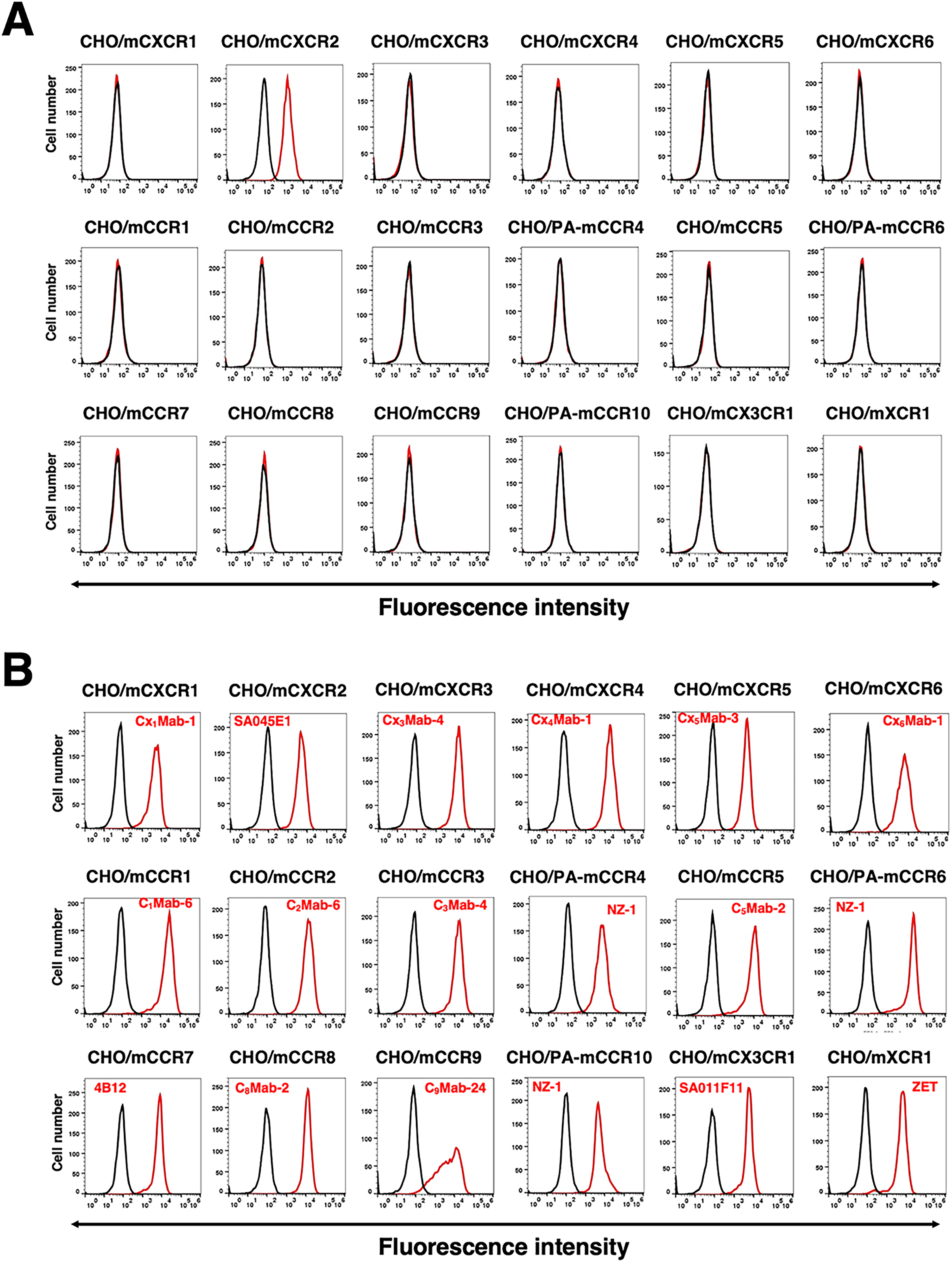
Cross-reactivity of Cx2Mab-5 in in mouse CC, CXC, CX3C, and XC chemokine receptor-overexpressed CHO-K1 cells.
We next evaluated antibody reactivity using a peptide-blocking assay with the mCXCR2 peptide. As shown in Figure 4, both Cx2Mab-5 and SA045E1 reacted with CHO/mCXCR2 and WEHI-3B cells. The reactivity of Cx2Mab-5 was completely blocked by the mCXCR2 peptide, indicating that its recognition is mediated through binding to the N-terminus of mCXCR2. Similarly, SA045E1’s reactivity was entirely neutralized by the mCXCR2 peptide. These findings demonstrate that the N-terminus of mCXCR2 is the epitope recognized by SA045E1. Taken together, Cx2Mab-5 is an anti-mCXCR2-specific mAb that recognizes the N-terminus of mCXCR2.
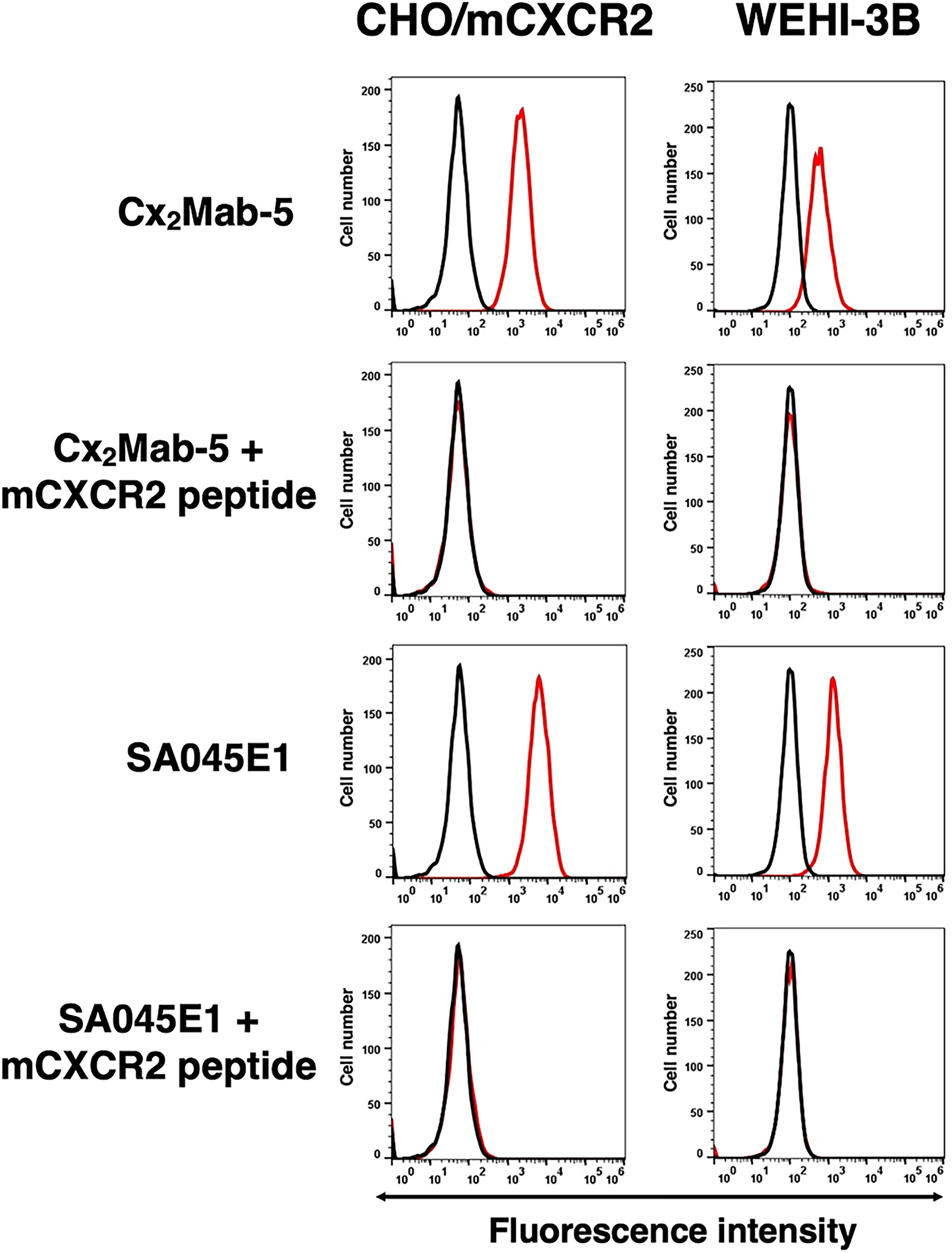
Peptide-blocking analysis using anti-mCXCR2 mAbs with mCXCR2 peptide. CHO/mCXCR2 and WEHI-3B cells were incubated with Cx2Mab-5 (1 μg/mL) plus control (0.1% DMSO in blocking buffer), Cx2Mab-5 plus mCXCR2 peptide (10 μg/mL), SA045E1 (1 μg/mL) plus control (0.1% DMSO in blocking buffer), or SA045E1 plus mCXCR2 peptide (10 μg/mL) for 30 minutes at 4°C. Cells were then treated with Alexa Fluor 488-conjugated anti-rat IgG. Fluorescence data were collected using the SA3800 Cell Analyzer. The black line represents the negative control (blocking buffer).
Determination of the binding affinity of Cx2Mab-5
The binding affinity of Cx2Mab-5 was assessed with CHO/mCXCR2 and WEHI-3B using flow cytometry. Results showed that the apparent KD values of Cx2Mab-5 and SA045E1 for CHO/mCXCR2 were 2.4 (±0.6) × 10−8 M and 1.5 (±0.1) × 10−8 M, respectively (Fig. 5A). The KD values of Cx2Mab-5 and SA045E1 for WEHI-3B were 1.8 (±1.0) × 10−8 M and 1.0 (±0.2) × 10−8 M, respectively (Fig. 5B). These results indicated that Cx2Mab-5 had a moderate affinity for CHO/mCXCR2 and WEHI-3B cells, almost equivalent to that of SA045E1.
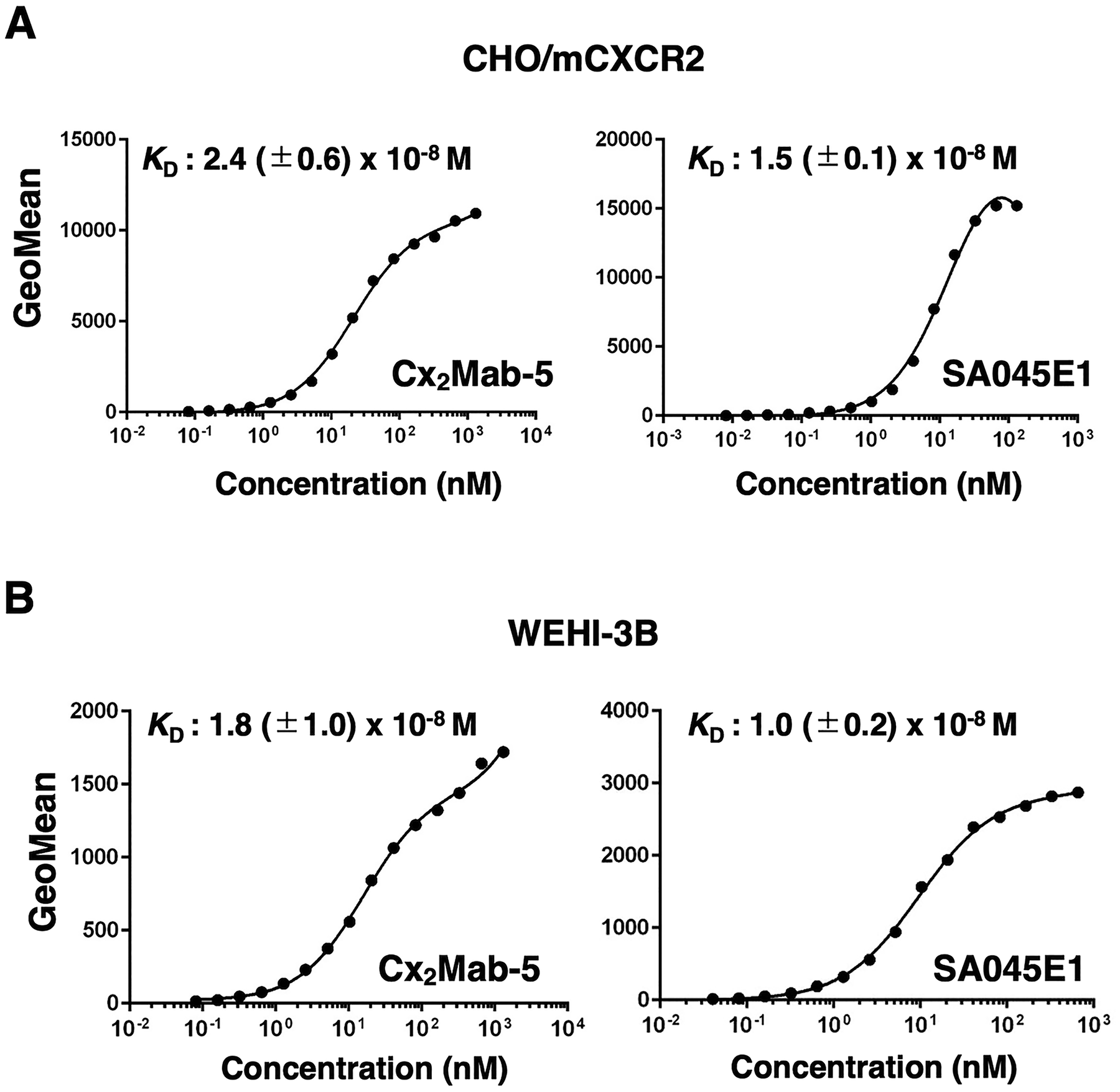
The determination of the apparent dissociation constant of Cx2Mab-5. CHO/mCXCR2
Western blot analyses using Cx2Mab-5
We investigated whether Cx2Mab-5 can be used for Western blot analysis by analyzing CHO-K1 and CHO/mCXCR2 cell lysates. As shown in Figure 6, Cx2Mab-5 could clearly detect mCXCR2 as around a 48–63 kDa band in CHO/mCXCR2 cell lysates, while no band around the predicted size was detected in parental CHO-K1 cells. In contrast, another anti-mCXCR2 mAb, SA045E1, could not detect any signals on the membrane. An anti-DYKDDDDK mAb (1E6) served as a positive control and detected a band in CHO/mCXCR2 cell lysates. An anti-IDH1 mAb (RcMab-1) was used for internal control. These results indicate that Cx2Mab-5 can detect mCXCR2 in Western blot analysis.

The detection of mCXCR2 by Western blot analysis. Cell lysates of CHO-K1 and CHO/mCXCR2 were electrophoresed and transferred onto polyvinylidene difluoride membranes. The membranes were incubated with 5 μg/mL of Cx2Mab-5, 5 μg/mL of SA045E1, 0.5 μg/mL of 1E6 (an anti-DYKDDDDK mAb), and 1 μg/mL of RcMab-1 and subsequently with horseradish peroxidase-conjugated anti-mouse or anti-rat immunoglobulins. Blots were developed using PierceTM ECL Plus Western Blotting Substrate or ImmunoStar LD and imaged with a Sayaca-Imager.
Immunohistochemistry using Cx2Mab-5
To investigate whether Cx2Mab-5 can be used for IHC, paraffin-embedded CHO-K1 and CHO/mCXCR2 sections were stained with Cx2Mab-5. Apparent membranous staining by Cx2Mab-5 was observed in CHO/mCXCR2 (Fig. 7, left upper). The SA045E1 did not stain CHO/mCXCR2 sections (Fig. 7, right upper). Neither Cx2Mab-5 nor SA045E1 reacted with the CHO-K1 section. (Fig. 7 lower). Additionally, Cx2Mab-5 slightly stained the WEHI-3B section (Supplementary Fig. S1). These results indicate that Cx2Mab-5 is suitable for IHC detection of mCXCR2-positive cells in paraffin-embedded tissue samples.
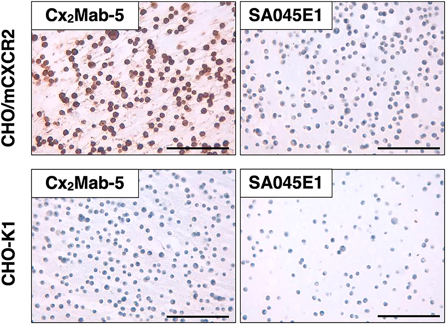
Immunohistochemical staining of a paraffin-embedded section of CHO/mCXCR2 and CHO-K1. The sections of CHO/mCXCR2 and CHO-K1 cells were treated with 5 μg/mL of Cx2Mab-5 or 5 μg/mL of SA045E1, followed by the Histofine Simple Stain Mouse MAX PO (rat). Color development was performed using DAB, and counterstaining was performed with hematoxylin (Merck KGaA). Scale bar = 100 μm.
Discussion
In this study, we developed a novel anti-mCXCR2 mAb (clone Cx2Mab-5) using N-terminal peptide immunization. Cx2Mab-5 is suitable for various experiments, including flow cytometry (Figs. 2–5), Western blotting (Fig. 6), and IHC (Fig. 7). In contrast, a commercially available anti-mCXCR2 mAb (clone SA045E1) was developed by immunizing rats with mCXCR2-transfected cells and is useful only for flow cytometry, not for Western blotting or IHC. Because the reaction of SA045E1 was neutralized by the N-terminal peptide (Fig. 4), the epitope of SA045E1 would be located in the N-terminal domains. We will determine the detailed binding epitope of Cx2Mab-5 and SA045E1 to clarify the difference between their applications.
Distinct surface-expressed and intracellular human CXCR2 glycoforms were identified in neutrophils. N-glycosylation is essential for maintaining receptor surface expression. 17 Western blot and immunoprecipitation analyses revealed that surface-expressed CXCR2 was an apparent size of 56 kDa with two N-linked 9-kDa carbohydrate moieties. 17 In addition, 38 and 40 kDa CXCR2 forms were found to be expressed intracellularly with N-glycosylation of high mannose or hybrid type. 17 The detection patterns of mCXCR2 by Cx2Mab-5 and 1E6 were different in Western blotting (Fig. 6). The modifications may affect the detection patterns observed by Western blotting.
CXCR2 is a key immunomodulatory factor with potential for next-generation cancer therapy. An immunosuppressive neutrophil subpopulation has been observed in human and mouse gliomas.18,19 The CXCR2+ neutrophils, recruited by glioma-secreting CXCL1, establish the immunosuppressive TME. Genetic depletion of these neutrophils improves antitumor immunity and prolongs survival. 19 MDSCs are also remarkable immunosuppressive cells in the TME. 20 The inhibition of CXCR2 is reportedly a promising treatment for controlling polymorphonuclear-MDSCs accumulation associated with tumor progression.21,22 Furthermore, the myeloid chemotaxis inhibition by CXCR2 blockade has an impact on reversing therapy resistance in a subset of patients with metastatic castration-resistant prostate cancer. 23
Cx2Mab-5 will be a valuable tool for elucidating the function of mCXCR2 in preclinical models. We will investigate whether the established Cx2Mab-5 exerts antitumor effects by inhibiting CXCR2+ neutrophils in mouse models. Since Cx2Mab-5 is rat IgG2a, the isotype changes to mouse IgG1 or IgG2a are required after degeminating the complementarity-determining regions. We developed Cx2Mab-5 via immunization with an N-terminal mCXCR2 peptide. Because the N-terminus of CXCR2 is necessary for ligand binding,7,24,25 Cx2Mab-5 is expected to be used in mouse models to determine whether Cx2Mab-5 inhibits mCXCR2 activation and exhibits antitumor efficacy.
Authors’ Contributions
H.T. and T.T. performed the experiments. T.T. analyzed the data. Y.K. and M.K.K. designed the experiments. T.T., H.S., and Y.K. wrote the article. All authors have read and agreed to the published version of the article.
Footnotes
Author Disclosure Statement
No competing financial interests exist.
Funding Information
This research was supported in part by Japan Agency for Medical Research and Development (AMED) under Grant Numbers: JP25am0521010 (to Y.K.), JP25ama121008 (to Y.K.), JP25ama221153 (to Y.K.), JP25ama221339 (to Y.K.), and JP25bm1123027 (to Y.K.), and by the Japan Society for the Promotion of Science (JSPS) Grants-in-Aid for Scientific Research (KAKENHI) grant nos. 24K18268 (to T.T.) and 25K10553 (to Y.K.).
Supplemental Material
References
Supplementary Material
Please find the following supplemental material available below.
For Open Access articles published under a Creative Commons License, all supplemental material carries the same license as the article it is associated with.
For non-Open Access articles published, all supplemental material carries a non-exclusive license, and permission requests for re-use of supplemental material or any part of supplemental material shall be sent directly to the copyright owner as specified in the copyright notice associated with the article.
