



Abstract

On behalf of the JALA scientific advisors and JALA editorial board, I am happy to present this year’s honorees of the prestigious JALA Ten. Each year, JALA seeks to highlight and honor the very best work of the year that will have a deep impact on how technology is used across a wide range of disciplines, including automation, life sciences and biomedical research, diagnostics, drug delivery, and regenerative medicine. Implementing the latest advances in microfluidics, nanotechnology, materials science, and other fields of research, this year’s JALA Ten honorees have and will continue to change the way research is performed and the way diseases are diagnosed and treated. As such, the work highlighted here should have far-reaching impact in our everyday lives. It demonstrates the promise that science brings toward a better future.
While a number of areas of research will feel the impact of this year’s JALA Ten, one highlight of the collection is the diverse ways in which the honorees have advanced biological molecule detection and established foundations for tomorrow’s biosensors in life sciences research and medical diagnostics.1–3 For example, Peter Lillehoj at Michigan State University (USA) and his collaborators have developed a microfluidic biosensor using immobilized antimicrobial peptides for highly specific, multiplexed detection of bacterial pathogens. 1 Showing both high pathogen detection specificity and accurate pathogen quantification, this work is an example of how microfluidic technology is changing how medical professionals will track and diagnose infectious diseases.
On the other end of the spectrum, Somin Eunice Lee at the University of Michigan (USA) has harnessed the power of nanotechnology to develop a gold nanoparticle–based plasmon ruler that is capable of single-molecule measurements. 2 Even more impressive is the application of this plasmon ruler toward the accurate detection and measurement of secreted single molecules in the cellular microenvironment. Understanding how cells communicate with their cellular microenvironment remains a challenge to study. Tools such as Lee’s plasmon ruler vastly improve life sciences researchers’ abilities to learn more about the interplay between cells and their microenvironments.
Another area of research highlighted this year is the advancement of technology toward customizable platforms for increased personalized clinical and research applications.4–7 The development of tunable injectable microporous gel scaffolds by researchers at the University of California, Los Angeles (USA) not only brings truly personalized medicine to regenerative medicine, but also allows researchers to quickly optimize culture conditions in research and drug development applications that require complex three-dimensional (3D) cell cultures. 4 The development of integrated platforms composed of modular technologies increases the diversity of clinical and research applications to which technology such as microfluidics can be applied and is highlighted here by work from Harvard University (USA) 6 and the University of Southern California (USA). 7
Beyond building better biosensors and more effective and cost-efficient devices, work featured this year includes improvements in automation of large-cargo intracellular delivery, as well as innovating the use of inorganic and biomaterials in medical applications.8–10 This year’s honorees represent the best in research that advances translational science and technology, and we are proud to honor these researchers for their amazing work. JALA and SLAS would like to thank all the nominees as well as those who nominated them. We would also like to thank everyone who worked to discuss and select the 2016 JALA Ten. The last 6 years have allowed us to highlight innovative and exciting research breakthroughs that will greatly impact our lives. This year is no different and continues to raise the bar for innovation. We look forward to seeing what next year brings.
Rapid Electrical Impedance Detection of Bacterial Pathogens Using Immobilized Antimicrobial Peptides 1
Current sensing technologies for quantifying microbial pathogens rely on highly specific antibodies for molecular recognition that suffer from limited stability in high-temperature environments and can be difficult to obtain for similar pathogen species. Using synthetic antimicrobial peptides (AMPs) with species-specific targeting and binding capabilities, Peter Lillehoj of Michigan State University (USA) and his collaborators have developed a microfluidic biosensor for rapid, multiplexed detection of bacterial pathogens. AMP-coated sensors demonstrate strong preferential binding to their corresponding targeted cells with negligible cross-binding ( Fig. 1 ) and impedance measurements correlated well to the cell concentration. With further development, this technology can provide a robust, portable platform for rapid pathogen detection.
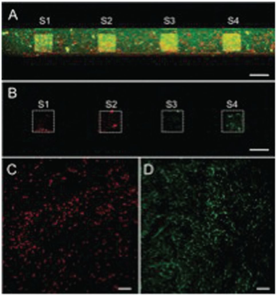
Pathogen-specific detection by microsensors with immobilized antimicrobial peptides. Reprinted by permission of SAGE Publications, Inc., 2016.
A Golden Ruler for Secreted Single-Molecule Measurements 2
Tracking and studying single molecules in living cells is of great interest to life sciences researchers; however, the tools to do so are highly limited. Even more difficult is studying the function of single molecules following cell secretion in order to better understand how these molecules function in cellular microenvironments. Somin Eunice Lee at the University of Michigan (USA) and her colleagues have developed an aptamer-gold plasmon ruler that now achieves reliable detection and study of secreted single molecules ( Fig. 2 ).
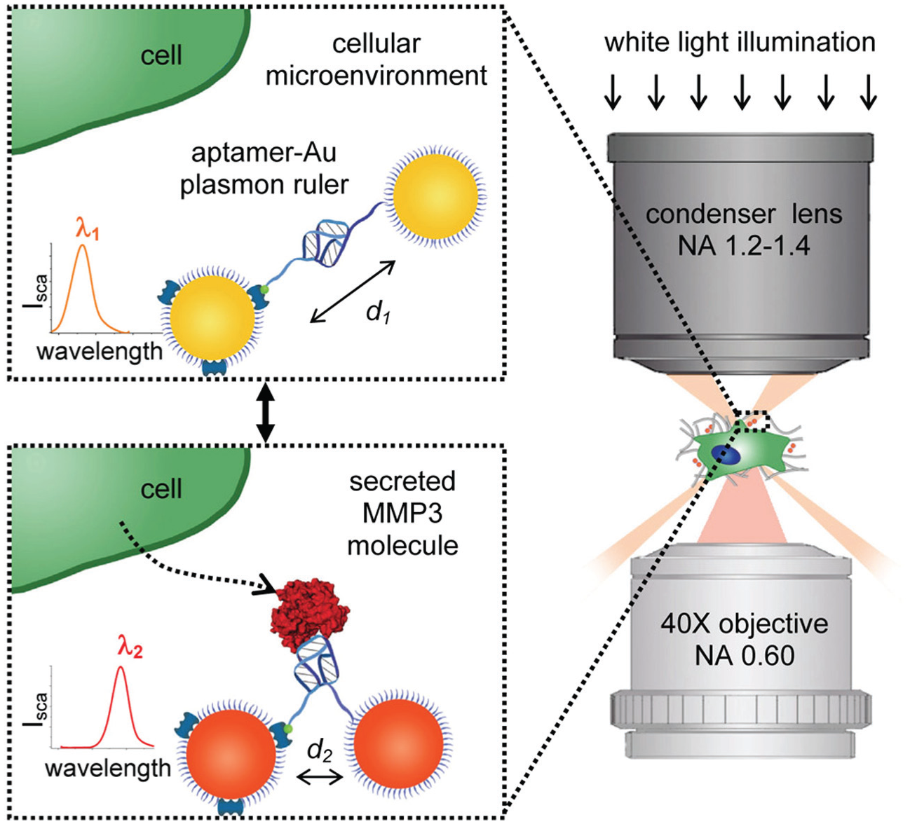
Schematic of secreted single-molecule measurement by reversible aptamer-Au plasmon ruler. Reprinted (adapted) with permission from Lee, S. E.; Chen, Q.; Bhat, R.; et al., Reversible Aptamer-Au Plasmon Rulers for Secreted Single Molecules, Nano Lett.
Furthermore, this plasmon ruler is reversible, allowing for detection of multiple events. High specificity of this reversible plasmon ruler is demonstrated with the specific detection of the matrix metalloproteinase MMP3 over its family member MMP9. Implementation of this plasmon ruler into life sciences and biomedical research should allow for greater understanding of the functions of secreted molecules in the microenvironment. These plasmon rulers should also find use in phenotypic drug discovery and other translational applications.
Using Nanotechnology to Track Cellular Functions in Real Time 3
Even with the increasing popularity of phenotypic research methods such as high-content applications and the emergence of cell-based therapies, there is a dearth of cell labeling and tracking reagents that can reliably and quantifiably sense key cellular functions. Chenjie Xu and his colleagues at Nanyang Technological University (Singapore) have developed a nanoparticle-based platform for just this purpose. Encapsulating specific biosensor molecules in biodegradable polymeric nanoparticles, these nanosensors are able to serve as an intracellular source of sensor molecules for up to 30 days within cells ( Fig. 3 ).
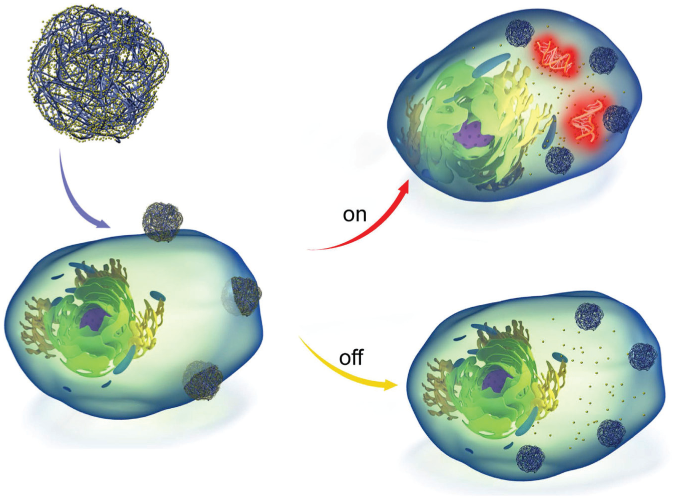
Schematic of real-time intracellular nanosensor. Reproduced under a Creative Commons CC-BY license, 2016.
This platform has a wide range of biomedical applications, as demonstrated by this team. Utilizing their nanosensors, they demonstrate real-time quantification of cellular processes such as nitric oxide production, as well as gene expression such as β-actin mRNA expression. The development of these nanosensors should serve as the foundation for a wide range of tools for molecular biology research, as well as phenotypic drug discovery.
Building Better Injectable Scaffolds for Accelerated Wound Healing 4
Scaffold materials for tissue engineering and 3D matrices for organ-on-a-chip technologies have been hampered by a fundamental limitation in the ability to control material porosity separately from mechanical properties. This limitation is especially exacerbated when considering flowable biomaterials, which are useful for in vivo delivery, filling of wounds, or encapsulation of cells. Dino Di Carlo and Tatiana Segura and their teams at the University of California, Los Angeles (USA) have overcome these limitations with the development of a tunable injectable microporous gel scaffold ( Fig. 4 ).
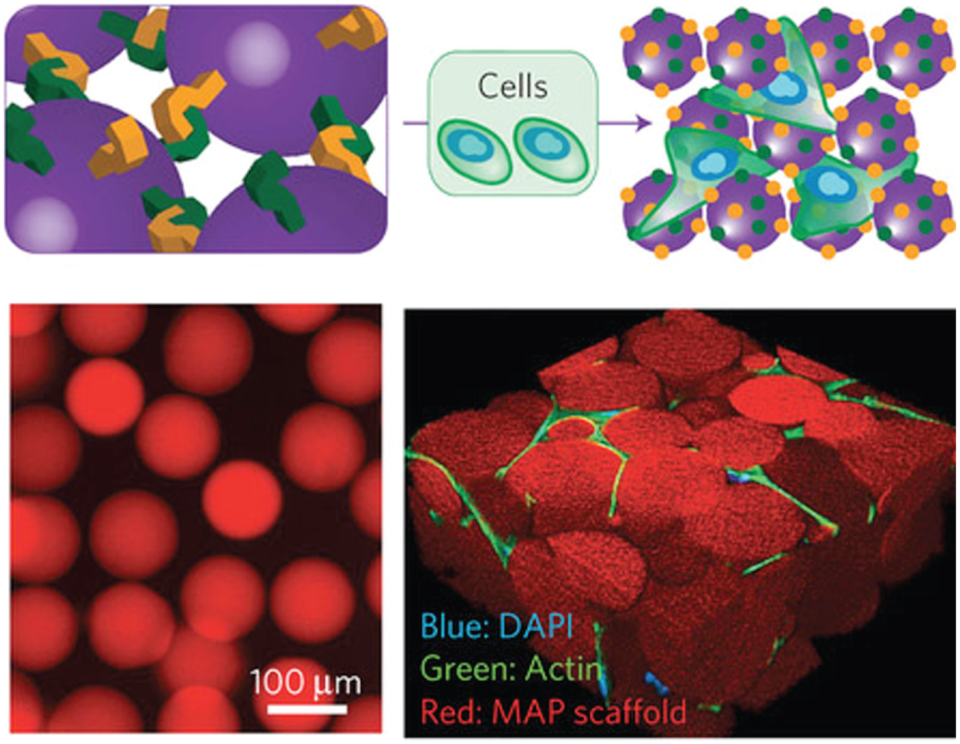
Schematic and representative image of injectable microporous gel scaffold. Adapted by permission from Macmillan Publishers Ltd., Nature Materials, copyright 2015.
Microporosity is important in a biomaterial, as it improves transport within a scaffold and enables rapid cellular ingrowth. This work demonstrates the first approach to achieve an injectable microporous material for tissue engineering, and this material promoted cellular growth in vitro and accelerated healing in vivo. Because of the modularity and tenability of these microporous annealed particle (MAP) gels, they should impact a wide range of regenerative medical applications, as well as drug development applications that require 3D cell culture or organ-on-a-chip methods of study.
A Simple and Reliable Micropump for Lab-on-a-Chip Devices 5
Tony Jun Huang’s research group at the Pennsylvania State University (USA) demonstrate a microfluidic pump, so-called sharp-edge-based acoustofluidic pump, that utilizes the acoustic streaming effects induced by acoustically oscillating tilted sharp-edge structures. Their pump is simply composed of a quarter-sized piezoelectric transducer and a microfluidic channel ( Fig. 5 ). Upon the oscillation, the tilted sharp-edge structures generate acoustic streaming effects, which in turn generate net forces in the direction that the sharp-edge structured oriented. Thus, fluid pumping takes place because the generated net forces push the bulk fluid to flow forward. By simply modulating the driving signals to the piezoelectric transducer, the pump can generate not only flow rates ranging from nanoliters to several microliters per minute, but also flow rates of various flow profiles. Thus, the acoustofluidic pump has the potential to be integrated into this kind of portable testing platform, because of its small size, easy fabrication, controllability, and tunability.
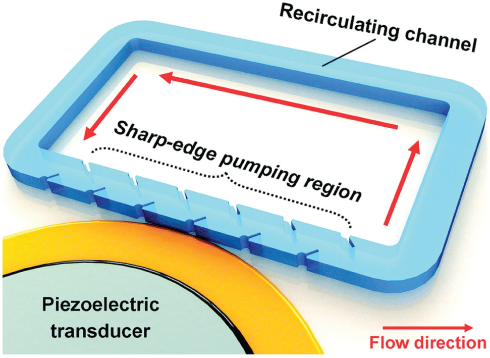
Schematic of acoustofluidic pump. Reproduced in part with permission of the Royal Society of Chemistry, 2016.
The Development of an Affordable Modular Mini-Microscope 6
Microscopes remain a key component of any life sciences research; however, they remain expensive and are often limited in specific functions when made more affordable. Y. S. Zhang from Harvard University (USA) has innovated a miniature microscope with built-in fluorescence capability for biomedical applications ( Fig. 6 ).
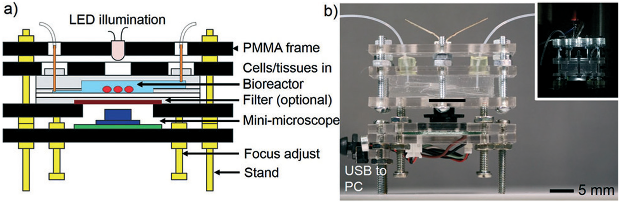
Schematic and photo of modular mini-microscope. Reproduced in part with permission of the Royal Society of Chemistry, 2016.
This mini-microscope has adjustable magnifications from 8× to 60× and achieves a high resolution of <2 μm. Using off-the-shelf components and a webcam, the mini-microscope system is inexpensive (<$10), and its modularity allows for convenient integration with a wide variety of preexisting platforms, such as cell culture plates and microfluidic devices. Therefore, this mini-microscope is likely to find widespread applications in cell biology, tissue engineering, biosensing, microfluidics, and organs-on-chips, which may potentially replace conventional benchtop microscopy where long-term in situ and high-throughput imaging and analysis are required.
A 3D Platform for Modular Microfluidic Circuits 7
Traditionally, microfluidic systems have been designed as monolithic devices, each with a single dedicated application. Noah Malmstadt and his team at the University of Southern California (USA) have developed a modular microfluidic platform where each module can be designed with discrete fluid handling, routing, or analysis function ( Fig. 7 ). These modules are assembled together to create an application-defined microfluidic circuit. A key benefit of this approach is design predictability. The behavior of the assembled system can be predicted by circuit design methodologies because each module has well-understood fluidic characteristics.
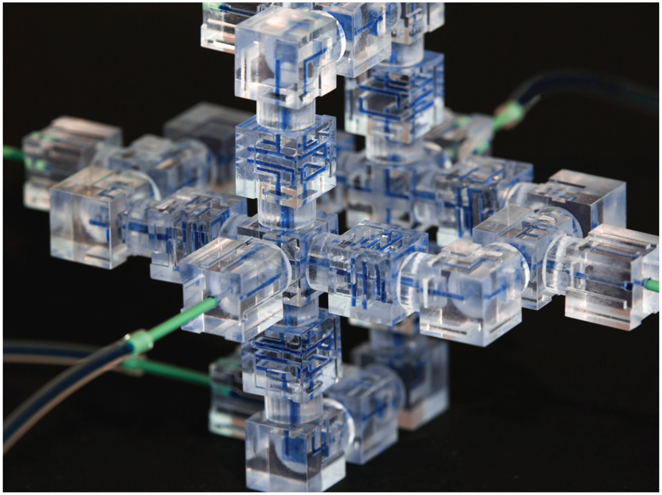
3D modular microfluidic circuit. Image courtesy of original authors.
This modular approach to microfluidic circuit design allows for a truly 3D microfluidic design rather than less efficient and more complex planar layer-by-layer arrangements of channels. Additionally, because individual models are manufactured by 3D printing, fabrication is simpler and cheaper. This work represents a shift in microfluidic design that should allow microfluidic devices to be incorporated in a wider range of commercial applications.
High-Throughput Photothermal BLAST Platform for Large-Cargo Intracellular Delivery 8
While there are a number of well-established methods for delivery of small-scale cargo, such as kilobit-sized nucleic acids, reliably delivering large cargo into cells has remained elusive. Developing a method for large-cargo delivery in a high-throughput manner is an even more difficult hurdle to overcome. Eric Pei-Yu Chiou and his team at the University of California, Los Angeles (USA) have accomplished such a feat with their new intracellular delivery platform called biophotonic laser-assisted surgery tool (BLAST) ( Fig. 8 ).
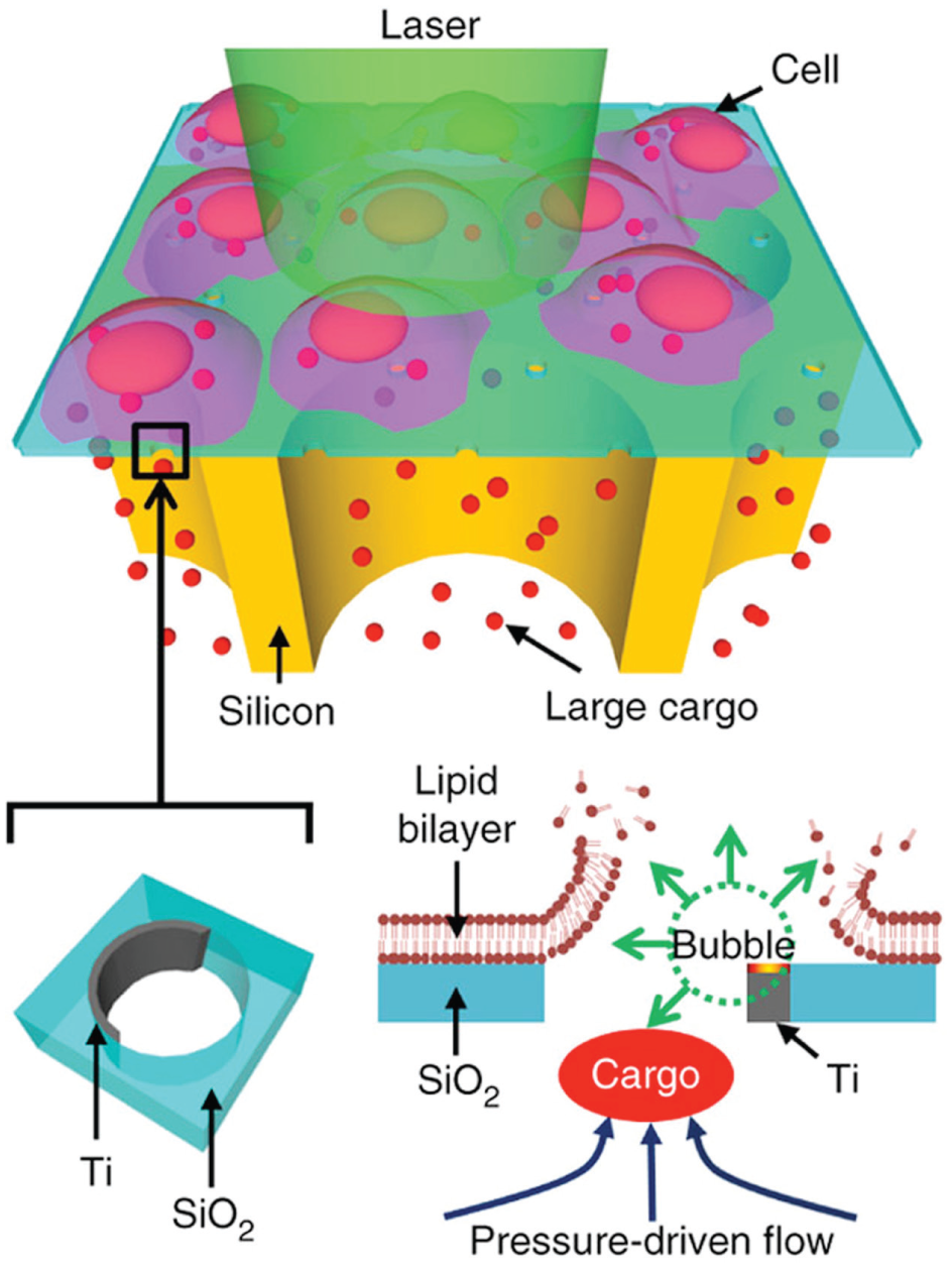
Schematic of BLAST-mediated large-cargo cellular delivery. Adapted by permission from Macmillan Publishers Ltd., Nature Methods, copyright 2015.
BLAST enables the delivery of cargo up to several microns in size into 100,000 cells in 1 min, which is five orders of magnitude faster than prior methods. Cargo is delivered into the cytosol of cells directly without undesirable endosome trapping. High efficiency, high cell viability, and nearly simultaneous delivery of diverse types of cargo into numerous cells under constant physiological conditions allow reliable measurements in a variety of biological settings.
Because of the reliability of the high-throughput platform and the increase in cargo size, BLAST can be applied to a broad range of applications, including delivery of large nanoparticle complexes, functional proteins, and large amounts of both DNA and RNA. As such, this platform should improve life sciences research, from basic molecular biological research to drug development and biomedical clinical research.
Slippery Rough Surfaces Inspired by Lotus Leaves and Nepenthes Pitcher Plants 9
The ability to repel liquids regardless of how they wet the surface has important technological implications for numerous industrial and biomedical processes, ranging from condensation heat transfer to water harvesting to antifouling of medical devices. However, maintaining liquid mobility on engineered surfaces under various conditions has been an engineering challenge for more than a decade. Now, researchers at the Pennsylvania State University (USA) led by Tak-Sing Wong have invented a new class of liquid-repellent surface, known as slippery rough surface, which can repel liquids in any state of wetness for the first time ( Fig. 9 ).
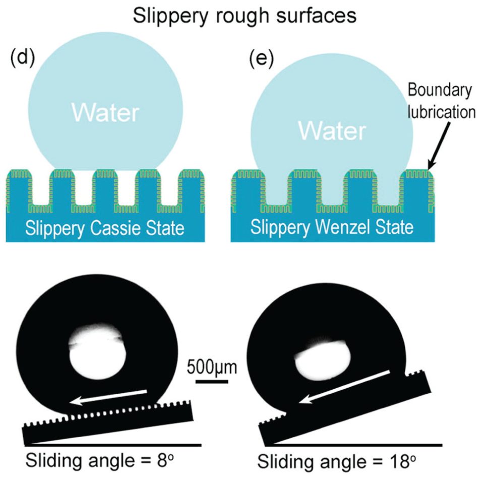
Nature-inspired slippery rough surfaces to repel liquids. Adapted under the terms of this standard ACS AuthorChoice/Editors’ Choice usage agreement. This is an unofficial adaptation of an article that appeared in an ACS publication. ACS has not endorsed the content of this adaptation or the context of its use.
Their surfaces, modeled after lotus leaves and the pitcher plant, have been developed by engineering hierarchical nano- and microscale textures and infusing liquid lubricant into the nanotextures to create a highly slippery rough surface. The new surface may open up new opportunities for scientific studies and engineering applications related to wetting, adhesion, transport phenomena, and biofouling.
Terminating Cancer with Liquid-Metal-Based Drug Delivery Systems 10
While the use of inorganic nanomaterials in drug delivery remains an area of active research in nanomedicine, clinical translation of inorganic material-based drug delivery systems has been difficult due to their toxicity and clearance failures. A promising new approach that overcomes these hurdles was developed by Zhen Gu and his team at the University of North Carolina at Chapel Hill (USA) and North Carolina State University (USA).
Utilizing a liquid-phase eutectic gallium-indium core and a thiolated polymeric shell, a transformable liquid-metal-based nanosphere drug delivery particle is formed ( Fig. 10 ). When functionalized with hyaluronic acid, these drug delivery complexes are capable of efficiently delivering chemotherapeutics and more effectively inhibiting tumor growth than conventional chemotherapeutics. Degradable in mildly acidic environments, this new liquid-metal-based approach opens new avenues to exploring the use of inorganic materials in a variety of nanomedical applications.
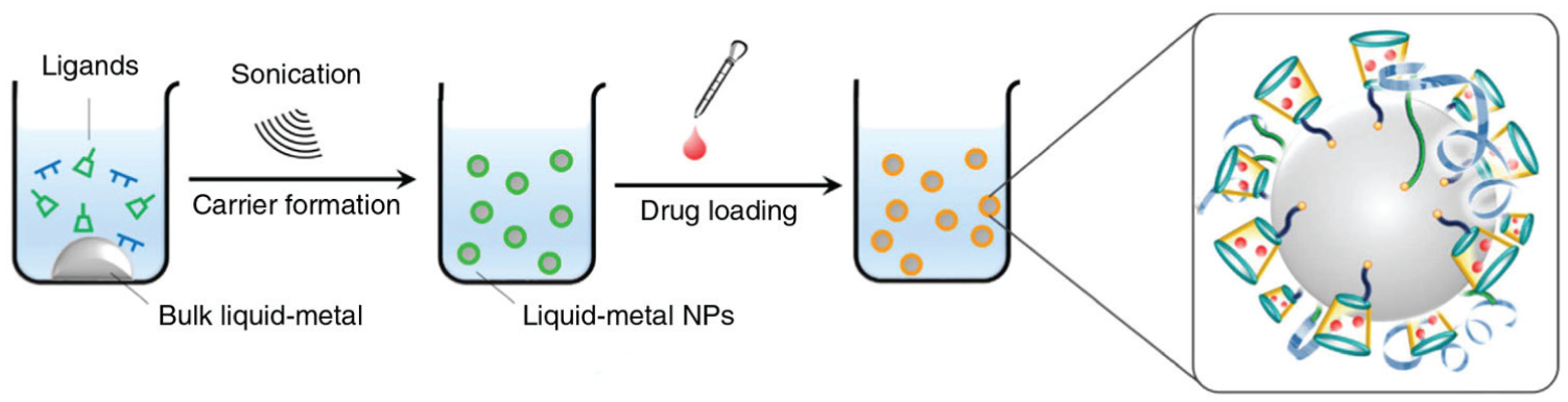
Schematic of liquid-metal-based drug delivery complex. Reproduced under a Creative Commons CC-BY license, 2016.