
Abstract
The study assessed the effectiveness of a novel plant-based collagen booster (PBCB) formulation in enhancing collagen synthesis in human dermal fibroblasts, comparing it to commercial hydrolyzed collagen products. Dermal fibroblasts are vital for skin health as they produce collagen, maintaining skin elasticity and strength. The new formulation combines amino acids, vitamins, and active ingredients from specific plant extracts to boost collagen production. In vitro experiments showed that this formulation increased collagen synthesis by approximately 5.42-fold compared to vitamin C and outperformed bovine and marine collagen products by 3.24 and 1.66 times, respectively and 4.19 times better than the PBCB product, at a 2.5 mg/mL concentration. Overall, the preliminary findings suggest that this novel formulation may have potential for supporting skin health and anti-aging applications; however, comprehensive molecular analyses, in vivo studies, and clinical trials are required for validation.
Introduction
Collagen and Skin Aging
Skin aging is a progressive, multifactorial process influenced by intrinsic and extrinsic factors. Intrinsic aging is primarily driven by genetic programming and hormonal changes, while extrinsic aging is accelerated by environmental and lifestyle factors such as prolonged UV exposure, pollution, and chemical irritants.1–3 These factors exacerbate visible signs of aging, including wrinkles, hyperpigmentation, and loss of elasticity, primarily by impairing fibroblast function, reducing the synthesis of collagen and elastin, and increasing melanin production in melanocytes. 3
Collagen is the most abundant structural protein in the skin, accounting for approximately 90% of dermal protein and 75% of the skin’s dry weight. 4 Of the more than 25 known collagen types, Types I (85%–90%), III (8%–11%), and V (2%–4%) are most prevalent in the skin. 4 Collagen plays a vital role in maintaining tissue architecture, wound repair, cell migration and adhesion, and platelet aggregation. 5 Structurally, it consists of three polypeptide chains wound into a triple helix, rich in glycine, proline or hydroxyproline, and alanine. 6
Fibroblasts are the primary collagen-producing cells and rely on mechanical tension within the extracellular matrix (ECM) to maintain collagen synthesis. With age, this mechanical signaling weakens, leading to reduced fibroblast activity and ECM remodeling. Simultaneously, matrix metalloproteinases (MMPs), particularly MMP-1, MMP-8, and MMP-13, are upregulated, contributing to collagen degradation. 7 Reactive oxygen species (ROS), often generated through UV exposure and environmental stressors, further enhance MMP expression, accelerating collagen breakdown.8,9 It is estimated that collagen production naturally declines by approximately 1% per year from early adulthood. 10 However, studies show that antioxidant supplementation can mitigate ROS-induced collagen damage and improve skin condition, especially in photoaged skin.11–14
Role of Amino Acids and Plant-based Supplements in Collagen Synthesis
Amino acids such as glycine, proline, and lysine serve as key building blocks for new collagen synthesis by dermal fibroblasts. 15 Hence, oral supplementation with collagen peptides or amino acid-rich formulations has gained popularity in addressing signs of aging. Nutraceuticals, defined as food-derived compounds offering therapeutic or preventive benefits, are increasingly employed to support skin health and delay photoaging. 16 The growing consumer demand for collagen supplementation has led to a rapidly expanding market with a compound annual growth rate of 5.5%, especially in the beauty and wellness sectors. 17 Traditional collagen supplements derived from animal sources, such as bovine, porcine, and marine, require enzymatic digestion to release usable amino acids. However, these sources may present challenges in terms of allergens, religious acceptability, and environmental sustainability.
Plant-based supplements, rich in bioavailable amino acids and antioxidants, offer a sustainable and inclusive alternative.18,19 Rather than delivering collagen directly, these formulations stimulate endogenous collagen production and protect existing ECM components. Given increasing consumer preference for clean-label, cruelty-free, and plant-based skincare solutions, botanical collagen precursors have emerged as promising candidates in nutricosmetic and functional food sectors. These alternatives aim to replicate the functional benefits of conventional collagen while addressing safety, ethical, and sustainability concerns. In the present study, the primary objective is to compare the performance of the novel plant-based collagen booster formulation (PBCB) against widely available commercial animal-derived (bovine and marine collagen) and PBCB products, as these represent the current benchmarks in the field. It evaluates the potential of PBCB to stimulate collagen production in human dermal fibroblasts (HDFs), which potentially might contribute to the development of novel anti-aging interventions.
Materials and Method
Equipment and Chemicals
Normal HDF cells (Cat No: CC-2511) were purchased from Lonza, Switzerland. Gibco Dulbecco’s Modified Eagle Media (DMEM) and Gibco fetal bovine serum (FBS) were from Thermo Fisher Scientific, Massachusetts, USA.
Chemicals such as sodium carbonate, Folin–Ciocalteu reagent, gallic acid, 2,2-diphenyl-1-picrylhydrazyl radicals (DPPH), ortho-phthalaldehyde (OPA) and fluorenylmethyloxycarbonyl chloride (FMOC-Cl), doxorubicin were procured from Sigma-Aldrich, Bangalore, India. Sodium ascorbate (Na Asc or vitamin C), hydroxyproline chloramine-T (prepared in 0.63 M sodium hydroxide [NaOH], 0.14 M citric acid, 0.453 M sodium acetate and 0.112 M acetic acid), and Ehrlich’s aldehyde reagent (1 M p-dimethylaminobenzaldehyde in 30:70 HCl and isopropanol) were procured from Sisco Research Laboratories (SRL), New Delhi, India. NaOH was from HiMedia, Mumbai, India.
The following instruments and materials were used: a POLARstar Omega plate reader (BMG Labtech, Ortenberg, Germany); F96 Maxisorp NUNC Immuno ELISA plates (Thermo Fisher Scientific, Massachusetts, USA); Falcon→ 6-well clear flat-bottom-tissue culture treated plates (Corning, New York, USA); an inverted phase contrast microscope (DMi1, Leica Microsystems, Wetzlar, Germany); a UVmini-1240 UV–VIS spectrophotometer (Shimadzu, Kyoto, Japan); and CellTiter-Glo→ reagent (Promega, Wisconsin, USA).
The variants of PBCB are TLGC_BF, TLGC_F1, and TLGC_F2. TLGC_BF is a base formulation containing amino acid and vitamin C mix (Essential, non-essential amino acids [0.5–5 mM] and vitamin C [1 mM]). The TLGC_F1 is one variant comprising base formulation (TLGC_BF) along with plant extracts of Adansonia digitata (Baobab) powder, Malpighia emarginata (Acerola cherry) extract, Myrciaria dubia (Camu Camu) powder, Rosa canina (Rosehip) extract, Lycium barbarum (Goji berry) powder, Terminalia ferdinandiana (Kakadu plum) extract, Phyllanthus emblica (Amla) powder, Hibiscus rosa-sinensis (Hibiscus) powder. The second variant, TLGC_F2, also contains the base formulation (TLGC_BF). In addition to this, it contains a blend of extracts from a different set of plants (Hippophae rhamnoides [Seabuckthorn] extract, Medicago sativa [Alfalfa] extract, Bambusa vulgaris [Bamboo] shoot extract, Vitis vinifera [Grape] seed extract, Punica granatum [Pomegranate] extract, Laminaria pallida [Kelp] powder, Arthrospira platensis [spirulina] powder, carrageenan and Chondrus crispus [Irish moss]). Each plant component in the formulations is mixed in equal proportion. While commercially available bovine collagen, marine collagen and commercial PBCB products are used for comparison purposes.
Proximate Analysis, Heavy Metals and Toxin, Microbiological Load Estimation
The proximate analysis of PBCB was conducted using standardized methods. The moisture content was determined through the gravimetric method as outlined in AOAC 935.29. 20 Crude protein levels were assessed using the Kjeldahl method according to IS 7219:1973, 21 while crude fiber content was analyzed via the enzymatic gravimetric method specified in AOAC 985.29. The crude fat content was measured gravimetrically using a Soxhlet extractor, following the guidelines of AOAC 932.06. Carbohydrate content was calculated using the difference method described in IS 1656:2007, and total sugar content was analyzed based on the procedures detailed in the FSSAI Manual of Fruits and Vegetables (2016).
For microbiological evaluation, various parameters of PBCB were assessed using established methods. The total plate count was determined using plate count agar and the pour plate technique, as referenced in IS 5402 (Part 1):2021. The presence of Escherichia coli was evaluated using modified tryptone soya broth with novobiocin, following ISO 16649-2:2001 standards. Staphylococcus aureus counts were established using Baird-Parker medium as per IS:5887 (Part-2):1976, 31 while Bacillus cereus was analyzed according to the methodology described in IS 5887:P6:RA2018. Additionally, the determination of total aflatoxins (B1, B2, G1, G2) and specifically Aflatoxin B1 was performed using a multitoxin method, as outlined in AOAC 2008.02.
Cell Cytotoxicity Assay
Cell cytotoxicity assay was performed as per the manufacturer’s protocol. 22 Briefly, PBCB variants and commercial collagen of concentration 2.5 mg/mL (maximum solubility concentration) were freshly prepared. It was serially diluted by twofold to achieve 1:2–1:128 dilutions (1.25–0.02 mg/mL) in complete DMEM media for the cytotoxicity test. A 20 mM stock solution of doxorubicin was prepared and used as a positive control at a concentration range of 10–0.5 μM. The HDF cells were seeded into a 96-well plate at a 1 × 10 4 cells/well density. The plate was incubated overnight in a humidified CO2 incubator at 37°C with 5% CO2. Test and control samples (100 µL) were transferred into respective wells in 96-well plates containing HDF and incubated for 24 h. After incubation, CTG Glo reagent was added to the plate containing cells and incubated for 1 h at RT. The luminescence was read using a plate reader to calculate the cytotoxicity of the test compound against doxorubicin toxicity. The cell imaging was performed using a bright-field microscope. Using a grid under the microscope, the plates were also observed for cell division, and the doubling time was calculated using Equation (1) given below.
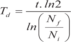
where Td is the doubling time, t is the culture duration, Ni is the initial cell number, and Nf is the final cell number.
Collagen Synthesis Estimation
The amount of collagen production is generally quantified by estimating the hydroxyproline content. Hence, the effect of test samples on collagen production was analyzed using the hydroxyproline assay. 23 Briefly, HDF cells in complete DMEM media were seeded into each well of a 6-well plate at 1 × 10 5 cell/well density and incubated overnight at 37°C and 5% CO2 in a humidified CO2 incubator. Post incubation, 2.5 mL test samples (TLGC_F1 and TLGC_F2) at 2.5 mg/mL concentration and Na Asc were freshly prepared in DMEM complete media and transferred into the respective wells of a 6-well plate containing HDF. The plate was incubated in a CO2 incubator for 72 h. Cell-free media was collected, to which equal volumes of 2 N NaOH were added and autoclaved at 121°C for 20 min. To 20 µL of this mixture, 90 µL of chloramine-T was added and incubated for 25 min at room temperature (RT). Next, 100 µL of Ehrlich’s aldehyde reagent was added, then incubated at 65°C for 20 min. The reaction mixture was measured spectrophotometrically at a wavelength of 550 nm.
The collagen content in the test samples was estimated from the hydroxyproline standard curve (50–250 µg/mL) and Equation (2) given below, and expressed as micrograms of collagen/milliliter (µg/mL).

where
To contextualize the efficacy of PBCB, the TLGC_F1 variant was compared with commercially available marine-based, bovine-based collagen and commercial PBCB booster products. Sodium ascorbate (vitamin C) is used as a positive control. All the samples were prepared at a 2.5 mg/mL concentration. The quantification of collagen synthesis was performed as explained above.
Amino Acid Profiling by HPLC
The amino acid (proline, hydroxyproline, glycine) composition was determined by high-performance liquid chromatography (HPLC) at an accredited third-party analytical laboratory. The TLGC_F1 was hydrolyzed with 6 N HCl, neutralized, and filtered through 0.45 µm membranes, followed by derivatization with ortho-phthalaldehyde (OPA) and fluorenylmethyloxycarbonyl chloride (FMOC-Cl). Primary amino acids were derivatized with OPA, while secondary amino acids (e.g., proline) were derivatized with FMOC-Cl. Chromatographic separation was performed on a C18 reverse-phase column (4.6 × 50 mm, 1.8–5 µm) maintained at 35°C, using a binary gradient of aqueous phosphate buffer (mobile phase A) and an organic solvent mixture of acetonitrile, methanol, and water (45:45:10; mobile phase B). The flow rate was set at 1 mL/min, with an injection volume of 10 µL. Detection was carried out using a diode array detector (DAD) at 338 nm for OPA derivatives and 262 nm for FMOC derivatives, and fluorescence detection (FLD) at excitation/emission wavelengths of 340/450 nm (OPA) and 260/325 nm (FMOC). Quantification was performed using calibration curves generated from amino acid standards, and peak integration was achieved using instrument-specific software.
Quantification of Vitamin C
The concentration of vitamin C in TLGC_F1 was determined by redox titration using a standardized iodine solution. 25 Briefly, 2 g of potassium iodide and 1.3 g of iodine crystals were dissolved in 50 mL of distilled water and diluted to 1 L to obtain a 0.005 mol/L iodine solution. A 5% soluble starch indicator was freshly prepared by dissolving 5 g of starch in 100 mL of boiling distilled water. Diluted sample solutions were titrated with the iodine reagent in the presence of starch until a persistent blue-black color appeared, indicating the endpoint. The reaction involved oxidation of ascorbic acid to dehydroascorbic acid with simultaneous reduction of iodine to iodide. Vitamin C concentration (mg/g sample) was calculated from the mean titration volume of concordant readings using stoichiometric relationships between iodine and ascorbic acid.
Determination of Total Phenolic Content
The total phenolic content TLGC_F1 was determined using the Folin–Ciocalteu method. 26 In brief, 0.1 mL of the TLGC_F1 (2.5 mg/mL) or standard (gallic acid) was mixed with 0.5 mL of 1 mol/L Folin–Ciocalteu reagent, 1.5 mL of 7.5% sodium carbonate (Sigma-Aldrich), and 7.9 mL of distilled water in a test tube. The mixture was thoroughly vortexed and incubated for 2 h in the dark at RT. The absorbance was then recorded at 765 nm using a UV-vis spectrophotometer. The TPC was expressed as milligrams of gallic acid equivalents (mg GAE) per gram of sample.
Quantification of Antioxidant Property
The antioxidant capacity of TLGC_F1 was estimated by DPPH assay. 26 A 0.05 mM DPPH working solution was prepared by diluting 2.5 mL of 10 mM DPPH stock solution with 500 mL of methanol. The stock solution was stored in an amber bottle to protect it from light, as DPPH is photosensitive and readily oxidized. For the assay, 0.1 mL of the sample or standard (vitamin C) was mixed with 2.9 mL of the 0.05 mM DPPH solution and incubated for 30 min at RT in the dark. The absorbance was then measured at 515 nm using a UV-vis spectrophotometer with methanol serving as the blank. The antioxidant activity was expressed as milligrams of ascorbic acid equivalents (mg AAE) per gram of sample.
Short-term Stability Study of the Principal Components of the Formulation
A preliminary stability study was conducted on the TLGC_F1 to evaluate the short-term stability of its key nutritional components. Samples from three production batches were stored under controlled ambient conditions (25°C ± 2°C temperature and relative humidity of 60% ± 5%) for 3 months. At the 3-month interval, the formulations were analyzed for the concentration of vitamin C, glycine, proline, and hydroxyproline using validated HPLC methods. Other physical and microbiological parameters were not evaluated at this stage, as this interim assessment focused solely on monitoring the short-term chemical stability of the principal active components before initiation of a comprehensive long-term stability program.
Statistical Analysis
All experiments were carried out in triplicate. The values are expressed as the mean value with standard deviation. Statistical analysis was carried out by using one-way ANOVA. The p value ≤ .05 was considered statistically significant. The 95% confidence intervals were determined. To account for the large variation between the treatment groups, the data were normalized using natural logarithmic conversion before calculating the effect size. Hedges’ g was calculated to quantify the effect size and assess the magnitude of differences between formulations.
Results
Proximate Composition
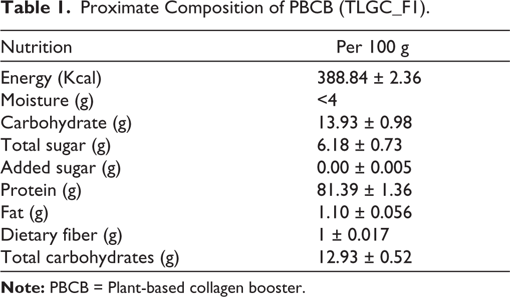
The proximate analysis of the PBCB revealed a nutrient profile dominated by energy content, followed by protein, carbohydrates, net carbs, total sugars, moisture, fat, and dietary fiber (Table 1). Protein, accounting for 81% of the dietary content, is essential for collagen biosynthesis, as collagen constitutes approximately 25% of the total protein in the human body. 4 The fine powder formulation is composed of selected plant extracts blended in specific ratios. Moisture analysis showed levels below 4%, consistent with Sharma et al., 18 who reported 4.02% in similar preparations. 18 Compared to porcine skin (45%), bovine hide (30%), and bones (23%), 27 this plant-only protein formulation offers a more efficient and ethical alternative for promoting endogenous collagen synthesis.
Proximate Composition of PBCB (TLGC_F1).
Safety and Quality Parameters
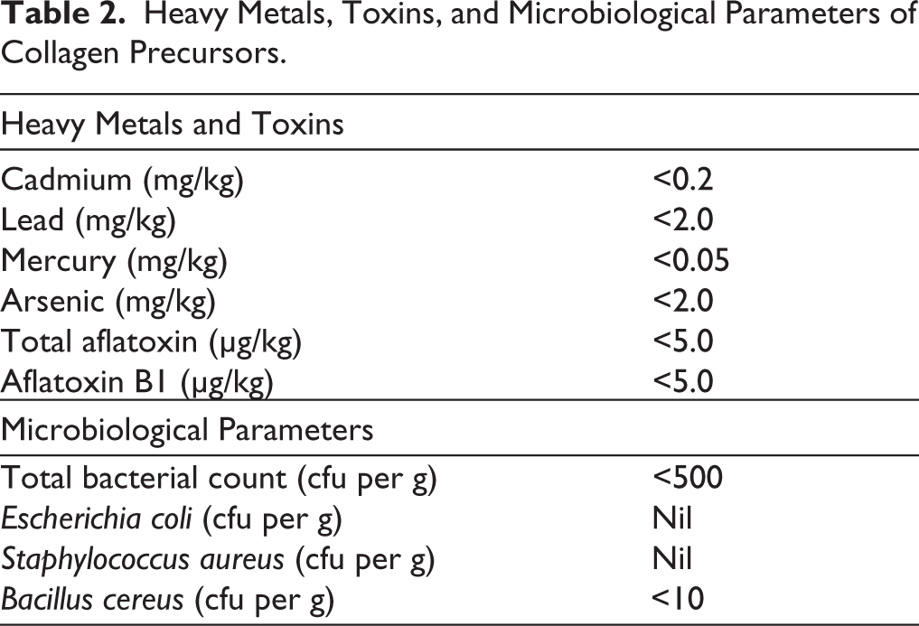
Safety analysis was conducted by evaluating heavy metals (arsenic, cadmium, lead, mercury) and microbial contamination. All tested heavy metal concentrations were within acceptable safety limits (Table 2), and microbial analysis confirmed the absence of E. coli, S. aureus, and B. cereus, indicating high microbiological quality. The total microbial count was comparable to a commercial product, supporting the stability of the formulation, safety, and suitability for consumption. 18
Heavy Metals, Toxins, and Microbiological Parameters of Collagen Precursors.
Cytotoxicity Profiling
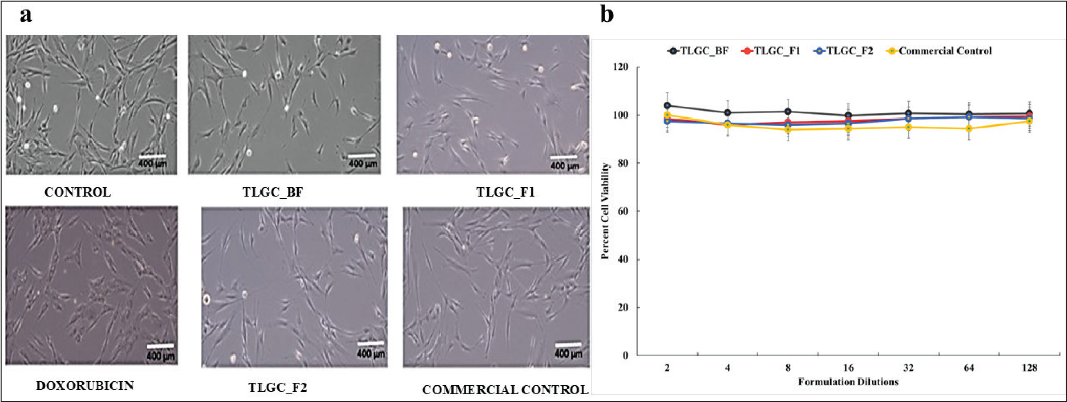
Cytotoxicity assessment in HDF cells demonstrated no adverse effects on cell morphology or viability. The formulation-treated cells showed 90%–94% viability, comparable to the untreated control (90%), even at a 2-fold dilution (Figure 1a and 1b). Cell doubling data indicated normal proliferation rates, with all treatments except doxorubicin (positive control) showing a 3-fold increase in 72 h, consistent with typical HDF doubling times of 18–24 h. Based on this, the 1:2 dilution (2.5 mg/mL) was chosen for further efficacy studies. Since TLGC_BF was only the base formulation used in the preparation of TLGC_F1 and TLGC_F2, it was not taken for further analysis.
(a) Human Dermal Fibroblast Cells Under the Microscope (at 10× Magnification) After Treatment with Formulations and Doxorubicin at a Scale of 400 µm. (b) HDF Viability in Various TLGC-formulations at Different Dilutions Compared to Commercial Products. The Error Bars Represent the Standard Deviation. HDF: Human Dermal Fibroblast.
Collagen Estimation (Hydroxyproline) Assay and Comparative Study
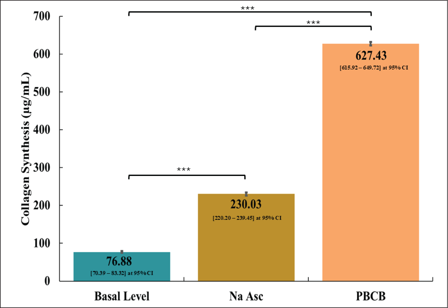
Collagen synthesis was assessed using a hydroxyproline assay. Sodium ascorbate (Na Asc, vitamin C), a known cofactor in collagen biosynthesis, served as the positive control. The formulation significantly enhanced collagen production (627 ± 5 µg/mL) compared to Na Asc (230 ± 4 µg/mL), with a Hedges’ g value of 3 and statistical significance at p = .0095 (Figure 2). Interestingly, increased Na Asc concentration did not further improve collagen synthesis, suggesting saturation of its biosynthetic role. The untreated cells (negative control), which served as the baseline, exhibited 77 μg/mL for collagen synthesis. As TLGC_F1 and TLGC_F2 exhibited comparable outcomes, only the data for TLGC_F1 are presented herein.
Collagen Synthesis Quantification in HDF Cells After the Treatment With PBCB Formulation at 95% Confidence Interval. Na Asc (Sodium Ascorbate) is a Vitamin C Positive Control. The Error Bars Represent the Standard Deviation. Statistical Significance is Indicated as *, with Exact Pairwise p values as Follows: Basal vs. Na Asc, p = .0008; Na Asc vs. PBCB, p = .0005; Basal vs. PBCB, p = .0001. HDF: Human Dermal Fibroblast; PBCB: Plant-based Collagen Booster.
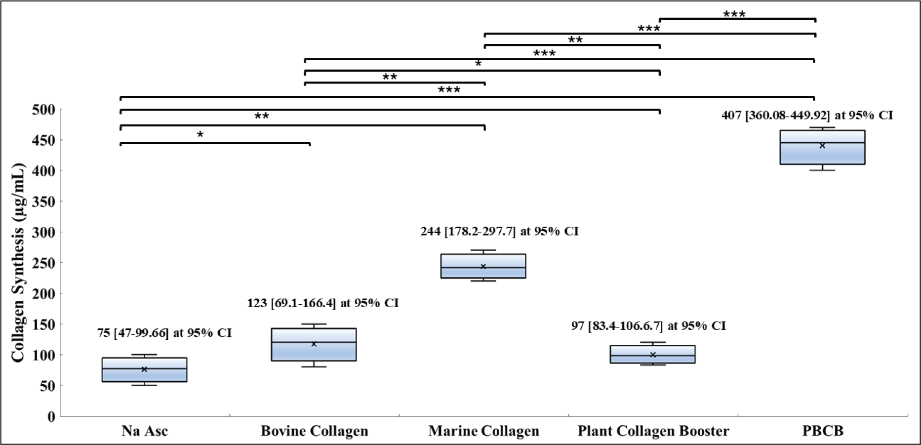
To benchmark efficacy, the TLGC_F1 was compared against commercial animal-based (bovine and marine collagen) and PBCB products. The commercial bovine collagen product showed a 1.64-fold increase in collagen synthesis (123 ± 15 µg/mL), the commercial marine collagen product showed a 3.25-fold increase (244 ± 65 µg/mL), and the commercial PBCB product showed a 1.29-fold increase (97 ± 14 µg/mL). In contrast, the novel formulation, PBCB (TLGC_F1), exhibited a 5.42-fold increase (407 ± 76 µg/mL) over the control (Figure 3). Thus, demonstrating that the novel PBCB formulation outperformed bovine and marine commercial products by 3.24 and 1.66 times, respectively and 4.19 times better than the commercial PBCB product. The differences were statistically significant (p = .0073), with Hedges’ g values of 2 (commercial bovine collagen product vs. TLGC_F1), 1 (commercial marine collagen product vs. TLGC_F1) and 4 (commercial PBCB vs. TLGC_F1), indicating a good effect size in comparison to other products compared. The animal-based collagen aids in restoring skin health either by directly supplying the collagen or via stimulation of collagen biosynthesis by collagen peptides. 28 Unlike animal-based collagen, PBCB not only trigger intrinsic collagen synthesis but also prevents collagen degradation by reducing inflammation, oxidative stress and inhibiting MMPs.10,29
Comparison of Collagen Synthesis Among Different Commercial Products (Bovine, Marine or PBCB) Against PBCB (TLGC_F1) at 95% Confidence Interval. Na Asc (Sodium Ascorbate) is a Vitamin C Positive Control. Statistical Significance is Indicated as *, with Exact Pairwise p Values as Follows: Na Asc vs. Bovine Collagen, p = .0418; Na Asc vs. Marine Collagen, p = .0038; Na Asc vs. Commercial PBCB, p = .075; Na Asc vs. PBCB (TLGC_F1), p < .0001; Bovine Collagen vs. Marine Collagen, p = .0027; Bovine Collagen vs. Commercial PBCB, p = .0491; Bovine Collagen vs. PBCB (TLGC_F1), p = .0006; Marine Collagen vs. Commercial PBCB, p = .0073; Marine Collagen vs. PBCB (TLGC_F1), p = .0009; and Commercial PBCB vs. PBCB (TLGC_F1), p = .0002. PBCB: Plant-based Collagen Booster.
Amino Acids, Vitamin C, Phenol Content and Antioxidant Property of the Formulation with Stability
Based on the chromatographic analysis, the amino acid profiling of TLGC_F1 revealed the presence of 800 ± 15 mg of glycine per gram of sample, while proline and hydroxyproline were quantified at 9 ± 2 mg/g and 30 ± 5 mg/g of sample, respectively. The vitamin C content, determined by the redox titration method, was found to be 3 ± 0.05 mg/g of sample. Furthermore, the total phenolic content of TLGC_F1, measured using the Folin–Ciocalteu assay, was 75 ± 3 mg GAE/g of sample, which corresponded to an antioxidant activity equivalent to 141 ± 18 mg AAE per gram of sample.
After 3 months of storage at 25°C ± 2°C and 60% ± 5% RH, the glycine content increased slightly from 800 ± 36 mg/g at day 0 to 821 ± 13 mg/g at 3 months. The slight increase in glycine content after 3 months is within the expected range of analytical variability and may additionally reflect minor changes in moisture content during storage, rather than indicating a true increase in glycine concentration. Proline and hydroxyproline concentrations were determined as 8.26 ± 1 and 33.5 ± 3 mg/g, respectively, at the end of 3 months. The vitamin C content, measured by the redox titration method, showed minimal change, increasing marginally from 3 ± 0.05 mg/g at day 0 to 3.06 ± 0.1 mg/g after 3 months. Overall, these results indicate that the key nutritional components of the formulation maintained their chemical stability over the 3-month evaluation period under controlled storage conditions.
Discussion
Traditional collagen peptides face limitations in bioavailability, as they are digested before systemic absorption and must be broken down to amino acids to be functional.30,31 This underscores the rationale for delivering amino acids directly, which are fundamental to collagen biosynthesis. Vitamin C enhances proline and lysine hydroxylation, stabilizing the collagen structure,32,33 while plant extracts rich in antioxidants and polyphenols, such as Centella asiatica, Aloe vera, and green tea, protect against oxidative stress, support fibroblast activity, and promote ECM regeneration.34–36 Furthermore, the plant extracts used in the formulation of PBCB are all well-studied for their antioxidant and anti-inflammatory properties. For instance, Baobab powder has anti-inflammatory activity and is reported to increase collagen due to its high vitamin C content. 37 Similarly, Acerola cherry, which is a rich source of vitamins, minerals, and bioactive compounds such as carotenoids and phenolic compounds, shows anti-aging, anti-inflammatory and antioxidant mechanisms. 38 Goji berries are a rich source of amino acids, among which proline and serine constitute about 30%. 39 Thus, PBCB formulation is proposed to synergistically modulate collagen biosynthesis pathways by neutralizing ROS, inhibiting collagen-degrading enzymes,10,29 thereby addressing multiple mechanisms involved in skin aging. This includes UV-induced inflammation, lipid peroxidation, and COX-2-driven matrix degradation. The synergy among the components offers a holistic, bioavailable, and sustainable solution for skin health. 40
Clinically, oral collagen or collagen booster supplements have been shown to improve skin hydration, elasticity and the prevention of wrinkles, thus effective against skin aging. For example, a double-blinded, placebo-controlled, randomized trial showed that the plant-based vegan collagen can increase collagen density and elasticity by 4.7% and 5.1% and decreased wrinkles, texture, and pores by 27.5%, 20.1%, and 12.3% compared to the placebo group. 15 In another trial, absolute collagen with vitamin C has shown clinical benefits for skin, hair and scalp. 41 Low-molecular-weight collagen peptides improved elasticity, hydration, and wrinkles in photoaged and dry skin individuals in 12 weeks. 42 However, the present findings are limited by the preliminary in vitro nature of the study. In vitro assays cannot account for factors such as bioavailability, metabolism, systemic interactions, or potential off-target effects, and the concentrations applied may not directly correspond to physiologically achievable levels. This study is limited by the absence of quantitative protein-level validation, such as Western blot or ELISA analyses for Type I and Type III collagen, which are necessary to directly confirm collagen modulation. Additionally, a dose-response evaluation using multiple concentrations was not performed, which restricts conclusions regarding concentration-dependent biological effects. We acknowledge that such a detailed stability assay, dose-response analysis and gene/protein-level expression studies would provide additional mechanistic insight into the effects of the formulation. Therefore, extensive molecular analysis and in vivo studies are planned to evaluate the pharmacokinetics, safety, and tissue-level responses of the formulation in subsequent investigations. Well-designed randomized clinical trials are also essential to confirm their efficacy on clinically relevant endpoints such as skin elasticity, hydration, and wrinkle reduction. Together, these studies will provide a more comprehensive understanding of the translational potential of the formulation.
Conclusion
PBCB significantly enhanced collagen synthesis in vitro and outperformed commercial marine and bovine collagen products. Its plant-based composition, free from toxic contaminants and microbiologically safe, positions it as a promising candidate for use in cosmeceutical and functional food applications. These findings are supported by existing evidence that individual amino acids may outperform hydrolyzed peptides in supporting collagen synthesis. 23 While the in vitro data are promising, we propose conducting in vivo studies to further validate the mechanistic findings, followed by well-designed randomized, double-blind, placebo-controlled clinical trials with objective skin health endpoints (e.g., elasticity, hydration, and wrinkle assessment) to establish translational relevance.
Footnotes
Acknowledgements
The authors acknowledge the management of Bangalore Bio Innovation Center, Bangalore, for extending support to the use of the cell culture facility.
Authors’ Contribution
All authors made substantial contributions to conception and design, acquisition of data, or analysis and interpretation of data; took part in drafting the article or revising it critically for important intellectual content; agreed to submit to the current journal; gave final approval of the version to be published; and agree to be accountable for all aspects of the work. All the authors are eligible to be author as per the International Committee of Medical Journal Editors (ICMJE) requirements/guidelines.
Consent to Participate
Not applicable.
Consent for Publication
Not applicable.
Data Availability Statement
All the data is available with the authors and shall be provided upon request.
Declaration of Conflicting Interests
The authors declared the following potential conflicts of interest with respect to the research, authorship, and/or publication of this article: All authors are employees of DE3PBIO, the company that developed the formulation and holds the associated intellectual property rights. DE3PBIO provided institutional support for this study, including research infrastructure, materials, facilities, and salary support for the authors as part of their regular employment. The author-employees were involved in the study design, execution of experiments, data collection, analysis, interpretation of results, and preparation of the manuscript. No external or third-party funding was received. The authors declare that they have no personal financial interests, equity holdings, or individual patent ownership related to this product beyond their employment and institutional affiliation with DE3PBIO.
Ethical Approval
Human dermal fibroblasts (HDFs) were purchased from Lonza (CC-2511). These cells were obtained by the supplier following informed donor consent and in compliance with applicable ethical regulations. The cells were de-identified and commercially available; therefore, their use in this study did not require additional institutional ethics committee approval.
Funding
The authors received no financial support for the research, authorship, and/or publication of this article.
Informed Consent
Informed donor consent details are given in the ethical approvals section.
Use of Artificial Intelligence-assisted Tools:
The authors declare that they have not used artificial intelligence (AI)-tools for writing and editing of the manuscript, and no images were manipulated using AI.