
Abstract
Developing safe and effective fibrinolytic enzymes for oral delivery is still a major challenge and concern in translational biotechnology. Although microbial proteases have gained increasing attention, the in vivo evidence of their systemic safety after oral administration remains limited. This study was designed to evaluate the systemic biocompatibility of a spray-dried microencapsulated fibrinolytic protease derived from Bacillus tequilensis Holothuria scabra Fermented Intestine 5 (HSFI-5) following repeated oral administration in mice. The strain was isolated from the fermented intestine of a sea cucumber captured from the seawaters of Nusa Tenggara, Indonesia. The extracellular enzyme from B. tequilensis HSFI-5 was partially purified and subsequently encapsulated via spray-drying using either maltodextrin (M) or Arabic gum (AG) as wall materials. Proteolytic activity was retained after microencapsulation (0.26–0.28 U/mL; ~5.52 units per milligram of protein [U/mg]). Thirty male C3H mice (n = 6 per group) received daily oral administration for four weeks. No significant differences were observed in body weight (BW), lipid profile, or endpoint inflammatory markers (IL–6, IL–1β, and C-reactive protein [CRP]) between treated and control groups (p > 0.05), although ΔIL-1β (change from baseline) showed an isolated statistical difference (p = .047). Hematological test results showed no consistent patterns suggestive of toxicity. Statistical analysis using one-way and two-way Analysis of Variance ( ANOVA) confirmed the absence of treatment-related effects (p > 0.05). In conclusion, the repeated oral administration of the tested microencapsulated protease formulation did not induce detectable systemic inflammatory or metabolic disturbances under the conditions of this study. This work provides initial in vivo evidence supporting the biological compatibility of orally delivered microbial protease formulations and contributes to the development of marine-derived bioactive enzymes for biotechnological applications.
Introduction
The need for effective antithrombotic agents continues to grow, driven by the global burden of thrombotic cardiovascular diseases. Proteolytic enzymes that degrade fibrin have emerged as promising candidates for thrombolytic and antithrombotic therapy. Well-known fibrinolytic enzymes such as nattokinase, urokinase, and lumbrokinase have demonstrated significant potential for promoting clot degradation and enhancing fibrinolytic activity. However, their production and purification remain costly, limiting broader accessibility, and encouraging the exploration of alternative microbial proteases with comparable biological activity.1–3
The development of stable oral enzyme formulations represents an important challenge in applied biochemistry and bioprocess engineering. Proteolytic enzymes may influence coagulation pathways, inflammatory signaling, and metabolic homeostasis, raising concerns regarding potential systemic effects during sustained administration. 4 Consequently, evaluating the physiological compatibility of emerging fibrinolytic enzymes is essential before considering their biomedical application. This concern is supported by reports from several widely used thrombolytic agents. For example, streptokinase and staphylokinase, two bacterial plasminogen activators widely investigated for thrombolytic therapy, are associated with immunogenic reactions and systemic fibrinolysis, which may increase the risk of hemorrhagic complications. 5 In addition, commonly used anticoagulant agents such as heparin act through relatively non-specific mechanisms and may lead to adverse outcomes, including excessive bleeding and heparin-induced thrombocytopenia. These limitations highlight the importance of identifying alternative fibrinolytic enzymes with improved physiological compatibility and reduced systemic adverse responses.5–9
Recent studies have reported that several Bacillus species, including Bacillus tequilensis, produce extracellular proteases with fibrinolytic and anticoagulant activity. These enzymes demonstrate promising catalytic properties and potential applications in thrombolytic biotechnology. However, most previous investigations have focused primarily on enzyme isolation, biochemical characterization, and in vitro activity assays.3,10–15 To date, there is no evidence that proteases derived from B. tequilensis have been developed or commercialized as antithrombotic agents. Furthermore, studies evaluating formulation strategies enabling oral delivery, such as microencapsulation, and experimental evidence describing systemic physiological compatibility following repeated exposure remain extremely limited.
Given the physiological limitations reported for several bacterial fibrinolytic agents, it is still important to determine whether proteases derived from B. tequilensis may trigger comparable adverse responses, such as gastrointestinal bleeding or inflammatory reactions. This is particularly true when administered repeatedly through the oral route. Proteases produced by Bacillus species exhibit broad catalytic activity and established biomedical relevance. A marine-derived strain, B. tequilensis Holothuria scabra Fermented Intestine 5 (HSFI-5), isolated from fermented sea cucumber, produces a fibrinolytic protease with reported thrombolytic activity.1,2 The sea cucumber was captured from the seawaters of Nusa Tenggara, Indonesia. The bacterial origin allows the classification of the enzyme produced in this study as marine-derived and bioactive. Although its enzymatic properties have been characterized, its safety profile following oral administration has not been established (Table 1). As shown in Table 1, although fibrinolytic enzymes from several Bacillus species have been extensively investigated, studies involving B. tequilensis remain largely limited to enzyme discovery and catalytic characterization.3,15 Translational studies addressing formulation development, systemic safety evaluation, and potential biomedical applications are still scarce.
Comparison of Bacterial Fibrinolytic Proteases Developed as Antithrombotic Agents from Different Bacillus Species.
This knowledge gap is particularly significant because the successful development of microbial fibrinolytic enzymes as antithrombotic agents requires not only catalytic efficacy but also evidence of physiological compatibility during sustained exposure. Despite the growing interest in Bacillus-derived proteases, experimental data describing the systemic safety of B. tequilensis protease formulations, especially following repeated oral administration, remain largely unavailable. Addressing this gap is essential to determine whether such enzymes can be safely advanced toward biomedical or biotechnological applications.
Oral delivery of protein enzymes poses additional challenges, including gastrointestinal degradation, structural modifications during processing, and potential host responses following repeated exposure. 1 Unlike plant-derived or digestive enzymes, bacterial fibrinolytic proteases have been rarely evaluated in oral formulations, leaving limited evidence regarding their systemic safety.11,12,16
Microencapsulation has been proposed as a strategy to enhance enzyme stability and regulate gastrointestinal exposure. Encapsulation matrices such as maltodextrin (M) and Arabic gum (AG) can preserve enzymatic activity while protecting proteins from environmental stress.17–19 However, data addressing systemic tolerance to orally administered microencapsulated microbial proteases remain scarce.
Systemic safety assessment is therefore an essential step in the biological evaluation of orally delivered proteases, particularly those with potential effects on coagulation pathways. Elevations in inflammatory biomarkers such as interleukin-6 (IL-6), interleukin-1β (IL-1β), and C-reactive protein (CRP), as well as alterations in lipid profile or body weight (BW), may indicate adverse physiological responses during sustained exposure.20,21
Therefore, this study evaluated the systemic effects of repeated oral administration of a microencapsulated extracellular protease derived from B. tequilensis HSFI-5. Hematological indices, lipid parameters, and inflammatory biomarkers (IL-6, IL-1β, and CRP) were assessed to determine potential toxicological effects following sustained exposure. By integrating enzyme semi-purification, scalable spray-drying microencapsulation, and biological compatibility evaluation, this study aims to advance the development of marine-derived protease formulations for applied biotechnology.
Materials and Methods
Sub-culture
B. tequilensis HSFI-5 was used in this study. The HSFI-5 culture was obtained from the Microbiology Laboratory at Universitas Muhammadiyah Semarang, Indonesia. Colonies were rejuvenated in Nutrient Broth (NB) medium, and morphology was assessed by Gram staining and microscopy, confirming Gram-positive rod-shaped bacterial cells consistent with Bacillus species (see Supplementary Figure S2). A confirmation test for extracellular fibrinolytic protease production by HSFI-5 was conducted as previously reported.3,4
Preparation of Extracellular Crude Fibrinolytic Protease
Following bacterial sub-culture, B. tequilensis HSFI-5 was cultivated for extracellular fibrinolytic protease production under previously established conditions as described in previous studies.3,13,14 After incubation, the culture broth was centrifuged at 8,000–10,000 × g for 15–20 minutes at 4 °C to remove bacterial cells and debris. The resulting cell-free supernatant was collected as a crude extracellular protease extract.13,14
The cell-free supernatant was concentrated and partially purified by ammonium sulfate precipitation, as previously reported.13,14 The resulting pellet was resuspended in buffer and dialyzed to remove residual salts. This preparation, which retained fibrinolytic activity while reducing low-molecular-weight impurities, 14 was used as the active core material for microencapsulation. Proteolytic activity and protein concentration were confirmed using caseinolytic and Bradford assays, respectively. In addition to total proteolytic activity (U/mL), the specific activity of the enzyme was calculated and expressed as units per milligram of protein (U/mg) based on protein concentration. The concentration was determined using the Bradford assay, providing an indicator of enzyme purity following partial purification. 1
Microencapsulation of Fibrinolytic Protease
Semi-purified fibrinolytic protease from B. tequilensis HSFI-5 was spray-dried with M and AG at 5% w/v each under previously optimized conditions.17–19 The spray-drying parameters were as follows: Inlet temperature, 110 °C–130 °C; outlet temperature, 60 °C–70 °C; and feed rate, 5–7 mL min⁻ 1 . 17 Spray-drying was conducted using a BÜCHI Mini Spray Dryer S-300 (BÜCHI Labortechnik AG, Flawil, Switzerland).
Before in vivo administration, the microencapsulated formulations were evaluated for retained proteolytic activity using a caseinolytic assay. M 5% microcapsules retained approximately 0.28 U/mL of activity, while AG 5% formulations showed slightly lower, yet still substantial, activity retention (~0.26 U/mL), confirming that the encapsulation process preserved enzyme function. This also reflects that M provided slightly better protection of enzymatic activity during spray-drying compared to AG, although both matrices demonstrated comparable preservation of enzyme functionality.
Experimental Animals and Study Design
The study was conducted as a preclinical repeated-dose oral safety evaluation, selected to represent escalating exposure levels within a practical oral formulation range. The selected doses (150 and 200 mg/kg BW) were determined based on preliminary range-finding considerations. They were intended to represent moderate-to-high exposure levels in repeated oral safety evaluations of enzyme-based formulations. These dose levels were chosen to ensure sufficient systemic exposure while remaining within physiologically tolerable limits during repeated administration.
Animals were maintained in accordance with standard welfare guidelines and monitored daily for behavioral and clinical signs of adverse effects. Representative images of experimental animals and oral administration procedures are provided in Supplementary Figure S1 to enhance transparency of the in vivo experimental workflow.
Thirty male C3H mice (one month old) were randomly allocated to experimental groups using a simple randomization method based on computer-generated random numbers. The animals were assigned to five groups: Negative control (NC), M-microencapsulated protease (150 or 200 mg/kg BW), and AG-microencapsulated protease (150 or 200 mg/kg BW). The NC group did not receive empty microcapsules; therefore, potential matrix-only effects were not independently evaluated in this preliminary study. Given the established safety profiles of M and AG, commonly used excipients, matrix-only effects were considered unlikely to confound systemic outcomes under the tested conditions. The NC group received no treatment because matrix-only controls were not included, and carrier-specific effects cannot be excluded.22,23
A group size of six animals per group was selected in accordance with standard practice in preliminary preclinical biocompatibility studies, where moderate effect sizes can be detected while minimizing animal use in accordance with ethical principles. Doses represent total microcapsule mass. Microcapsules were administered orally once daily for four weeks. Animals were housed under standard laboratory conditions (22 °C ± 2 °C, 12-hour light/dark cycle) with ad libitum access to standard pellet feed and water. Animals were monitored daily for systemic tolerance following sustained exposure. Male mice were selected to minimize hormonal variability that may influence inflammatory biomarker responses during repeated-dose evaluation. 23
Body Weight and Routine Blood Tests
BW was monitored weekly. Blood was collected at study completion and at 24 and 48 hours after the final dose. Samples were analyzed using an automated hematology analyzer (Sysmex KX-21). Parameters included erythrocyte indices, leukocyte distribution, and platelet markers. Measured parameters included erythrocyte indices (red blood cell count [RBC], hemoglobin concentration [HGB], Hematocrit [HCT], mean corpuscular volume [MCV], Mean corpuscular hemoglobin [MCH], Mean corpuscular hemoglobin concentration [MCHC]), leukocyte profiles (white blood cell count [WBC], lymphocyte percentage [LYM%], neutrophil percentage [NEUT%], absolute lymphocyte count [LYM#], and absolute neutrophil count [NEUT#], and platelet parameters (platelet count [PLT], platelet distribution width [PDW], mean platelet volume [MPV], and platelet large cell ratio [P-LCR]). Platelet-related parameters included PLT and PDW at both observation time points, whereas MPV and P-LCR were available only for the 24-hour assessment. Analyses were conducted according to the manufacturer’s standard procedures to assess hematological compatibility and potential systemic effects. 24
Lipid Profile Analysis
Lipid profiles, including total cholesterol, high-density lipoprotein (HDL), low-density lipoprotein (LDL), and triglycerides, were measured pre- and post-treatment using standard biochemical assays. 25
Inflammatory Marker Assessment
Serum IL-6, IL-1β, and CRP concentrations, as inflammation biomarkers, 26 were quantified in mice using Enzyme-Linked Immunosorbent Assay (ELISA) kits according to the manufacturer’s protocols (Abbkine cat. no. KTE7009 and KTE7005; Elabscience E-cat. no. EL-M0053) using a Bio-Rad ELISA reader.
Data and Statistical Analysis
Hematological subsets (n = 3 per group) were analyzed descriptively, whereas lipid and cytokine datasets (n = 6 per group) were subjected to inferential analysis. These hematological subsets were analyzed descriptively rather than inferentially, as the limited sample size does not provide sufficient statistical power for robust hypothesis testing. Therefore, interpretation focused on identifying consistent trends or coordinated deviations indicative of potential toxicological effects. It emphasized potential dose-dependent or coordinated pathological patterns rather than statistical significance testing.
Given the study’s safety-oriented objective, statistical analysis was conducted to detect treatment-related deviations in physiological parameters rather than to assess therapeutic efficacy. Laboratory personnel performing biochemical and hematological analyses were not blinded to group allocation; however, automated analytical instruments were used to minimize operator bias.
Data organization and graphical visualization were performed using Python (version 3.10; Python Software Foundation) employing the pandas and matplotlib libraries for data handling and figure generation. 27 Data are displayed as mean ± SD. A one-way Analysis of Variance (ANOVA) was used to evaluate potential differences among experimental groups (p < 0.05). For BW progression, a two-way ANOVA (treatment × time) was performed.
Results
Previous investigations of fibrinolytic proteases have primarily focused on in vitro activity or parenteral delivery. Data regarding oral exposure are still limited. This study evaluated spray-dried microencapsulation of a fibrinolytic protease and its short-term oral biocompatibility in mice. The aim was to assess the feasibility and biological tolerance of an orally deliverable fibrinolytic protease derived from B. tequilensis HSFI-5.
Confirmation of Fibrinolytic Activity and Microcapsule Formation
Fibrinolytic activity was confirmed using a fibrin plate assay before formulation. After ammonium sulfate precipitation and dialysis, the enzyme was successfully spray-dried with M or AG. Proteolytic activity was retained following encapsulation (0.26–0.28 U/mL). A slight difference in retained activity was observed among the encapsulating agents, with M showing marginally higher retention than AG. However, the difference was not substantial, implying that both matrices were effective in preserving enzyme functionality. The corresponding specific activity was approximately 5.52 U/mg protein. This may indicate that catalytic efficiency per unit protein was preserved following partial purification and spray-drying encapsulation. The consistency of specific activity values across samples suggests that the encapsulation process did not alter the intrinsic enzymatic properties. These data indicate that enzymatic activity was preserved after spray-drying and that the formulation remained biologically stable under the tested conditions. Powder recovery confirmed successful microcapsule formation suitable for in vivo testing.
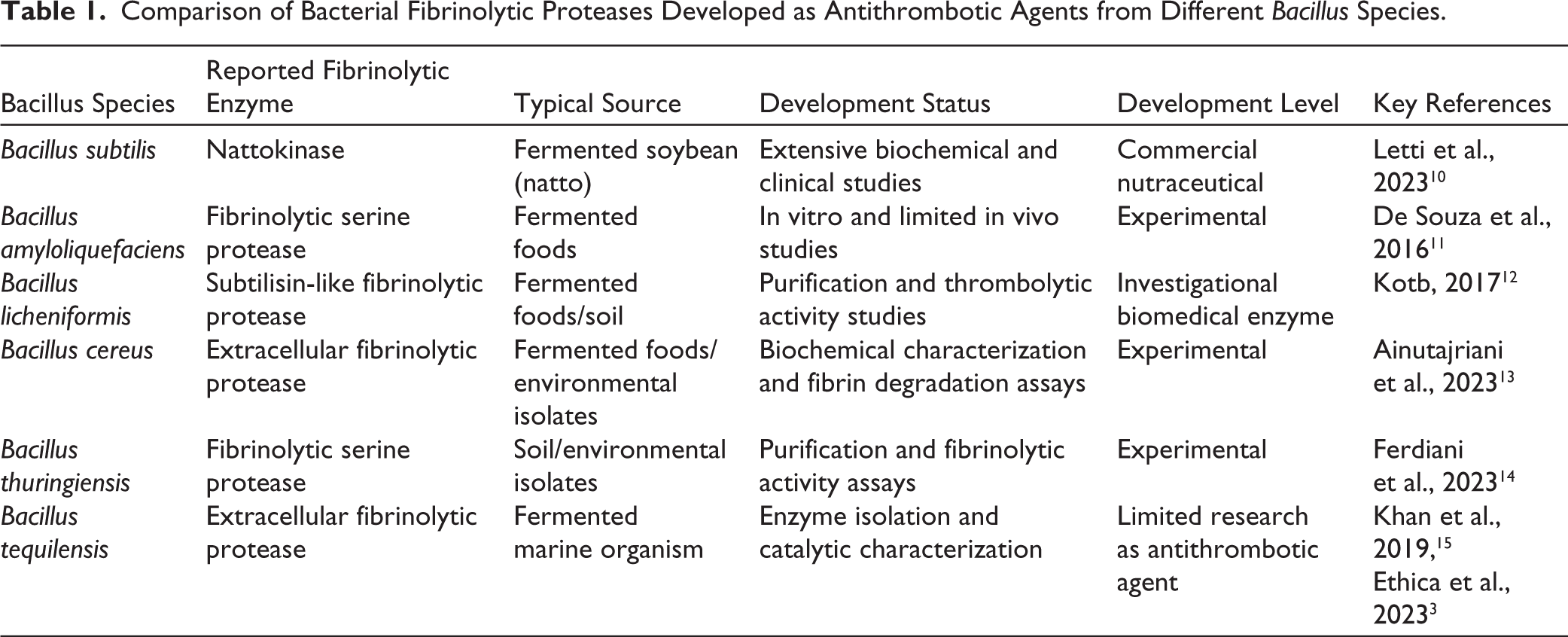
Representative photographs of the recovered spray-dried powders are shown in Figure 1 to qualitatively confirm successful microcapsule formation before in vivo administration. Although detailed morphological characterization is reported separately, preliminary observations verified the formation of discrete microcapsules with diameters predominantly within the expected 1–10 µm range, consistent with stable spray-drying encapsulation. These results confirm the successful preparation of an active, structurally suitable enzyme formulation for subsequent biocompatibility evaluation.
Representative Photographic Documentation of Microcapsules Produced from Bacillus tequilensis HSFI-5 Extracellular Protease Using M (left) and AG (right) as Encapsulating Matrices (5% w/v). Differences in Powder Texture and Aggregation Behavior Reflect Carrier-dependent Physicochemical Properties Following Spray-drying. The Images Provide Qualitative Confirmation of Successful Powder Recovery Before Biological Evaluation.
Body Weight Evaluation
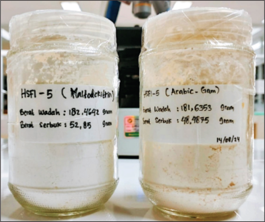
Table 2 shows the BW trajectories of C3H mice receiving microencapsulated extracellular protease. As shown in Table 2, all experimental groups exhibited gradual physiological increases in BW over the four-week study period. No excessive or abnormal weight gain was observed in mice receiving either M- or AG-microencapsulated protease compared with the NC. The data indicate that oral administration of microencapsulated protease did not induce abnormal weight gain or signs of metabolic imbalance.
Weekly BW of C3H Mice (g) Across Experimental Groups.
All animals remained clinically normal throughout the experimental period, with no observable signs of distress, behavioral abnormalities, or treatment-related adverse effects. BW increased progressively across all groups during the four-week observation period, reflecting normal physiological growth. Comparable weight gain trajectories were observed between the NC and treatment groups (M and AG), indicating that oral administration of microencapsulated fibrinolytic protease did not interfere with general metabolic status or feeding behavior (Table 2). Two-factor analysis, including treatment and time, showed no significant effect of treatment on BW progression (p > 0.05).
Hematological Parameters at 24 and 48 Hours Post-administration
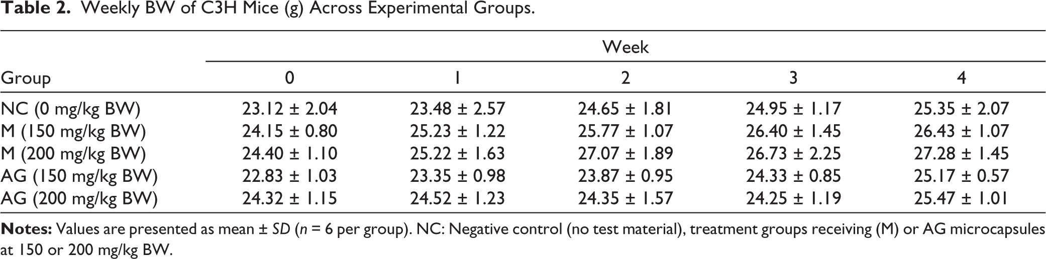
Hematological parameters evaluated 24 hours after the final administration following repeated dosing of microencapsulated B. tequilensis HSFI-5 protease are shown in Table 3. Erythrocyte indices, including RBC, HGB, HCT, MCV, MCH, and MCHC, remained within expected murine physiological ranges across all treatment groups. Although minor inter-group variability was observed, no coordinated pattern indicative of hemolysis, acute anemia, or impaired erythropoiesis was detected. Due to the limited number of samples available for hematological analysis (n = 3 per group at each time point), the data are presented descriptively and were not subjected to inferential statistical testing.
Hematological Parameters of C3H Mice 24 Hours After the Final Administration Following Repeated Dosing of Microencapsulated Bacillus tequilensis HSFI-5 Protease.
Leukocyte parameters demonstrated variability typical of short-term immune redistribution following oral exposure. In particular, relative shifts between lymphocyte and neutrophil percentages were observed in some groups; however, these changes were not accompanied by consistent alterations in absolute leukocyte counts, suggesting physiological fluctuation rather than acute inflammatory activation.
Platelet-related indices (PLT, PDW, MPV, and P-LCR) likewise remained within the range of biological variability. While individual values differed among groups, no coherent trend suggestive of thrombocytopenia, platelet activation, or coagulation disturbance was evident at this early time point. Overall, the 24-hour hematological profile supports short-term systemic compatibility of the microencapsulated protease formulations under the experimental conditions tested.
Hematological profiles evaluated 48 hours after the final dose following four weeks of repeated oral administration are presented in Table 4. Erythrocyte indices (RBC, HGB, HCT, MCV, MCH, and MCHC) remained within expected murine physiological ranges across all treatment groups.
Hematological Parameters of Mice at 48 Hours After the Final Administration Following Repeated Dosing of Microencapsulated Bacillus tequilensis HSFI-5 Protease.
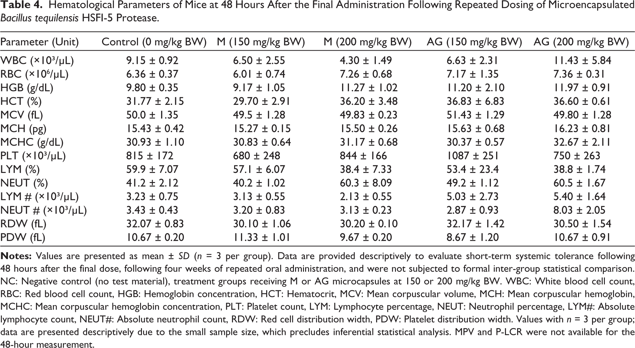
Although mean RBC, HGB, and HCT values were numerically higher in certain treatment groups, all values remained within established physiological ranges for murine hematological parameters. These variations were not accompanied by consistent dose-dependent trends or parallel changes in related hematological indices, indicating that they do not reflect pathological erythrocytosis or hemoconcentration but rather normal biological variability. Isolated increases in erythrocyte-related parameters (RBC, HGB, HCT) are commonly observed in small-animal cohorts and are generally considered physiologically normal when not accompanied by coordinated pathological changes. Under the conditions tested, the absence of consistent trends across groups further supports the interpretation that these results are not treatment-related.
As shown in Table 4, leukocyte distributions varied between groups. Relative increases in neutrophil percentage were observed in higher dose groups (M 200 mg/kg and AG 200 mg/kg); however, corresponding absolute neutrophil counts did not show a consistent treatment-related escalation. Similarly, lymphocyte percentages varied without corresponding changes in absolute counts, suggesting physiological variability rather than sustained immune activation. Platelet counts and related indices (PLT, PDW) showed inter-group variability but no coherent trend indicating thrombocytopenia, platelet activation, or coagulation disturbance. MPV and P-LCR measurements were available only at the 24-hour observation point and are therefore not presented in Table 4. Due to the limited sample size (n = 3), hematological test results are presented descriptively and require confirmation in larger studies.
Overall, group-wise hematological evaluation at both 24 and 48 hours post-administration did not reveal coordinated suppression of erythrocyte indices or consistent leukocyte expansion patterns suggestive of acute inflammatory toxicity. Although numerical variations were observed across groups, particularly in neutrophil distribution at higher doses, these were not accompanied by parallel pathological shifts in absolute counts or platelet parameters. Nevertheless, the limited sample size per subgroup restricts statistical inference and underscores the need for larger-scale toxicological assessment in future studies.
Serum Lipid Profiles
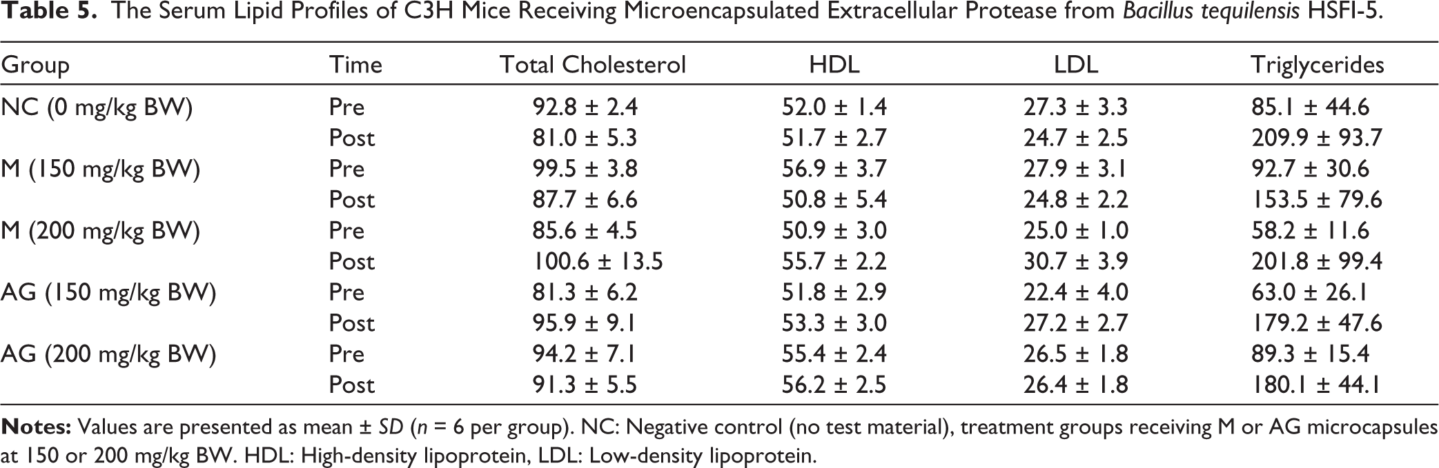
To evaluate potential metabolic disturbances, lipid profiles were assessed before and after treatment, including total cholesterol, HDL, LDL, and triglycerides. Table 5 displays the serum lipid profiles of C3H mice receiving microencapsulated extracellular protease from B. tequilensis HSFI-5. As seen in Table 5, after treatment, there was variation among individuals in all groups, including the NC. Triglyceride levels increased across several groups, including the control group, indicating that the variation was unlikely to be treatment-related. There was no clear pattern of dyslipidemia linked to M- or AG-microencapsulated protease. Total cholesterol, HDL, and LDL levels stayed about the same in all groups. These results suggest that the tested formulations did not affect lipid metabolism during the study.
Across all treatment groups, post-treatment lipid parameters remained within physiological ranges and showed patterns similar to those of the NC group. One-way ANOVA revealed no statistically significant differences among groups for total cholesterol, HDL, LDL, or triglycerides (p > 0.05).
The Serum Lipid Profiles of C3H Mice Receiving Microencapsulated Extracellular Protease from Bacillus tequilensis HSFI-5.
Systemic Inflammatory Markers
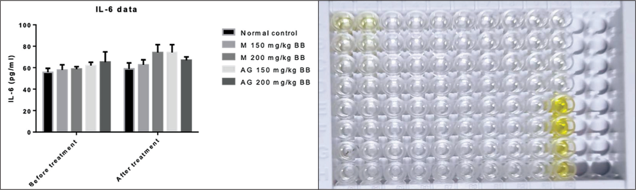
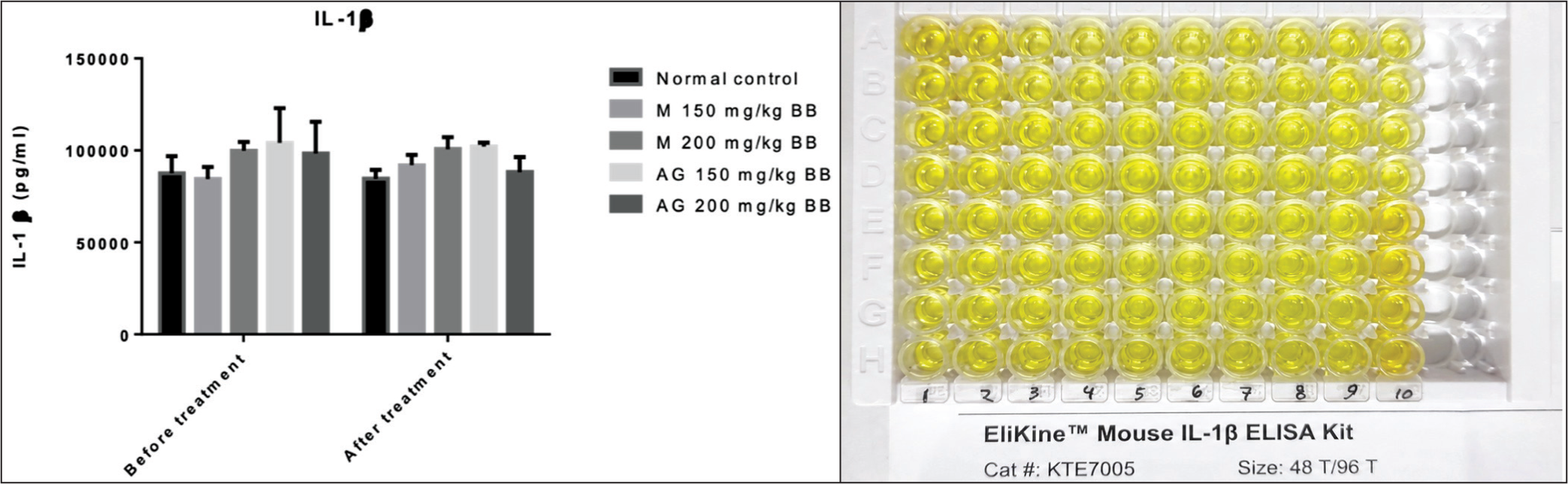
Serum IL-6 and IL-1β levels did not differ significantly among groups (Figures 2 and 3). CRP concentrations showed variability across all groups, including controls, with no dose-dependent pattern (Table 6). Analysis of ΔCRP (post–pre) demonstrated substantial overlap between groups. Overall, repeated oral administration did not induce consistent systemic inflammatory responses.
Serum IL-6 Concentrations in Mice After Four Weeks of Oral Administration of Extracellular Protease from Bacillus tequilensis HSFI-5 Microencapsulated With M or AG at Doses of 150 and 200 mg/kg BW. One-way ANOVA Showed: F(4,25) = 1.09, p = .38 (ns).
Serum IL-1β Concentrations in Mice After Four Weeks of Oral Administration of Extracellular Protease from Bacillus tequilensis HSFI-5 Microencapsulated With M or AG at Doses of 150 and 200 mg/kg BW. Bars Represent Group Means With Individual Values Shown for Each Experimental Group. NC Denotes the NC Group without Test Material. One-way ANOVA Showed: F(4,25) = 1.21, p = .33 (ns).
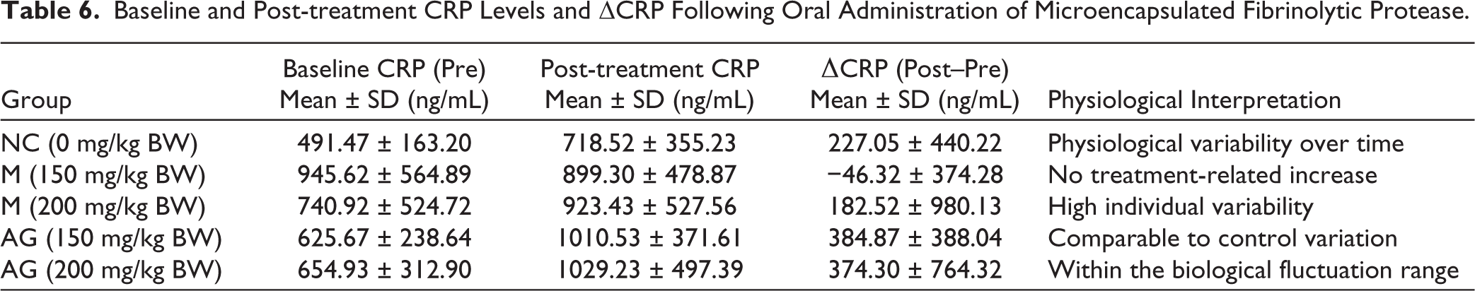
Baseline and Post-treatment CRP Levels and ΔCRP Following Oral Administration of Microencapsulated Fibrinolytic Protease.
Evaluation of systemic inflammatory markers indicated that microencapsulation with either M or AG was not associated with consistent increases in IL-6, IL-1β, or CRP when compared with the control group. Variations in IL-6 concentrations were observed across all experimental groups, including untreated animals (Figure 2). It appears that these fluctuations were not directly related to protease administration.
Figure 3 shows the serum IL-1β response in each experimental group after administering M- and AG-microencapsulated protease. IL-1β levels in treated animals remained close to those in the NC group, indicating that oral exposure to the microencapsulated protease did not elicit a significant pro-inflammatory response via IL-1β. As seen in Figure 3, there was some variation among individual animals, as expected for cytokine responses. Mice that received the higher dose (200 mg/kg BW) of AG-microencapsulated protease had lower IL-1β levels after treatment than the other groups. This trend should be interpreted cautiously and does not establish an anti-inflammatory effect.
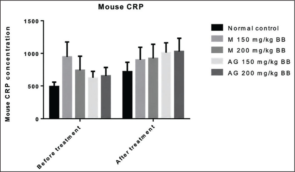
CRP levels were analyzed as a marker of systemic inflammatory stress. CRP varied across groups but showed no consistent treatment-dependent pattern. CRP values across all treatment groups were comparable to baseline values observed in the NC animals (see Figure 4 and Table 6). Statistical evaluation using one-way ANOVA showed no significant differences among experimental groups (p > 0.05). CRP did not show a treatment-dependent increase, suggesting that repeated oral administration of microencapsulated fibrinolytic protease did not provoke a systemic inflammatory response.
CRP Marker Values (in ng/mL) Following Oral Administration of Microencapsulated Extracellular Protease from Bacillus tequilensis HSFI-5. Serum Levels Were Measured After Four Weeks of Treatment. One-way ANOVA Showed: F(4,25) = 1.48, p = .24 (ns).
Baseline CRP measurements (pre-test) were obtained before treatment to account for inter-individual variability (see Table 6). Although CRP values increased in several groups at the endpoint, comparable changes were also observed in the NC group. Analysis of ΔCRP (post–pre) showed substantial overlap among groups, with no consistent treatment-dependent pattern. The observed CRP fluctuations were comparable to those in controls, suggesting normal physiological variation rather than treatment-related inflammation.
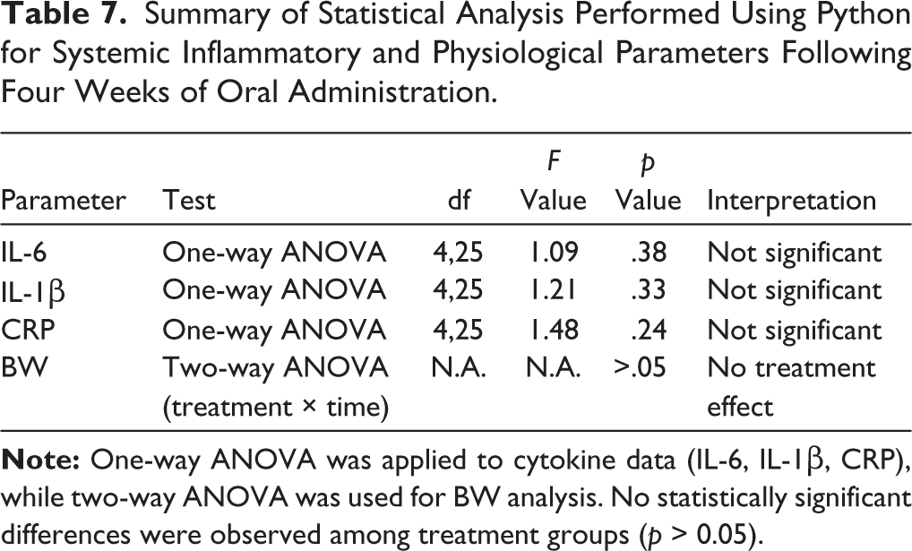
To improve clarity and transparency in statistical analysis, a summary of Python-generated ANOVA results is presented in Table 7. The results confirm that no statistically significant differences were observed across treatment groups for all evaluated parameters (p > 0.05).
Summary of Statistical Analysis Performed Using Python for Systemic Inflammatory and Physiological Parameters Following Four Weeks of Oral Administration.
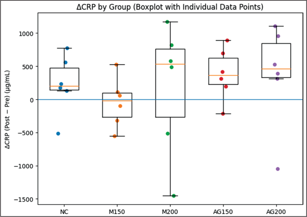
There was variability across all experimental groups, including the controls, suggesting that the changes observed are likely due to normal biological variation rather than the treatment. Notably, animals given higher doses of protease did not show increased inflammatory markers. The AG-microencapsulated formulation at the higher dose had values similar to or lower than those of other groups, suggesting it did not cause inflammatory intolerance. Visualization of ΔCRP distributions further demonstrated substantial overlap among groups (Figure 5), supporting the absence of treatment-related inflammatory effects.
Distribution of ΔCRP (Post–pre) Values Across Experimental Groups Following Oral Administration of Microencapsulated Bacterial Fibrinolytic Protease. Boxplots Show the Median and Interquartile Range, and Individual Data Points are Overlaid to Illustrate Biological Variability. The Horizontal Line Shows No Change from Baseline (Δ = 0).
Analysis of ΔIL-1β showed a statistically significant inter-group difference (p = .047); however, this isolated finding was not accompanied by parallel changes in IL-6 or CRP. Therefore, its biological relevance appears limited and should be interpreted cautiously, particularly given the small sample size, absence of matrix-only controls, and use of healthy animals. These findings should be considered exploratory rather than evidence of anti-inflammatory efficacy.
Discussion
This work evaluated the systemic biocompatibility of a spray-dried microencapsulated fibrinolytic protease derived from B. tequilensis HSFI-5 following repeated oral administration in mice. Overall, the results indicate that oral exposure to the microencapsulated enzyme did not induce detectable systemic disturbances in hematological parameters, lipid metabolism, or inflammatory biomarkers. Although not all parameters showed complete uniformity, no consistent treatment-related deviation emerged. These data, therefore, support the physiological compatibility of the tested formulation under the experimental conditions and suggest that the developed preparation may be suitable for further translational investigation.
Safety of Orally Delivered Fibrinolytic Proteases
Our findings indicate that microencapsulation enables oral administration of fibrinolytic protease without detectable systemic disturbance under the tested conditions. Endpoint inflammatory biomarkers, including IL-6, IL-1β, and CRP, remained comparable to those observed in untreated control animals. This suggests that repeated oral exposure did not trigger measurable immune activation.
Observed CRP variability across experimental groups was consistent with physiological variation commonly reported in murine inflammatory assays. Previous studies have shown that serum CRP concentrations in healthy mice generally range from 0 to 2.3 mg/L, reflecting normal biological fluctuations rather than pathological inflammatory responses. 24 The absence of coordinated elevations in inflammatory biomarkers indicates that the tested formulation was well tolerated systemically during the four-week exposure period.
These findings are in line with previous reports on fibrinolytic enzymes derived from Bacillus species, particularly nattokinase from Bacillus subtilis. Both exhibited minimal systemic toxicity and stable hematological profiles following oral administration in both animal and human studies.28–30 In these studies, no significant alterations in inflammatory markers or lipid metabolism were observed. It aligned with the absence of significant changes in IL-6, IL-1β, CRP, and lipid parameters in the present work. All of these supported the hypothesis that fibrinolytic proteases, when properly formulated, can be administered orally without inducing adverse systemic effects. However, most previous studies have evaluated fibrinolytic enzymes in native or fermented forms, whereas the present study specifically investigates a spray-dried microencapsulated formulation. This distinction is important, as encapsulation processes may influence enzyme stability, bioavailability, and physiological interactions. Thus, the absence of detectable systemic disturbances in the present study provides additional evidence that the encapsulation strategy itself does not introduce adverse biological effects.
Comparison with Previously Reported Bacillus Fibrinolytic Enzymes
Our data are consistent with previous reports on the safety of fibrinolytic enzymes derived from Bacillus species. Several clinical and experimental studies have demonstrated that orally administered fibrinolytic enzymes, particularly nattokinase produced by Bacillus subtilis, demonstrate minimal systemic toxicity and maintain stable hematological parameters. This has been observed in both animal models and human subjects.28–30 For example, recent clinical investigations have reported that long-term nattokinase supplementation does not produce adverse hematological or inflammatory effects and may be safely administered even in patients with cardiovascular conditions. A 90-day randomized controlled trial involving individuals with coronary artery disease also reported no significant adverse outcomes following nattokinase supplementation. 30
However, most previous studies have examined fibrinolytic enzymes as native enzyme preparations or fermented food products. In contrast, this work specifically evaluated a spray-dried microencapsulated formulation. This distinction is important because encapsulation processes and carrier materials can potentially alter enzyme stability, bioavailability, or biological compatibility. The absence of detectable systemic disturbances in the present study, therefore, provides additional evidence that the encapsulation strategy and the selected wall materials do not cause adverse physiological effects.
Role of Microencapsulation in Oral Enzyme Stability
Microencapsulation has emerged as an important strategy for improving the stability and delivery of bioactive proteins intended for oral administration. Proteolytic enzymes are particularly susceptible to denaturation or degradation during processing and gastrointestinal exposure. Spray-drying encapsulation using carbohydrate-based matrices such as M and AG has been widely applied to protect enzyme structure while preserving catalytic activity during storage and delivery.
In the current study, the spray-drying process successfully preserved measurable proteolytic activity following encapsulation, indicating that the enzyme retained functional integrity during formulation. The observed constant specific activity (~5.52 U/mg protein) further implies that spray-drying encapsulation preserved the enzyme’s intrinsic catalytic efficiency. Then the differences in retained activity (U/mL) between M and AG formulations are attributable to carrier-dependent protection rather than changes in enzyme quality. The observed stability is consistent with previous studies demonstrating that polysaccharide-based matrices can form protective barriers that reduce thermal and oxidative damage to encapsulated enzymes during drying and storage.17–19
The slightly higher retained activity observed in M-based microcapsules may be attributed to its film-forming properties and lower viscosity, which can enhance encapsulation efficiency during spray-drying. In contrast, AG, while effective, may result in slightly reduced activity retention due to differences in matrix structure and drying behavior. Nevertheless, both carriers had maintained enzyme stability within a comparable range, supporting their suitability for oral enzyme formulation.
Beyond protecting enzymatic activity, encapsulation may also regulate the enzyme’s interactions with gastrointestinal environments and host tissues. It potentially improves tolerability during oral administration. Our results, therefore, support the feasibility of using spray-dry microencapsulation as a scalable strategy for the development of orally deliverable enzyme formulations derived from marine microbial sources.
Study Limitations and Future Research
Several limitations should be considered when interpreting our findings. One important limitation of the experimental design is the absence of matrix-only control groups receiving empty M or AG microcapsules. Consequently, potential effects attributable solely to the encapsulation materials cannot be completely separated from those of the embedded protease.
The descriptive nature of hematological data represents a limitation of the study, as the small sample size precludes robust statistical inference. Future studies with larger cohorts are needed to confirm these observations. In addition, histopathological evaluation of major organs, particularly the gastrointestinal tract and liver, was not performed in the present study and represents an important limitation. Given that oral administration initially exposes the enzyme to the gastrointestinal mucosa before potential systemic absorption, it should be incorporated in future investigations. It means the next studies should incorporate tissue-level analysis to provide a more comprehensive toxicological assessment. However, the absence of significant alterations in systemic inflammatory biomarkers and hematological parameters suggests that no overt systemic or inflammatory toxicity occurred under the tested conditions.
Although both M and AG are well regarded as safe excipients commonly used in oral formulations, their independent biological effects under the specific dosing conditions applied here were not directly evaluated. Future studies should therefore include matrix-only controls, larger experimental cohorts, and histopathological examination of target organs. Furthermore, longer-term exposure studies and pharmacodynamic evaluations will be necessary to determine whether the encapsulated enzyme maintains therapeutic fibrinolytic activity following oral administration. Such investigations will be essential to support the further development of marine-derived fibrinolytic enzymes as potential biotechnological or biomedical products.
Although this work demonstrates systemic biocompatibility following repeated oral administration, it does not directly establish whether the encapsulated protease is absorbed into the systemic circulation in an active form. Oral delivery of protein-based enzymes is often limited by gastrointestinal degradation and enzymatic digestion. Therefore, further pharmacokinetic and bioavailability studies are needed to determine the extent of gastrointestinal absorption, systemic distribution, and functional persistence of the protease following oral administration. Nevertheless, the absence of systemic inflammatory and hematological disturbances. It suggests that, if absorption occurs, it does not induce detectable adverse physiological responses under the tested conditions.
Overall, the present data provide one of the first experimental indications that a marine-derived fibrinolytic protease from B. tequilensis can be formulated as a stable, orally deliverable enzyme preparation without detectable systemic intolerance during repeated exposure. While most previous studies on bacterial fibrinolytic enzymes have focused primarily on enzymatic characterization or in vitro thrombolytic activity, the current work integrates enzyme semi-purification, scalable spray-dry microencapsulation, and in vivo systemic biocompatibility evaluation. This was done within a single translational framework. This approach moves beyond enzyme discovery toward practical formulation development, an essential step for future biomedical or nutraceutical applications. In this context, the results help bridge a critical gap between microbial enzyme discovery and the development of physiologically compatible, orally administrable fibrinolytic bioproducts derived from marine microbial resources.
Conclusions
This study demonstrates that spray-dried microencapsulated fibrinolytic protease derived from B. tequilensis retains measurable enzymatic activity (0.26–0.28 U/mL; ~5.52 U/mg protein) following encapsulation and can be administered orally without inducing consistent or biologically relevant alterations in hematological, metabolic, or inflammatory parameters (p > 0.05) in a four-week repeated-dose model. These findings are in line with the formulation’s short-term systemic biocompatibility and indicate its potential as a platform for developing orally deliverable enzyme-based biotechnological products. Further studies are needed to evaluate long-term safety and bioavailability to support its progression toward biomedical applications.
Supplementary Material
Supplementary material for this article is available online.
Supplementary Material
Supplementary material for this article is available online.
Footnotes
Acknowledgements
The authors thank Universitas Muhammadiyah Semarang (UNIMUS), the National Research and Innovation Agency (BRIN), and Universitas Gadjah Mada (UGM) for their research support and collaboration. We also acknowledge the technical assistance from the laboratory staff involved in this study.
Authors’ Contribution
All authors made substantial contributions to conception and design, acquisition of data, or analysis, and interpretation of data; took part in drafting the article or revising it critically for important intellectual content; agreed to submit to the current journal; gave final approval of the version to be published; and agreed to be accountable for all aspects of the work. All the authors are eligible to be authors as per the International Committee of Medical Journal Editors’ requirements/guidelines.
Consent to Participate
Not applicable.
Consent for Publication
Not applicable.
Data Availability Statement
All the data is available to the authors and shall be provided upon request.
Declaration of Conflicting Interests
The authors declared no potential conflicts of interest with respect to the research, authorship, and/or publication of this article.
Ethical Approval
This study was approved by the Health Research Ethics Commission, LPPT (Laboratorium Penelitian dan Pengujian Terpadu) Universitas Gadjah Mada, Indonesia (Approval No.: 00070/XI/UN1/LPPT/EC/2024), and all procedures were conducted in accordance with the approved protocol.
Funding
The authors received no financial support for the research, authorship, and/or publication of this article.
Informed Consent
Not applicable.
Use of Artificial Intelligence-assisted Tools
AI tools were used solely for language editing and clarity improvement during manuscript preparation. AI was not involved in study design, data analysis, or interpretation of results. All content was carefully reviewed and validated by the authors, who take full responsibility for the integrity of the work.
References
Supplementary Material
Please find the following supplemental material available below.
For Open Access articles published under a Creative Commons License, all supplemental material carries the same license as the article it is associated with.
For non-Open Access articles published, all supplemental material carries a non-exclusive license, and permission requests for re-use of supplemental material or any part of supplemental material shall be sent directly to the copyright owner as specified in the copyright notice associated with the article.