
Abstract
Background
Zinc nanoparticles (ZnNPs) have emerged as significant products in nanotechnology, serving as promising candidates for cancer research.
Objectives
In this study, Artemisia scoparia (A. scoparia) extract was utilized to synthesize biogenic ZnNPs and evaluate their inhibitory effects on cell proliferation and K-Ras gene expression in the A549 cell line.
Methods
Biogenic ZnNPs were synthesized using the leaf extract of A. scoparia. The synthesized ZnNPs were characterized using Field Emission Scanning Electron Microscopy (FE-SEM), Transmission Electron Microscopy (TEM), Fourier Transform Infrared Spectroscopy (FT-IR), Energy Dispersive X-ray Spectroscopy (EDS), and X-ray Diffraction (XRD). The cytotoxic effects of the ZnNPs on the A549 cell line were assessed using the MTT assay after 24 h of treatment. K-Ras gene expression relative to β-actin was analyzed using real-time PCR.
Results
The particle size of the ZnNPs ranged from 62 to 103 nm. Cytotoxicity results revealed a dose-dependent reduction in the viability of A549 cells after 24 h treatment with A. scoparia extract and ZnNPs synthesized with the extract. The Half Maximal Inhibitory Concentration value (IC50) for the ZnNPs was calculated as 10.26 µg/mL compared to the control group (p < 0.005). After 24 h of treatment, K-Ras gene expression decreased by 0.6-fold (p < 0.001).
Conclusions
The findings indicate that the synthesized ZnNPs exhibit cytotoxic and antiproliferative effects on A549 lung cancer cells.
Keywords
Introduction
Nanotechnology has revolutionized various fields of science and medicine, enabling the development of nanoscale materials with unique physicochemical properties.1,2 Among these, zinc nanoparticles (ZnNPs) have garnered significant attention due to their biocompatibility and versatile applications, particularly in drug delivery systems. 3 ZnNPs serve as effective carriers for small drug molecules and large biomolecules such as DNA, RNA, and proteins, enhancing therapeutic efficacy through targeted delivery. 4
Recent studies indicate that modifying nanoparticles with bioactive compounds extracted from medicinal plants can significantly improve their biological functions.5,6 Artemisia scoparia (A. scoparia), a plant with well-documented pharmacological properties, presents a promising candidate for the green synthesis of biogenic nanoparticles, offering an eco-friendly alternative to conventional chemical methods.7–10 The biofabrication of ZnNPs using A. scoparia extract not only reduces the need for toxic reagents but also enhances the biological activity of the nanoparticles. 11
Green synthesis methods have gained recognition as a sustainable and environmentally friendly approach in anticancer research.12,13 Conventional nanoparticle synthesis often involves hazardous chemicals, high energy consumption, and environmental concerns, whereas green synthesis utilizes biological sources such as plants, bacteria, and fungi to produce nanoparticles.14–16 This method enhances biocompatibility, reduces toxicity compared to chemically synthesized counterparts, and improves the biological activity of nanoparticles. 17 The potential of these biogenic nanoparticles in cancer treatment makes green nanotechnology an essential avenue for future biomedical applications, reinforcing the importance of sustainable methods in developing novel cancer therapies.18,19
In green synthesis, plant extracts act as natural reducing, chelating, and stabilizing agents during nanoparticle formation. A. scoparia contains various bioactive compounds such as phenolics, flavonoids, terpenoids, and proteins, which possess functional groups including hydroxyl (–OH), carbonyl (C=O), and amine (–NH2). These groups can interact with zinc ions (Zn2+) through coordination and electrostatic interactions.
During synthesis, zinc nitrate provides Zn2+ ions, which react with hydroxide ions generated by NaOH to form zinc hydroxide intermediates
Under heating conditions, zinc hydroxide is converted into ZnO NPs (ZnNPs)
Phytochemicals present in the plant extract help regulate nucleation and growth of nanoparticles and act as capping agents, preventing aggregation and stabilizing the formed ZnNPs. This plant-mediated process eliminates the need for toxic chemical reducing agents and enhances the biocompatibility of the synthesized nanoparticles. 17
Lung cancer is one of the leading causes of cancer-related mortality worldwide, characterized by uncontrolled cell growth in lung tissue. 20 It is broadly classified into two major types: non-small cell lung cancer (NSCLC) and small cell lung cancer (SCLC), with NSCLC accounting for approximately 85% of cases. 21 Among NSCLC subtypes, adenocarcinoma, squamous cell carcinoma, and large cell carcinoma are the most prevalent. 22
Traditional treatment options for lung cancer include surgery, chemotherapy, radiation therapy, and targeted therapies. 23
K-Ras is one of the most frequently mutated oncogenes in human cancers, particularly in lung adenocarcinoma. 24 This gene encodes a GTPase protein involved in cell signaling pathways that regulate growth, proliferation, and survival. 25 When mutated, K-Ras becomes constitutively active, leading to uncontrolled cell division and resistance to apoptosis—two hallmarks of cancer. 26 Inhibiting K-Ras is considered a major challenge in cancer research. 27 Recent advances in drug development have led to the emergence of K-Ras inhibitors, aiming to block its signaling cascade and restore normal cellular function. 28 Effective suppression of K-Ras expression or activity has the potential to halt tumor progression, reduce metastasis, and improve patient survival rates. 29 Studies, including those investigating nanomaterials, suggest that nanoparticles can contribute to K-Ras regulation by enhancing drug delivery and exerting direct cytotoxic effects, making nanotechnology-based approaches an exciting prospect for future cancer therapies. 30
The development of green-synthesized nanoparticles, particularly those derived from plant extracts, presents a promising avenue for anticancer therapy, offering improved biocompatibility, reduced toxicity, and enhanced therapeutic potential. 31
In this study, biogenic ZnNPs were synthesized using A. scoparia leaf extract, and their cytotoxic and antiproliferative effects were evaluated on the A549 lung cancer cell line. The aim of this research is to assess the inhibitory potential of the synthesized nanoparticles on cell proliferation and their ability to modulate K-Ras gene expression, providing new insights into the biomedical applications of ZnNPs in cancer treatment.
Materials and methods
Preparation of A. scoparia extract
A. scoparia was purchased from the National Plant Bank of the Iranian National Genetic and Biological Resource Center, where the plant material had been collected in spring 2025. Five grams of dried leaves were obtained from 100 g of fresh plant material, corresponding to a 1:20 ratio of dried powder to fresh sample. For extract preparation, 5 g of stem powder and 5 g of leaf powder were immersed in 100 mL of distilled water, boiled, and then centrifuged at 4000 rpm for 4 min. The resulting extract was further purified by filtration through Whatman filter paper. 14
Synthesis of ZnNPs
For green synthesis of high-purity ZnNPs, 200 mL of zinc nitrate solution (1.5 mM) was mixed with 20 mL of A. scoparia leaf extract and 10 mL of sodium hydroxide (1 M). The mixture was stirred in the dark at 60°C for 24 h, leading to the formation of a white zinc oxide precipitate. The nanoparticles were purified by centrifugation, washed sequentially with distilled water and ethanol, and dried. The final powdered nanoparticles were analyzed using Field Emission Scanning Electron Microscopy (FE-SEM), Transmission Electron Microscopy (TEM), Fourier Transform Infrared Spectroscopy (FT-IR), Energy Dispersive X-ray Spectroscopy (EDS), and X-ray Diffraction (XRD). 17
Characterization of ZnNPs
The morphology and structural properties of nanoparticles were validated using FE-SEM, XRD, TEM, FT-IR, and EDS. FE-SEM imaging was conducted using the MIRA3 system (TE-SCAN), and TEM images were acquired using the EM10C-100 KV Zeiss system. FT-IR analysis was performed using the Spectrum Two system (PerkinElmer), and XRD patterns were obtained using the X'Pert Pro Panalytical system. Additionally, nanoparticles were synthesized using the sol-gel method with zinc acetate and analyzed through X-ray diffraction, infrared spectroscopy, UV-Vis spectroscopy, and EDS. 17
Cell culture and treatment
Human foreskin fibroblasts (HFF; control cell line) and human lung cancer A549 cells were procured from the Iranian National Genetic and Biological Resource Center. Cells were cultured in RPMI 1640 medium supplemented with 10% fetal bovine serum (FBS) and 1% penicillin-streptomycin (100 U/mL penicillin and 100 µg/mL streptomycin) in a humidified incubator at 37°C with 5% CO2. 8 Cells were seeded in 96-well plates at a density of 10,000 cells per well and allowed to adhere overnight before treatment with nanoparticles at concentrations of 3.125, 6.25, 12.5, 25, 50, and 100 µg/mL. 17
Before biological experiments, dried ZnNPs were freshly dispersed in sterile distilled water and sonicated for 20 min using an ultrasonic bath to improve dispersion and reduce aggregation. The suspensions were vortexed immediately before addition to the culture medium to ensure uniform distribution. Fresh suspensions were prepared for each experiment to minimize time-dependent agglomeration.
Cytotoxicity assay
The MTT assay was performed to evaluate cytotoxic effects. A 5 mg/mL MTT stock solution was prepared. After 24 h of treatment, 100 µL of MTT solution was added to each well and incubated for 3 h. The resulting formazan crystals were dissolved in 100 µL of DMSO, and absorbance was measured at 570 nm using an ELISA reader. Cell viability and cytotoxicity were calculated as
19


The Half Maximal Inhibitory Concentraion (IC50) values were determined using the Pharm-PCS statistical package (version 5.0).
Results
Validation of ZnNPs synthesis using A. scoparia extract
The successful synthesis of ZnNPs was visually confirmed by a distinct color change in the solution. After 24 h, the initial light-colored solution transitioned to a darker shade, indicating nanoparticle formation (Figure 1). Formation of ZnNPs. (A) Initial solution of zinc nitrate precursor before addition of A. scoparia extract. (B) Visual change in the solution indicating the effect of A. scoparia extract on the biosynthesis of ZnNPs.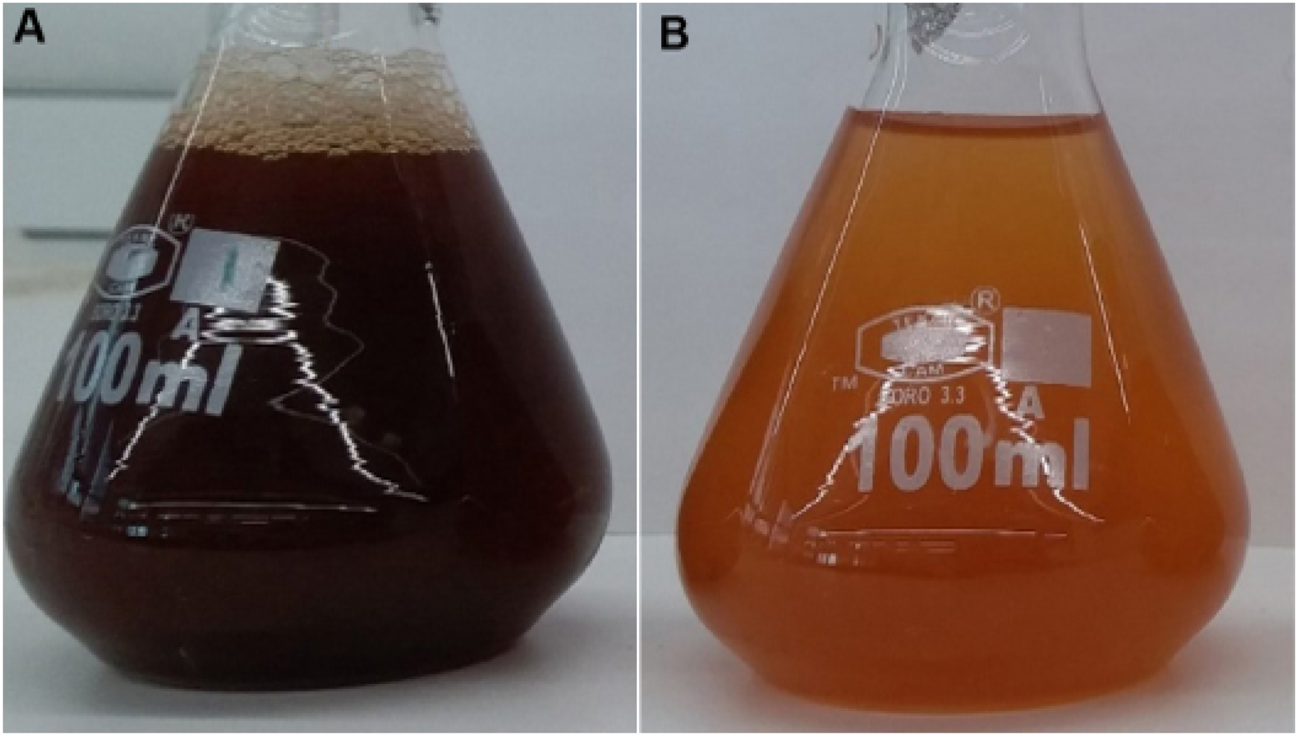
FT-IR spectral analysis confirmed the presence of biomolecules involved in nanoparticle synthesis. A broad and intense peak observed between 3548 cm-1 and 3239 cm-1 corresponds to the stretching vibrations of alcoholic and phenolic O–H groups present in the plant extract. Absorption peaks at 3031 cm-1 indicate C–H stretching vibrations of aromatic benzene rings, characteristic of carvacrol compounds. The presence of an amide C=O stretching band at 1619 cm-1, along with a confirming peak at 1481 cm-1, further supports biomolecular interactions. The peak at 1395 cm-1 corresponds to CH2 bending vibrations, while C–O phenolic stretching is observed at 1084 cm-1. Additionally, C–H bending vibrations associated with C=C–H groups appear at 618 cm-1 and 480 cm-1 (Figure 2(A)). Characterization of biosynthesized ZnNPs. (A) FT-IR spectrum of A. scoparia extract showing functional groups involved in nanoparticle synthesis. (B) FT-IR spectrum of biogenically synthesized ZnNPs indicating capping and stabilizing interactions. (C) XRD pattern of biosynthesized ZnNPs demonstrating crystalline structure. (D) EDS spectrum of ZnNPs synthesized using A. scoparia extract, confirming elemental composition. (E) FE-SEM image showing surface morphology and size distribution of ZnNPs. (F) TEM image showing spherical morphology and primary particle size of ZnNPs.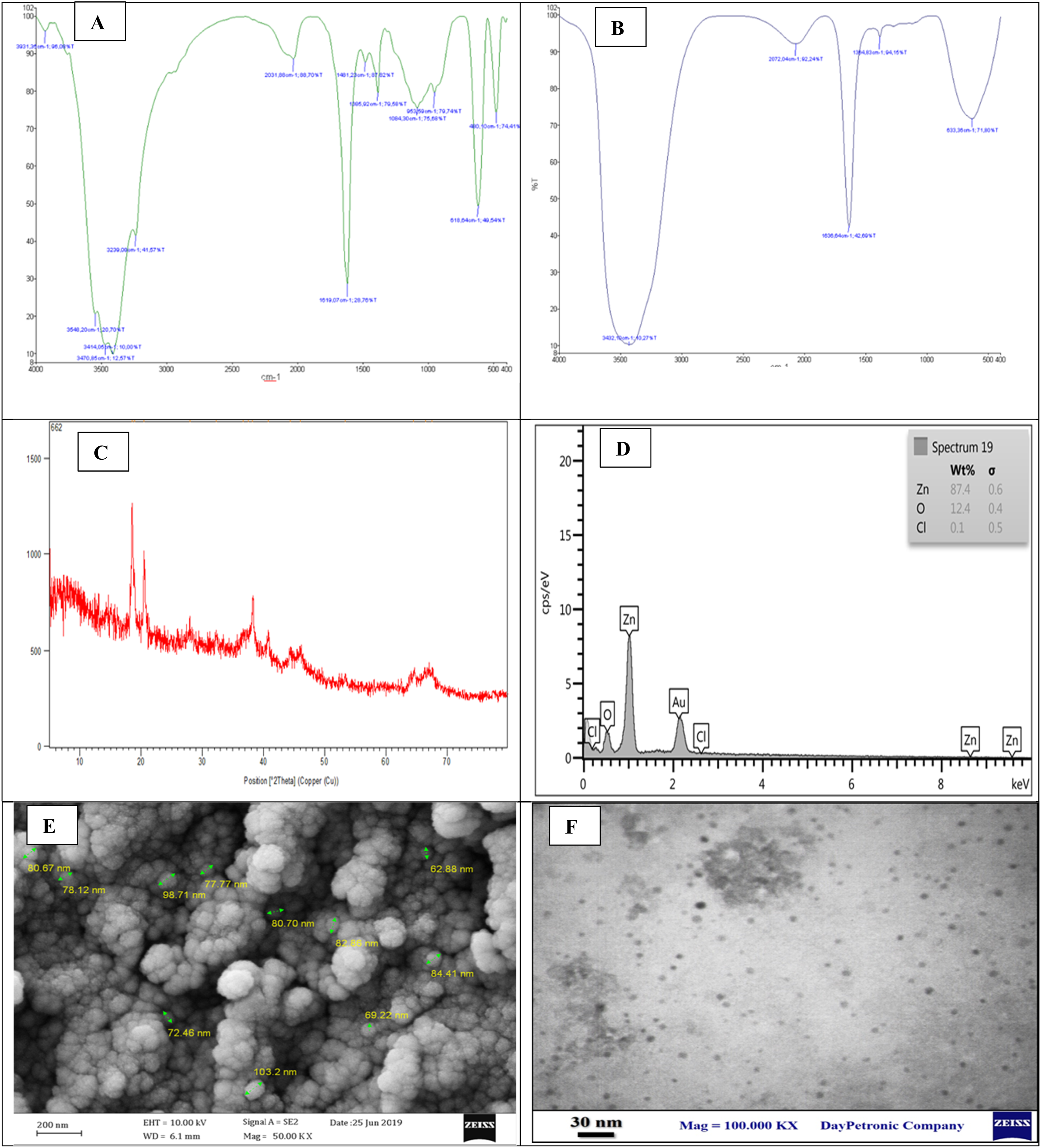
FT-IR analysis of the synthesized ZnNPs (Figure 2(B)) showed a strong 3432 cm-1 band, attributed to symmetric stretching of water molecules and O–H functional groups, indicating moisture absorption onto the nanoparticles. The 1636 cm-1 peak corresponds to N–H and C=C vibrations, likely associated with amide and vinyl groups. The characteristic ZnO stretching vibration was detected at 633 cm-1, confirming the presence of ZnNPs.
XRD results (Figure 2(C)) revealed sharp diffraction peaks, indicative of the crystalline nature of the synthesized nanoparticles. EDS analysis (Figure 2(D)) confirmed the presence of zinc, oxygen, and chlorine, with zinc being the predominant element (87.4%).
FE-SEM imaging (Figure 2(E)) estimated the particle size within the range of 62–103 nm, while TEM analysis (Figure 2(F)) confirmed the spherical morphology of the synthesized ZnNPs. TEM demonstrated predominantly spherical nanoparticles in the nanometer range (62–103 nm) with no evidence of large micrometer-scale aggregates in the observed fields. Minor clustering may occur due to drying during sample preparation; however, no substantial agglomeration was detected.
Cytotoxic effects of A. scoparia extract and its biosynthesized ZnNPs on A549 cells
After 24 h of treatment, control HFF cells exposed to biosynthesized ZnNPs, A. scoparia extract, and untreated cells showed no cytotoxic effects, indicating that the plant extract and the nanoparticles containing the extract are safe for normal cells (Figure 3(A)). Cytotoxic effects of A. scoparia extract and biosynthesized ZnNPs on cell lines. (A) HFF as normal control cells. (B) A549 cells. Cells were treated with varying concentrations of A. scoparia extract and ZnNPs for 24 h. Data are presented as mean ± SD. Statistical significance: *p < 0.05, **p < 0.01, ***p < 0.001.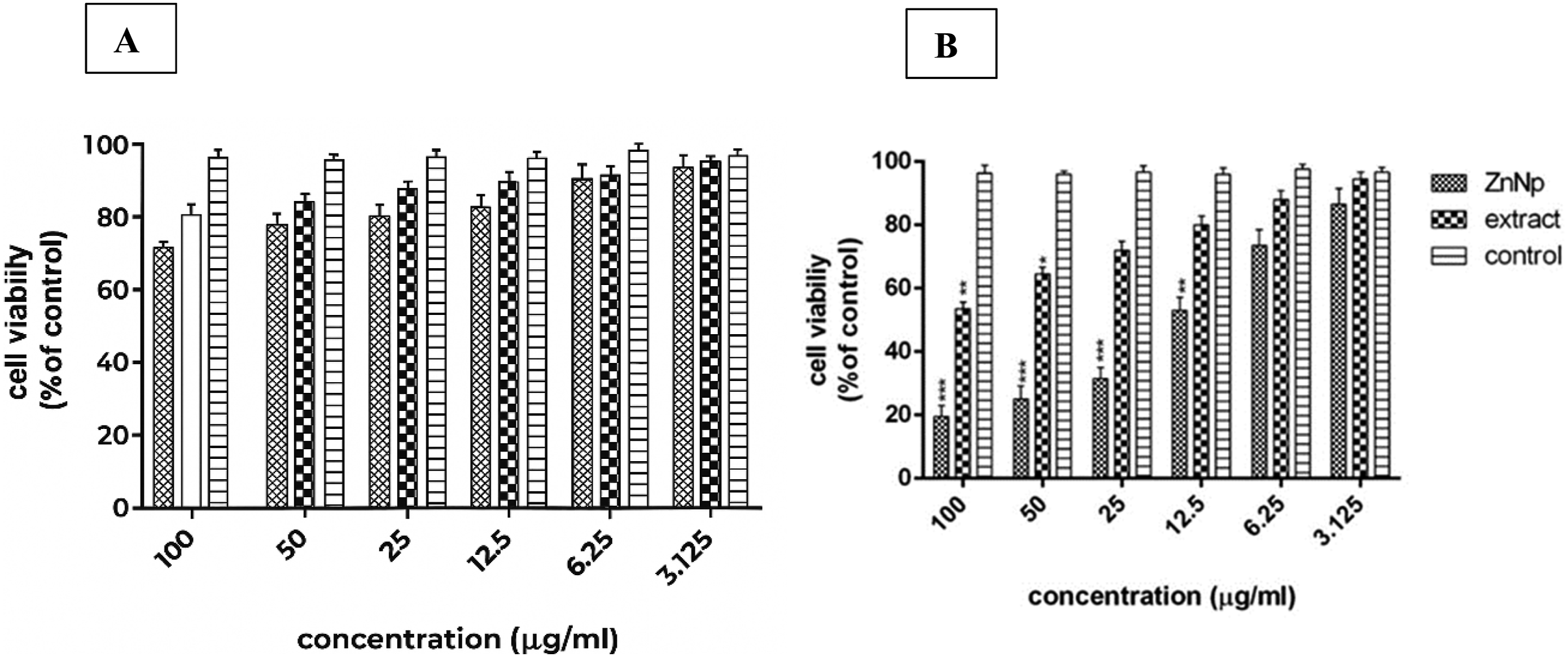
Both A. scoparia extract and its biosynthesized ZnNPs inhibited A549 cell survival in a concentration-dependent manner (Figure 3(B)). The reduction in cancer cell viability was statistically significant at concentrations above 12.5 µg/mL compared to the control group (p < 0.001 and p < 0.01). The IC50 value of the nanoparticles synthesized with A. scoparia extract was 10.26 µg/mL, lower than the IC50 of the extract alone, indicating stronger cytotoxic effects.
Downregulation of K-Ras gene expression in A549 cells following treatment
K-Ras gene expression in A549 cells after treatment with IC50 concentrations of A. scoparia extract and its biosynthesized ZnNPs is illustrated in Figure 4. A significant reduction in K-Ras expression was observed, indicating effective gene modulation by the nanoparticles. Downregulation of K-Ras gene expression in A549 cells. Expression of K-Ras relative to β-actin in A549 cells after 24-h treatment with IC50 concentrations of A. scoparia extract and biosynthesized ZnNPs. Data are presented as mean ± SD. Statistical significance: *p < 0.05, **p < 0.01, ***p < 0.001.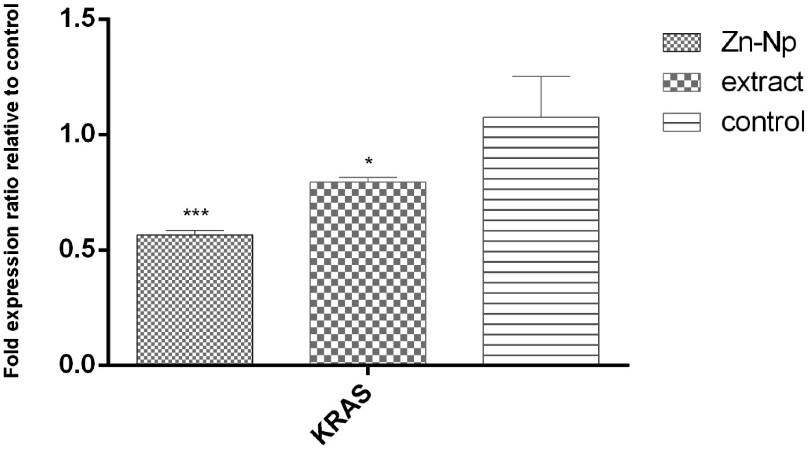
Discussion
Sequences of forward and reverse primers used for real-time PCR.

Several previous studies support the efficacy of biosynthesized ZnNPs against lung cancer cells. For example, ZnNPs synthesized using Curcuma longa extract exhibited significant cytotoxicity against A549 cells, consistent with our findings. 32 Similarly, ZnNPs produced with Withania somnifera extract inhibited cancer cell proliferation by modulating oncogenic gene expression. 33 These studies, alongside our results, emphasize the role of plant-derived bioactive compounds in enhancing nanoparticle therapeutic activity, confirming the superior anticancer properties of biosynthesized ZnNPs. 34 Moreover, nanoparticles derived from Moringa oleifera were reported to display variable cytotoxicity depending on the cancer model used, 35 highlighting the influence of plant source, nanoparticle size, surface properties, and synthesis method on biological efficacy.36,37
Our results further align with reports showing that biosynthesized nanoparticles can downregulate oncogenes such as K-Ras, reinforcing the potential molecular impact of plant-mediated ZnNPs. 38 The specificity of these effects in A549 cells, compared to normal HFF cells, demonstrates a degree of selective cytotoxicity, which is critical for therapeutic applications.
While the MTT assay confirmed cytotoxicity by assessing mitochondrial activity, complementary assays such as lactate dehydrogenase (LDH) release could provide additional insight into membrane integrity and biocompatibility. Although LDH assays were not performed in this study due to laboratory limitations, future investigations will incorporate such assessments along with apoptosis markers to comprehensively evaluate nanoparticle-induced cytotoxic effects.
It is noteworthy that metal oxide nanoparticles can partially aggregate in aqueous or biological media due to van der Waals interactions and ionic strength effects. Complete prevention of aggregation under physiological conditions is challenging. However, our microscopic analyses (FE-SEM and TEM) confirmed primary particle sizes within the nanometer range (62–103 nm) without micrometer-scale structures. Sonication was applied prior to cellular exposure to enhance dispersion. Therefore, while minor nanoscale clustering may occur dynamically in culture medium, the absence of large aggregates suggests that the observed biological effects are primarily attributable to individual nanoscale ZnO structures.39,40
In summary, the present study demonstrates that A. scoparia-mediated ZnNPs possess potent cytotoxic and gene-modulatory effects on A549 lung cancer cells. These findings are supported and reinforced by previous studies on plant-derived nanoparticles, confirming both the relevance of plant bioactive compounds in nanoparticle synthesis and their applicability in targeted anticancer strategies.
Conclusion
In this study, we successfully synthesized biogenic ZnNPs using A. scoparia leaf extract, demonstrating a green, eco-friendly, and cost-effective approach for nanoparticle production. The synthesized ZnNPs exhibited controlled size (62–103 nm), spherical morphology, and high stability, confirmed through FE-SEM, TEM, XRD, FT-IR, and EDS analyses.
Biological evaluation revealed that these ZnNPs exert strong cytotoxic and antiproliferative effects on A549 lung cancer cells, with an IC50 value of 10.26 µg/mL, significantly lower than the plant extract alone, highlighting the enhanced therapeutic potential of plant-mediated nanoparticle synthesis. Moreover, treatment with ZnNPs led to a notable downregulation of K-Ras gene expression (0.6-fold decrease), suggesting a mechanistic basis for their anticancer activity and reinforcing their potential as targeted therapeutic agents.
These findings demonstrate that A. scoparia-mediated ZnNPs not only inhibit cancer cell proliferation but also modulate oncogenic pathways, providing a dual mode of action that can be harnessed for future cancer therapy development. The present study provides novel insights into the role of plant-derived phytochemicals in nanoparticle stabilization, cytotoxic enhancement, and gene regulation, offering a foundation for further preclinical and mechanistic studies.
Overall, this work highlights the significant potential of biogenic ZnNPs as biocompatible, effective, and sustainable nanomedicine candidates for lung cancer treatment, contributing valuable knowledge to the field of green nanotechnology and cancer therapeutics.
Footnotes
Acknowledgment
The authors sincerely thank the Research Laboratory staff at Tehran Branch of Islamic Azad University for their technical assistance.
Funding
The authors received no financial support for the research, authorship, and/or publication of this article.
Declaration of conflicting interests
The authors declared no potential conflicts of interest with respect to the research, authorship, and/or publication of this article.