



Abstract
Background and Objectives
Sickle cell anemia (SCA) is associated with recurrent acute inflammatory processes. These inflammatory processes could lead to elevation of Factor VIII and Von Willebrand Factor levels, thereby increasing the risk of stroke in SCA children. This study aims to determine vWF/FVIII levels in children with SCA and their association with abnormal transcranial Doppler (TCD).
Subjects and Methods
This study enrolled 75 children, including 24 SCA cases with normal TCD, 27 SCA cases with abnormal TCD, and 24 Hb AA controls, all aged between 2 and 16 years. Transcranial Doppler (TCD) ultrasound was performed to measure the cerebral blood velocity. Venous blood drawn from each participant was used to determine the levels of von Willebrand Factor Antigen (vWF: Ag) and Factor VIII (FVIII) and the complete blood count (CBC). Relationships among the measured parameters were determined using SPSS version 25. Statistical significance was set at P < .05.
Results
FVIII and vWF levels were significantly higher among children with SCA compared to the Hb AA controls (P < .001). Although SCA patients with abnormal TCD tended to have higher levels of FVIII and vWF, this result did not attain statistical significance (P > .05). There was a moderate negative correlation between the left middle cerebral artery and FVIII, (r = −0.332; P = .017). Children with SCA showing an abnormal TCD velocity had significantly higher platelet count compared to those with normal TCD (P = .018).
Conclusion
Children with SCA have elevated levels of FVIII and vWF, and an abnormal TCD velocity is associated with elevated platelet count.
Introduction
Sickle cell disease (SCD) is one of the most prevailing severe monogenic disorders worldwide. 1 The most severe and frequently observed variant of SCD is sickle cell anemia (SCA) characterized by a homozygous point mutation in the β globin gene (GAG- GTG) at the sixth codon of β globin gene. The manifestation of SCD includes hemolytic anemia, recurrent vaso-occlusive crisis, recurrent infections, and stroke. 2 These manifestations are accompanied by inflammatory processes which may influence the status of some hematological parameters and other biological markers in SCD patients.
Patients with SCD have an increased risk of cerebrovascular accident, which is a leading cause of death in both children and adults. 3 The reported age-adjusted incidence of stroke in SCD is 0.61–0.76 per 100 patient-years during the first 20 years of life. 4 The incidence of the ischemic variant, which constitutes 54% of all cerebrovascular accidents (CVAs), is highest during the first decade of life and after the age of 30 years. 5 Between the ages of 20 and 30 years, ischemic CVA is replaced by hemorrhagic CVA. 5 The prevalence of stroke in patients with SCD in a multi-centre study in Nigeria was 12.4%, 6 thus affirming that the occurrence of stroke as a complication of SCD is common among Nigerian patients.
SCA is associated with the activation of the coagulation system from childhood leading to hypercoagulability, which in turn has been reported to play a role in the development of vasculopathy. 7 Therefore, it is speculated that stroke and abnormal transcranial Doppler (TCD) velocity may be linked to derangements in clotting factors, such as von Willebrand Factor (vWF) and Factor VIII (FVIII), in children with SCD.
vWF has been shown to play an important role in thrombus formation at the site of vascular damage, and an increased level of vWF is a marker of endothelial activation and dysfunction. 8 It renders the endothelium more adhesive to blood cells, especially sickled red blood cells, causing resistance to the blood flow, which may favor fibrin formation and subsequently lead to the formation of occlusive thrombi and the incidence of stroke. While the association between vWF and coronary heart disease has been well studied, there is limited information on the role of vWF in the development of stroke 8 among children with SCD. Similarly, high levels of FVIII are associated with increased risk of deep vein thrombosis, 8 arterial thrombosis in coronary heart disease, 9 and stroke. 10 However, little is known about the contribution of FVIII to abnormal TCD velocity and stroke development in children with SCD.
Nevertheless, it is established in vivo factors such as reduced blood flow, vessel wall damage, and hypercoagulability of blood may alter the delicate balance that exists between fibrin formation and fibrinolysis.8–11 These disturbances favor fibrin formation that could lead to the development of occlusive thrombi, which may be venous or arterial. Arterial thrombosis results from the formation of platelet aggregates at sites of vessel-wall injury under high shear rates, whereas venous thromboembolism results from clot formation in a vein at sites of reduced blood flow.
High FVIII levels may stimulate the formation of thrombin, resulting in increased platelet activation and fibrin formation, which may further contribute to the development of large occlusive thrombi from the microthrombi on the damaged endothelium of SCD patients, thereby leading to abnormal TCD velocity or stroke.
TCD ultrasound scan is a noninvasive method that has been adopted by the SCD program globally to screen children below the age of 17 years and identify those at risk of stroke. This screening is performed because there is a high risk of stroke in the first decade of life, and the incidence of ischemic stroke has an average of 1.02% per year between 2 and 5 years of age. Moreover, the incidence of stroke ranges from 0.61–0.76 per 100 patient-years during the first 20 years of life with a high risk of stroke in this period. Therefore, TCD screening is targeted at these age groups for primary stroke prevention in children with SCD.4,5,12,13
Some studies have established links between markers of hemolysis (hematocrit, hemoglobin concentration, reticulocyte count, and bilirubin and LDH levels) and TCD velocities in patients with SCD.14–17 However, little is known about the roles of procoagulant factors like vWF, FVIII, platelet, and WBC and their relationships with TCD in SCD patients in the sub-Saharan Africa. Assuming that healthy Hb AA participants are unlikely to have conditions that influence the above parameters, this study was conducted to assess the levels of vWF/FVIII, complete blood count (CBC), and their associations with TCD velocity among children with SCA and their healthy Hb AA counterparts attending a tertiary health facility in Nigeria.
Subjects and Method
Study Design and Location
This comparative cross-sectional study was conducted among children with SCA and their healthy Hb AA counterparts between January and May 2018. The study was conducted at the Lagos University Teaching Hospital (LUTH), Lagos and the Sickle Cell Foundation Centre Lagos, both located adjacent to each other in Idi Araba, Lagos State, South – West Nigeria. LUTH is a 750-bed tertiary hospital, with referrals from all levels of health care facilities within Lagos and the surrounding environs. The hospital has a Paediatric Department which runs in-patient services alongside Paediatric Outpatient Clinics and a Paediatric Haematology Clinic where SCD patients are examined. The Paediatric Haematology Clinic operates on a weekly basis, and Hb electrophoresis is the routine method of SCD diagnosis. Apart from the routine clinical care and therapeutic interventions offered at this clinic, routine TCD is a normal protocol for all patients with SCD within the age bracket of 2–16 years as part of the primary stroke prevention strategy. The Sickle Cell Foundation Centre is a non-governmental organization that is involved in the care and welfare of patients with SCD across Nigeria. TCD ultrasonography screening, prenatal diagnosis, and genetic counseling and testing are conducted routinely for SCD patients at this foundation. In addition, the foundation offers major diagnostic and research facilities/supports for SCD.
Ethical Considerations
Ethical approval for this study was obtained from the Lagos University Teaching Hospital's Health Research and Ethics Committee prior to the commencement of the study (NHREC:19/12/2008a). Written informed consent and assent were obtained from the parents or guardians and the participants as applicable.
Study Participants
Study participants comprised children with SCA and HbAA within 2–16 years of age. Children in the Hb AA group were recruited from a wellness clinic and among those that came for preschool admission medical checkups at the Paediatric Outpatient Department of the hospital. In total, 75 children comprising 24 SCA patients with normal (standard risk) TCD, 27 SCA patients with abnormal (high risk) TCD, and 24 Hb AA controls were enrolled in this study. All participants were matched for age and sex. All participants with SCA were in steady state which was defined as the absence of any acute complication, crisis, or infection for at least one month and the absence of any blood transfusion in the preceding three months prior to recruitment respectively. All children with SCA included in the study were hydroxyurea naïve and not on chronic blood transfusion therapy. Children with SCA who were not in steady state were excluded along with those with leg ulcer, avascular necrosis, and stroke. In addition, children with SCA having conditional TCD velocity (TAMMV 170-199cm/sec) were excluded from the study. All participants fulfilling the inclusion criteria were serially recruited, and a structured interviewer-administered questionnaire was used to obtain relevant socio-biological data. All participants identified with abnormal TCD velocities in the study were counseled for chronic blood transfusion and or Hydroxyurea therapy as these were their first recorded abnormal values.
Sample Size Determination
The minimum sample size was determined using the formula designed to demonstrate a significant difference in the comparison of two means.
For this study, the mean of the serum level of vWF for Hb AA and Hb SS subjects as documented in the literature 7 was used.
The formula is represented below.
18

The power that was used in this study was 90%; therefore, u = 1.28. 18
V is the percentage point of the normal distribution corresponding to the two-sided significance level. Our significance level was chosen to be 5%; therefore, V = 1.96. 18
μ0 = Mean of serum vWF: Ag in Hb AA children (from literature by coagulometer) = 92.39%. 7
μ1 = Mean of serum vWF: Ag in children with SCD (from literature by coagulometer) = 150.05%. 7
μ1−μ0 = difference between the means of serum vWF: Ag level in children with SCD and Hb AA controls.
σ1 = standard deviation of the serum vWF: Ag level in children with Hb SS obtained (from literature) = 49.75. 7
σ0 = standard deviation of the serum vWF: Ag level in Hb AA controls (from literature) = 24.94. 7
Thus, the minimum sample size
Although the minimum calculated sample size was 10, the sample size was increased to 24 to increase the overall power of the study.
Diagnosis of SCA and Determination of the HbAA Genotype
The diagnoses of SCA and HbAA were confirmed by Hb electrophoresis in an alkaline buffer. The complete procedure has been described in detail in the Supplemental Material.
TCD Screening Test
TCD was performed by an expert at the Sickle Cell Foundation Centre using a 2 MHz pulsed hand-held probe with a Doppler box (Scan Med product). Patients were not sedated during the procedure. The highest velocity, ie, the time average mean of maximum velocity (TAMMV), was measured at the middle cerebral artery, internal carotid artery, and anterior cerebral artery on both sides of the temporal region. Patients were divided into two groups depending on the TCD result. According to the Stroke Prevention Trial in Sickle Cell Anemia (STOP), patients with SCD with a TAMMV of less than 170 cm/sec were classified as standard risk (normal TCD), while those with at least one TAMMV value ≥200 cm/sec were classified as high risk (abnormal TCD).14–17
Blood Specimen Collection, Preparation, Storage, and Analysis
Participants were allowed to rest for at least five minutes before collecting samples for the coagulation factors. Samples were collected between 9 am and 12 pm. Tourniquet was tied for less than one minute and released immediately after the first collection tube started to fill.
Five milliliter of venous blood was collected from each participant. First, samples for coagulation factors were collected: 2.5 mL of blood was drawn into a specimen bottle containing 0.25 mL of 3.2% buffered sodium citrate and was used to determine the levels of vWF & FVIII. In addition, 2.5 mL of blood was collected into commercially prepared Ethylene diamine tetraacetic acid [EDTA] bottle and used to determine the CBC. The specimen in the citrated bottle was centrifuged at 2000 × g for 15 min, and plasma was separated within 2 h of collection and transferred to plain cryotubes. Plasma aliquots were stored at −80 °C at the Central Research Laboratory, LUTH, until further analysis was performed.
The separated plasma was used for the analysis of vWF Ag and Factor VIII. Factor VIII activity was determined using a semi-automated Sysmex coagulometer (CA-101). The vWF Ag was analysed by the ELISA method using the ASSAYPRO Human vWF ELISA Kit, Lot No 01751728, USA, according to the manufacturers` specifications. The reference value of VWF was 0.3-1.57 IU/mL, and the reference value of FVIII percentage activity was 50%-100%. Complete details of the procedures are available in the Supplemental Materials.
The sample in the EDTA specimen bottle was used to determine the CBC. CBC analysis was performed within 2 h of sample collection using an Auto Haematology Analyzer BC 3200 manufactured by Shenzhen Mindray Biomedical Electronic Technology, China, according to manufacturer's specification.19,20 The reference value of the platelet was 150-450 × 109/L, whereas that of WBC was 4-11 × 109/L. Complete details of the procedures are available in the Supplemental Materials.
Data Analysis
Data obtained from the questionnaires, along with the results of TCD screenings and samples analyses, were entered into a Microsoft Excel 2007 spreadsheet. Data were imputed into the spreadsheet and analysed using SPSS version 25. Categorical variables were presented using frequency distribution tables. Test of normality was conducted for continuous variables using the Kolmogorov–Smirnov test and Shapiro–Wilk test. The continuous variables were presented as medians and interquartile range because they were not normally distributed. Statistical differences between medians for continuous variables were tested using the Kruskal–Wallis H test (test for several independent samples). Spearman's rank correlation was used to determine the relationship between the level of vWF: Ag, FVIII, and TCD. Statistical significance was set at P < .05.
Results
In total, 75 children comprising 24 SCA cases with normal (standard risk) TCD, 27 SCA cases with abnormal (high risk) TCD, and 24 Hb AA controls were recruited. The participants were aged between 2 and 16 years with a mean of 8.4 ± 3.4 years.
As shown in Table 1, there were significant differences in laboratory parameters, except for the MCH, between SCA cohorts and the controls.
VWF, FVIII, and Hematological Profile of the Study Participants Median (25th to 75th Percentile) of Laboratory Values.
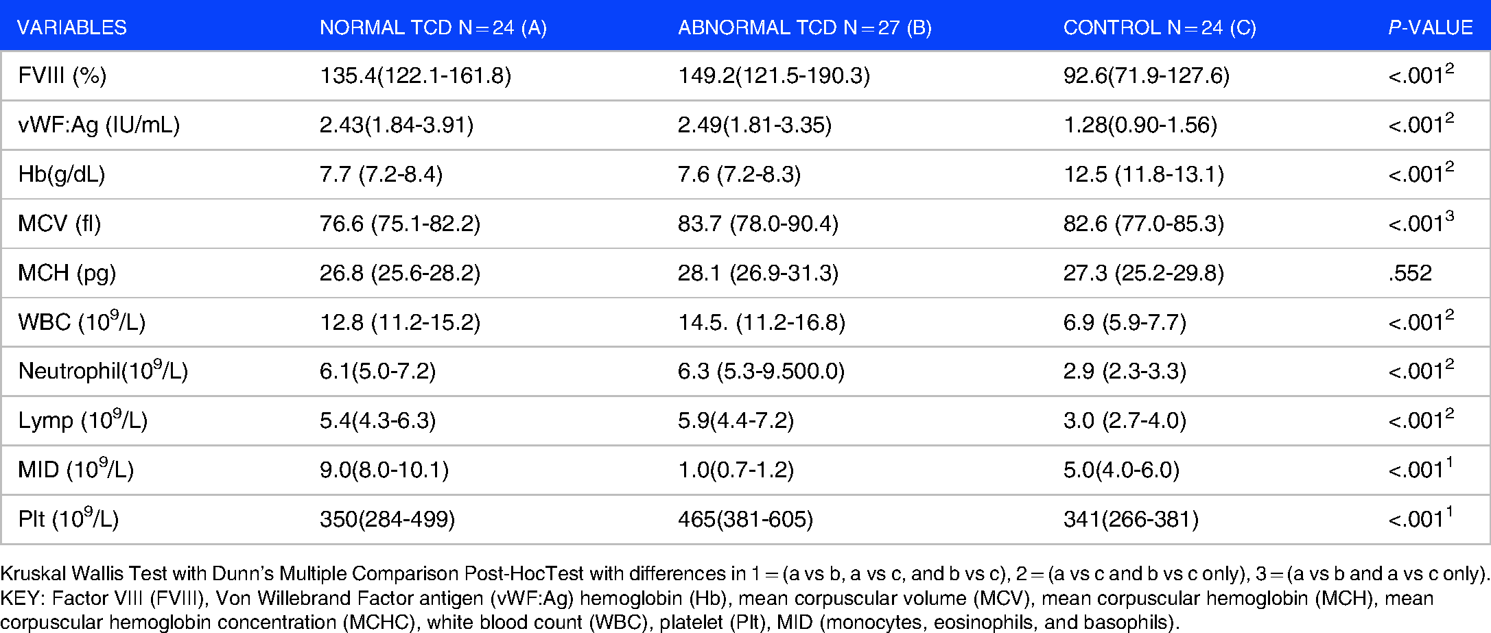
Kruskal Wallis Test with Dunn’s Multiple Comparison Post-HocTest with differences in 1 = (a vs b, a vs c, and b vs c), 2 = (a vs c and b vs c only), 3 = (a vs b and a vs c only).
KEY: Factor VIII (FVIII), Von Willebrand Factor antigen (vWF:Ag) hemoglobin (Hb), mean corpuscular volume (MCV), mean corpuscular hemoglobin (MCH), mean corpuscular hemoglobin concentration (MCHC), white blood count (WBC), platelet (Plt), MID (monocytes, eosinophils, and basophils).
Comparison of VWF, FVIII, and Hematological Parameters Between SCA Cohort and Hb AA Control
As shown in Table 2, there was a significant difference in the median value of TAMMV in patients with SCA who had a normal TCD [147 cm/s (140-157 cm/s)] when compared with the median value of those who had an abnormal TCD [217 cm/s (206-230 cm/s), P < .001].
Comparison of Median Haematological Parameters, vWF:Ag, FVIII, and Time Average Mean of Maximum Velocity Between Patients with Hb SS with Normal and Abnormal TCD.

**Statistically significant P < .05.
There were no statistically significant differences in Hb and WBC between children with SCA with normal (7.7 g/dL and 12.8 × 10⁹/L) and abnormal TCD (7.6 g/dL and 14.5 × 10⁹/L; P = .895 and .412). However, SCA children with abnormal TCD had a significantly higher mean platelet count [465 × 109/L (381-605 × 109/L)] compared to those with normal TCD [350 × 109/L (284-499 × 109/L)]; P = .018. Although SCA participants with abnormal TCD tended to have higher values of FVIII and vWF, there were no statistically significant differences in the median values of FVIII and vWF: Ag between children with SCA with normal (135.4%, 2.43 IU/mL) and abnormal TCD (149.2%, 2.49 IU/mL) (P = .583 and .571, respectively) (Table 2).
As shown in Table 3, there was a significant negative correlation between FVIII and time average mean of maximum velocity in the left middle cerebral artery (LMCA). However, there was no correlation between vWF: Ag, FVIII, and the time-average mean of maximum velocities in the other blood vessels.
Correlation Between vWF: Ag, FVIII and the Time-Average Mean of the Maximum Velocity (TAMMvel) in the Middle Cerebral Artery (MCA), Internal Carotid Artery (ICA) and Anterior Cerebral Artery (ACA) of SCA Patients.

*Statistically significant P < .05.
As shown in Table 4, the levels of FVIII, VWF, and WBC did not differ between HB SS participants with normal or abnormal TCD.
Association of FVIII, VWF, Plt, and WBC with TCD Velocity of SCA Patients.
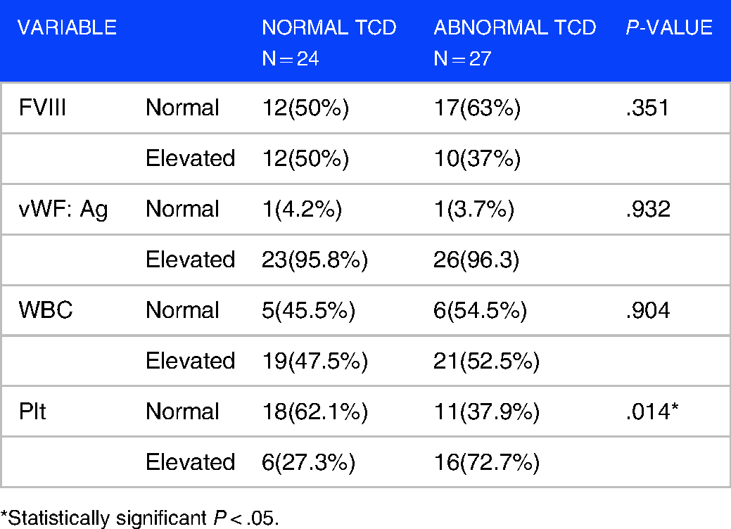
*Statistically significant P < .05.
However, the proportion of SCA participants who had normal platelet count along with normal TCD (62.1%) was greater than those with abnormal TCD (37.9%). Meanwhile, the proportion of SCA participants who had an elevated platelet count along with abnormal TCD (72.7%) was greater than those with normal TCD (27.3%). This difference was statistically significant (P = .014).
The proportion of SCA participants with normal WBC and normal vWF (51.4%) was greater than those who had normal WBC and elevated vWF (48.6%), while a greater proportion of participants with elevated WBC also had elevated vWF (95%), (P < .001).
Moreover, a greater proportion (95.7%) of SCA participants with elevated platelet count also had elevated vWF (P = .004). There were no statistically significant relationships between FVIII and WBC or platelet count (Table 5)
Association of White Blood Cells and Platelet Counts with vWF and FVIII of SCA Patients.
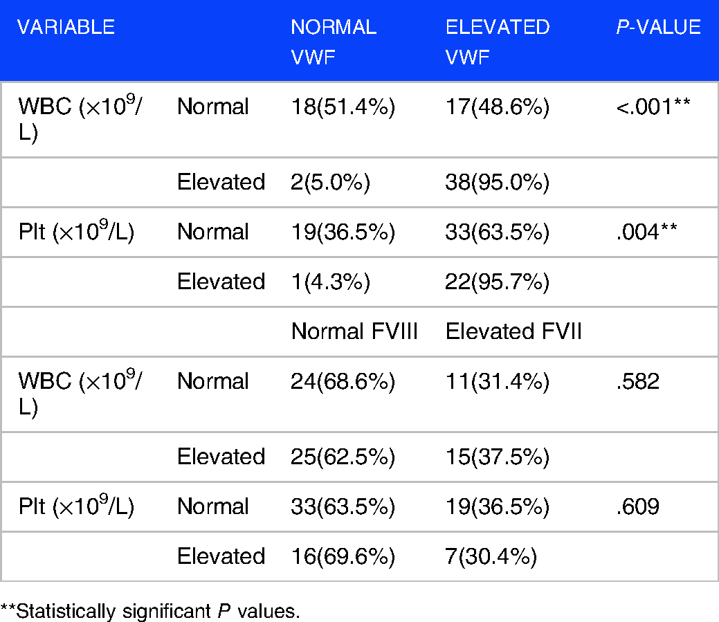
**Statistically significant P values.
Discussion
Stroke is a common cause of death and poor life-quality among children with SCD. The risk of stroke incidence has been found to increase with abnormal TCD velocity and a hypercoagulable state which is characterized by elevated procoagulant factors, such as vWF and FVIII. There are limited studies comparing vWF and FVIII levels in patients with SCA along with their TCD velocities. Therefore, this study aimed to determine the role of the levels of procoagulant factors in the development of abnormal TCD velocity which is highly predictive of stroke in children with SCA.
In this study, FVIII and vWF: Ag levels were significantly higher in children with SCA than in controls. This finding is consistent with reports from other studies.7,21–24 The elevated levels of FVIII and vWF may be attributed to the constant hemolysis of sickled red cells and vascular blockage by sickled erythrocytes which leads to vascular endothelial activation and secretion of coagulation proteins such as large quantities of ultra large and hyper-adhesive vWF molecules. In addition, stasis, combined with increase FVIII levels, may lead to thrombotic complication including interruption of blood flow in the cerebral circulations, thereby leading to increased TCD velocity. High TCD velocity is associated with stroke risk. This was also demonstrated via the correlation of FVIII with LMCA in this study. Nevertheless, this association appears to be an isolated finding as none of the other cerebral vessels assessed in this study showed any significant relationship with either FVIII or vWF. Hence, we do not know why Left and not Right side was affected with FVIII. Nevertheless, our observation may suggest possible differential effects of these procoagulant factors on the various segments of the cerebral circulation. Therefore, more studies are required to investigate whether there is any relationship between particular segment(s) of cerebral circulation and the levels of FVIII or vWF.
Moreover, children with SCA enrolled in this study had a lower PCV, higher WBC, and higher platelet count when compared with the controls. Similar reports have been documented in earlier studies conducted in Nigeria.25–28 The low PCV is expected because patients with SCD usually have chronic hemolysis resulting from sickling-induced damages to the red cell membrane. The increase in WBC is also expected because sickled RBC interact with and adhere to leucocytes, thereby increasing leucocytes recruitment to the vessel wall and mediating increased inflammatory response. 29 In addition, leukocytosis has been implicated in the pathogenesis of silent cerebral infarction in SCD. 30 The correlations among WBC and the levels of FVIII and vWF point to possible synergies in their procoagulant and proinflammatory roles. However, these did not seem to translate to vascular abnormalities as no relationship was found between WBC and abnormal TCD velocity. This raises the question as to whether there are other epigenetic factors involved in the genesis of abnormal TCD velocity other than those mentioned above.
The observed relationship between abnormal TCD velocity and platelet count in this study may be attributed to the increased platelet activation that occurs in patients with SCD. 31 Our finding is similar to reports from a previous study conducted in Brazil where patients with SCA who had abnormal TCD also had increased platelet counts. 32 Studies have shown that platelet count greater than 450 000 /µL is associated with stroke in SCD.33,34 The median platelet count of our study participants with abnormal TCD velocity was 465 000 /µL and greater than the recommended cutoff values for stroke risk. This further affirms the possible roles of platelets in thrombotic complications and their relationship with TCD velocity among our SCA cohort. These observations indicate that platelet count may have a stronger correlation with abnormal TCD velocity among our cohort than with WBC.
Although it was anticipated that the procoagulant markers would have a stronger correlation with higher and abnormal TCD velocities due to increased endothelial activation seen in SCA, this was not the case in this study. Possible explanations for our findings could be the roles of some epigenetic factors that could be masking the effects of these markers. For example, certain polymorphisms in vWF have been found to significantly raise the risk of ischemic stroke. 35 Furthermore, other mechanisms such as increased activity of vWF could also contribute to the risk of abnormal TCD velocity and stroke. ADAMTS13 is a major down-regulator of vWF activity; thus, low levels of ADAMTS13 are associated with an increased risk of ischemic stroke. 36 vWF activity was not measured in this study because this could perhaps show better correlation with abnormal TCD velocities and stroke risk.
Although a few earlier studies have examined the roles of vWF in stroke development among other patients without SCD,37–39 there are limited studies comparing vWF and FVIII levels in patients with SCA with abnormal TCD velocity, thereby highlighting the contribution of the current study towards understanding the roles of these factors in TCD abnormality. Similar to our finding, a study conducted in Nigeria documented no statistically significant differences in the levels of vWF: Ag of patients with hypertension with stroke compared with those without stroke. 37 In contrast, some other non-local studies have documented increased vWF in symptomatic patients who had carotid artery stenosis when compared with asymptomatic patients.38,39 further affirming the possible roles of variable and local epigenetic factors. These observations raise the need for more local studies on the roles of procoagulant markers (vWF and FVIII) as stroke phenotype markers in patients with SCA across different localities.
Conclusion
This study showed that children with SCD had elevated vWF and FVIII levels and those with abnormal TCD velocity had elevated platelet count, thus showing that a relationship exists between elevated platelet count and abnormal TCD velocity. Therefore, SCD children with elevated platelet count are to be closely monitored for abnormal TCD velocity and increased risk of developing stroke. The isolated finding of the correlation between TCD velocity and FVIII levels in the left middle cerebral artery raises the need for further studies to explore the roles of FVIII and vWF as stroke phenotype markers among pediatric SCA patients.
Study Limitations
Owing to the lack of facilities/resources, we did not assay the activity of vWF nor determine the presence of some genetic influencers of SCD, such as HbF and Alpha Thalassemia, which could impact several parameters in patients with SCA including the TCD velocity.40,41 Despite these limitations, this study was able to establish, for the first time in our locality, that children with SCA had higher levels of FVIII and vWF. Furthermore, our study established that there exists a relationship between abnormal TCD velocity, platelet count, and left middle cerebral artery.
Supplemental Material
sj-docx-1-bdx-10.1177_26348535221130289 - Supplemental material for Relationships Between Transcranial Doppler Velocity, Von Willebrand Factor, Factor VIII, and Hematological Parameters in Children with Sickle Cell Anemia: A Comparative Cross-Sectional Study
Supplemental material, sj-docx-1-bdx-10.1177_26348535221130289 for Relationships Between Transcranial Doppler Velocity, Von Willebrand Factor, Factor VIII, and Hematological Parameters in Children with Sickle Cell Anemia: A Comparative Cross-Sectional Study by Eniola Kehinde Enifeni, Ann Abiola Ogbenna, Alaba Olanrewaju Daramola, Ademola Samson Adewoyin, Oladele Simeon Olatunya and Edamisan Temiye in Plasmatology
Footnotes
Author Contributions
Availability of Data
The data used for this study is available from the corresponding author upon reasonable request.
Ethics Approval
The study was approved by the Ethics and Research Committee of the Lagos University Teaching Hospital (NHREC:19/12/2008).
Supplemental Material
Supplemental material for this article is available online.

References
Supplementary Material
Please find the following supplemental material available below.
For Open Access articles published under a Creative Commons License, all supplemental material carries the same license as the article it is associated with.
For non-Open Access articles published, all supplemental material carries a non-exclusive license, and permission requests for re-use of supplemental material or any part of supplemental material shall be sent directly to the copyright owner as specified in the copyright notice associated with the article.
