
Abstract
Background
Uterine leiomyosarcomas are rare and aggressive malignancies of the uterine corpus with high recurrence rates and poor prognoses. The current recommendation for detection of recurrent uterine leiomyosarcoma involves periodic physical examination and conventional imaging such as CT or MRI. The role of fluorine-18-fluorodeoxyglucose positron emission tomography with integrated computed tomography (FDG-PET/CT) in the detection of recurrent uterine leiomyosarcomas is not yet established.
Purpose
To evaluate the use of FDG-PET/CT as a single integrated modality for the evaluation of suspected recurrent uterine leiomyosarcomas.
Material and Methods
A retrospective study was performed on patients who underwent FDG-PET/CT scans for suspected recurrent uterine leiomyosarcoma. Only patients with follow-up data were included in the study. FDG-PET/CT was evaluated as a single integrated imaging modality. A positive lesion on FDG-PET/CT was defined as a focal abnormality detected on either the PET or CT components, or both.
Results
Sixteen consecutive patients over 5 years underwent FDG-PET/CT for suspected recurrent uterine leiomyosarcoma. Five patients were excluded due to incomplete follow-up data. The remaining 11 patients were aged 36–58 years (mean age 48). FDG-PET/CT had a sensitivity of 100% (95% CI 63–100) and specificity of 100% (95% CI 20–100) for the detection of recurrent uterine leiomyosarcomas. Sites of metastases include lungs, peritoneum, liver, pancreas and breast, of which lungs and peritoneum were the most common. Two (18%) patients had discordant findings: FDG-PET negative metastatic nodules in the breast and lung detected on the CT component. The maximum standardized uptake value (SUVmax) of metastatic lesions ranged from 2.0 to 16.0 (mean 7.6).
Conclusion
FDG-PET/CT as a single integrated modality may be a useful for the evaluation of suspected recurrent uterine leiomyosarcomas. FDG-PET negative discordant nodules detected on the CT component may represent metastases and should be followed up closely.
Uterine sarcomas are a heterogeneous group of rare but aggressive tumors, accounting for only 3–7% of all uterine corpus malignancies (1, 2). They have a tendency for local recurrence, distant metastasis and generally have a poor prognosis (1, 2). Uterine leiomyosarcomas are one of the common subtypes of uterine sarcomas (3). The current recommendation for detection of recurrent uterine leiomyosarcoma involves periodic physical examination with conventional imaging such as computed tomography (CT) or magnetic resonance imaging (MRI) (4).
Fluorine-18-fluorodeoxyglucose (FDG) positron emission tomography with integrated computed tomography (PET/CT) is a hybrid functional and anatomical imaging modality based on the observation that high-grade malignancies exhibit higher glucose metabolism relative to non-malignant tissue (5). Fluorine-18 is a cyclotron-produced positron emitter with physical half-life of 110 minutes. It is incorporated into the glucose molecule to form FDG, a glucose analogue radiopharmaceutical. FDG is intravenously injected and undergoes cellular uptake similar to glucose. Upon phosphorylation by hexokinase into FDG-6-phosphate, its metabolism is arrested leading to intra-cellular accumulation. By this principle, FDG-PET/CT has emerged as a highly accurate functional imaging modality in the detection of regional and metastatic disease for many types of malignancies such as lymphomas and non-small cell lung cancer (5).
Due to the rarity of uterine leiomyosarcomas, there is little published literature describing the clinical role of FDG-PET/CT for its detection. Hence, the overall utility of FDG-PET/CT in uterine leiomyosarcomas has not yet been established (6). The purpose of this study was to evaluate the use of FDG-PET/CT for suspected recurrent uterine leiomyosarcomas.
Material and Methods
A retrospective review of scan reports was performed on patients who underwent FDG-PET/CT scans at the Department of Nuclear Medicine and PET, Singapore General Hospital from February 2004 to February 2009 for suspected recurrent uterine leiomyosarcoma. Institutional ethics approval was not required. Only patients with follow-up data were included in the study. Follow-up data involved either histopathological correlation of positive FDG-PET/CT findings or surveillance CT imaging for a minimum of 1 year after FDG-PET/CT.
All scans were performed on a dedicated hybrid PET/CT scanner (Siemens Biograph LSO, Siemens Medical Solutions, Erlangen, Germany). Whole-body FDG-PET imaging was acquired from the skull vertex to the femur over seven bed positions at 3 minutes each. Unenhanced CT scan (single-slice, 110 mAs, 130 kV) were obtained for attenuation correction and anatomical correlation. Patient preparation involved 6 hours of fasting and a serum glucose level below 180 mg/dL (10 mmol/L) prior to FDG injection. The administered FDG activity ranged between 333–444 MBq (9–12 mCi). FDG-PET/CT images were reviewed in the coronal, transaxial and saggital planes by two Nuclear Medicine physicians on a Syngo workstation (Siemens Medical Solutions, Erlangen, Germany) and any disagreement was resolved by consensus. The CT component was interpreted in cooperation with a diagnostic radiologist.
FDG-PET/CT was evaluated as a single integrated imaging modality. A positive lesion on FDG-PET/CT was defined as a focal abnormality detected on either the PET or CT components, or both. A FDG-avid lesion was defined as a focus of increased FDG uptake above the surrounding background FDG activity, regardless of the maximum standardized uptake value (SUVmax). The reported SUVmax of FDG-avid metastatic lesions were collected. Results were tabulated and analyzed by standard non-commercial statistical software.
Results
A total of 16 consecutive patients underwent FDG-PET/CT for suspected recurrent uterine leiomyosarcoma over a 5-year period. Five patients were excluded due to incomplete follow-up data. The remaining 11 patients had adequate follow-up data and were included into the study. Patients were aged 36–58 years (mean age 48). All 11 patients underwent FDG-PET/CT to investigate suspected recurrent uterine leiomyosarcoma. All had total hysterectomy with bilateral salpingo-oophorectomy as primary therapy. Of these, three patients had pelvic lymph node dissection, one patient had adjuvant radiotherapy and one patient had adjuvant chemo-radiotherapy. Nine patients with positive FDG-PET/CT findings were followed up with correlative histopathology. Two patients with negative FDG-PET/CT findings were followed up with surveillance CT for at least 1 year.
A wide range of FDG-avid lesions were detected in the nine patients with positive FDG-PET/CT findings, which included lungs, peritoneum, liver, pancreas and breast (Figs. 1–3). All were subsequently proven histologically to be metastatic uterine leiomyosarcomas. Lungs and peritoneum were the most common sites of metastases. The SUVmax of these lesions ranged from 2.0 to 16.0 (mean 7.6). The results are summarized in Table 1.
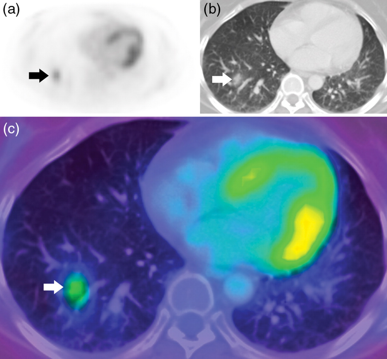
Transaxial view of a FDG-avid (SUVmax 3.5) lung nodule in the right lower lobe depicted in (a) PET, (b) CT and (c) fusion PET/CT views. The patient subsequently underwent right lower lobe wedge excision and histopathology confirmed metastatic uterine leiomyosarcoma
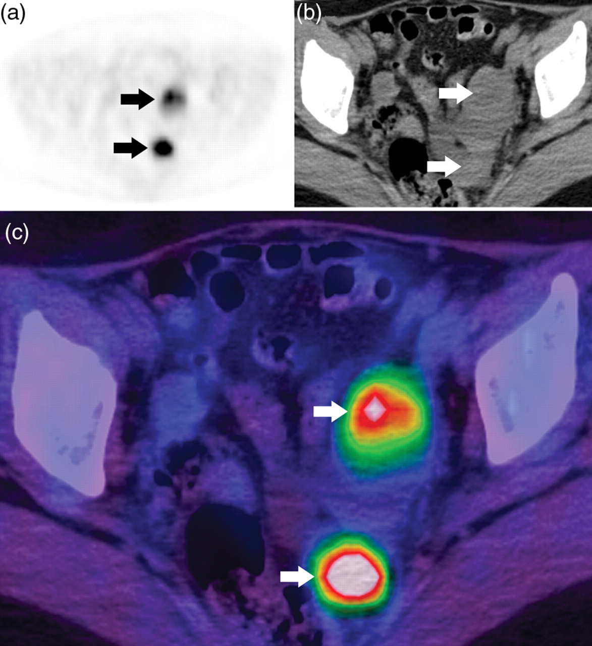
Transaxial view of two FDG-avid (SUVmax 16.0) peritoneal nodules in the pelvis depicted in (a) PET, (b) CT and (c) fusion PET/CT views. The patient subsequently underwent surgical excision of the peritoneal nodules and histopathology confirmed metastatic uterine leiomyosarcoma
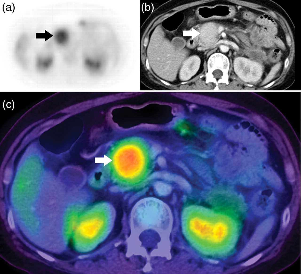
Transaxial view of a FDG-avid (SUVmax 6.4) mass in the pancreatic head depicted in (a) PET, (b) CT and (c) fusion PET/CT views. The patient subsequently underwent Whipple's operation and histopathology confirmed metastatic uterine leiomyosarcoma
Summary of 11 patients with suspected recurrent uterine leiomyosarcoma
*Follow-up CT scan
†Correlative histopathology
‡FDG-PET negative but detected on CT component
SUVmax = maximum standardized uptake value, FDG-PET/CT = fluorine-18-fluorodeoxyglucose positron emission tomography with integrated computed tomography, N/A = not applicable
There were two (18%) patients with discordant FDG-PET/CT findings. One patient with FDG-PET positive metastases also had a 0.9 cm FDG-PET negative right breast nodule detected on the CT component, which was later excised and histologically proven to be metastatic uterine leiomyosarcoma. Another patient had a 1.4 cm FDG-PET negative pulmonary nodule at the base of the right lower lobe detected on the CT component, which was excised and histologically proven to be metastatic uterine leiomyosarcoma (Fig. 4). Two patients with negative FDG-PET/CT findings were followed up by CT imaging for a minimum of 1 year which showed no scan evidence of recurrent disease.
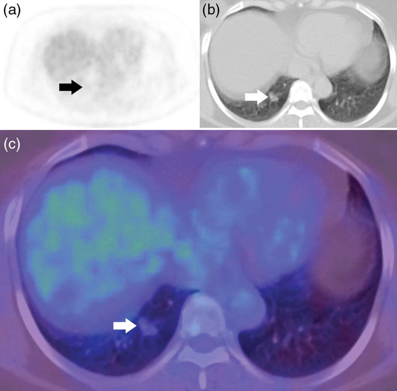
Transaxial view of a 1.4 cm non-FDG avid lung nodule at the right lung base depicted in (a) PET, (b) CT and (c) fusion PET/CT views. The patient subsequently underwent right lower lobe wedge resection and histopathology confirmed metastatic uterine leiomyosarcoma
Overall, FDG-PET/CT as a single integrated modality had a sensitivity of 100% (95% CI 63–100) and specificity of 100% (95% CI 20–100) for the detection of recurrent uterine leiomyosarcomas. As this study had evaluated FDG-PET/CT as a single integrated modality, the two cases of FDG-PET negative metastases did not decrease the overall accuracy as they were detected on CT component as discordant nodules.
Discussion
The aim of this study was to evaluate the use of FDG-PET/CT for suspected recurrent uterine leiomyosarcomas. We found that FDG-PET/CT as a single integrated modality had good sensitivity and specificity for its detection. Careful evaluation of the CT component may circumvent cases of FDG-PET negative discordant nodules and improve the overall accuracy of FDG-PET/CT as a single integrated modality.
Uterine leiomyosarcomas are rare but aggressive malignancies of the uterine corpus with high recurrence rates and poor prognoses (1, 2, 7). To date, the clinical utility of FDG-PET/CT for the detection of recurrent uterine leiomyosarcomas has yet to be defined (6). FDG-PET/CT is a functional study of relative glucose metabolism between malignant and non-malignant tissue (5). Many cancers demonstrate increased FDG uptake by means of GLUT-1 glucose transporter up-regulation, which is detectable on FDG-PET/CT (8). In uterine leiomyosarcomas, Nagamatsu et al. reported a significant difference in GLUT-1 expression score between uterine leiomyosarcomas and benign uterine leiomyomas (9). They also found a strong correlation between GLUT-1 expression score and the SUVmax of uterine tumors on FDG-PET (9). At a cellular level, Rao et al. reported that up to 50% of uterine leiomyosarcomas were positive for GLUT-1, which also correlated with aggressive behavior (10).
There are few published studies on the clinical utility of FDG-PET/CT in detecting recurrent uterine leiomyosarcomas. It had been suggested that FDG-PET/CT may have a role in improving the early detection of locally recurrent and metastatic disease, thereby enabling earlier therapeutic intervention (11, 12). Our study found that FDG-PET/CT as a single integrated modality had a sensitivity and specificity of 100% in detecting recurrent uterine leiomyosarcomas, in concordance with other studies (11, 12). Murakami et al. evaluated the use of FDG-PET without integrated CT to detect disease recurrence in four patients with treated uterine leiomyosarcomas and found 100% sensitivity and specificity (11). Park et al. found the same results in a study of 14 asymptomatic patients evaluated with either FDG-PET or FDG-PET/CT for surveillance of treated uterine leiomyosarcomas (12).
The two FDG-PET negative breast (0.9 cm) and lung (1.4 cm) discordant nodules detected on the CT component were histologically proven metastases. The GLUT-1 expressions in these discordant nodules were not examined, but they are likely to represent low-grade metastases. This highlights the importance of careful evaluation of the CT component for the detection of FDG-PET negative nodules, which may represent metastatic uterine leiomyosarcoma.
A limitation of this study was the small sample size of only 11 patients, accounting for the wide 95% confidence interval in the statistical results. There was also a referral bias, as all the patients who were referred for FDG-PET/CT had high pre-test probabilities for recurrent disease. These factors preclude any definitive conclusions to be drawn from this study.
In conclusion, FDG-PET/CT as a single integrated modality could be a useful imaging modality for the evaluation of suspected recurrent uterine leiomyosarcomas. Larger studies on this topic are recommended. FDG-PET negative discordant nodules detected on the CT component may represent metastases and should be followed-up closely.