
Abstract
Background
Simplifying a postoperative surveillance protocol for endovascular aneurysm repair (EVAR) requires quality control comparing computerized tomography (CT) and ultrasound (US) results of abdominal aortic aneurysm (AAA) diameter measurements and endoleaks.
Purpose
To test if US is comparable to CT, then assess a simplified follow-up with our conventional surveillance to assess patient safety.
Material and Methods
During 2001-2006, data on 56 patients treated with Talent stent graft were prospectively registered. Median follow-up was 41.5 months (range, 2-94 months), with CT, US, and plain film abdomen X-rays (PFA) at 1, 6, and 12 months, then yearly. Bland-Altman plot was used to assess the agreement between CT and US measuring the AAA diameters and mixed model by the time effect to assess the difference in diameter over time. Sensitivity and specificity for detection of endoleaks by US, with CT as ‘gold standard’ were calculated. A simplified surveillance protocol with US/PFA at 6 and 8 weeks, CT/US/PFA at 1 year, and yearly US/PFA thereafter, was evaluated. CT was carried out when poor visibility, endoleak detected, AAA diameter increase (≥5 mm) on US or migration (≥10 mm) on PFA. This regime was compared with our conventional follow-up protocol.
Results
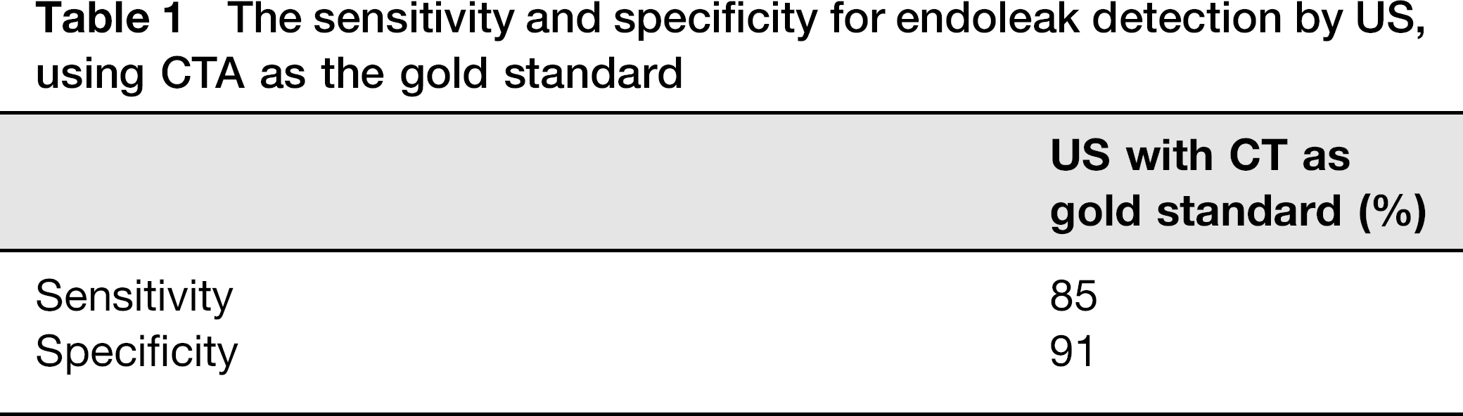
Diameter measurements on US appear comparable to CT with 91% specificity and 85% sensitivity for endoleaks detected by US. Using the simplified surveillance protocol no endoleaks, migrations, or endotension requiring treatment were overlooked. The simplified protocol generated 53 selective CT scans, avoiding approximately 144 CT scans. If further simplified by omitting the 1-year CT scan, one type II endoleak would be missed with a 1-year delay, eliminating a further 45 CT scans.
Conclusion
US appears comparable to CT in the follow-up of Talent stent grafts in our institution. The proposed simplified surveillance protocol seems safe and can lead to a significant reduction in the number of CT scans.
Endovascular aneurysm repair (EVAR) of abdominal aortic aneurysms (AAA) has the advantage of lower operative systemic morbidity and mortality, balanced by the need for lifelong follow-up and risk of re-interventions (1). The detection of endoleak, aneurysmal growth, stent graft migration, structural damage, and graft-limb occlusions pose a considerable challenge and expense (2). Computerized tomography angiography (CTA) is considered the gold standard for detection of endoleak and postoperative aneurysmal growth. The drawbacks, however, include radiation exposure, the risk of contrast-induced nephropathy, allergic reactions, and indeed the considerable expense (3, 4). Duplex ultrasound scan (US) is non-invasive, safe, and inexpensive. Nonetheless it is operator-dependent and visualization may be challenging (5, 6). Several institutions have advocated the benefits of a simplified surveillance protocol with duplex ultrasound and plain film of abdomen (PFA), given that the aneurysm is shrinking or has been stable over 2 years (7, 8). Postoperative shrinkage or stable AAA diameter has become an indicator of successful treatment following EVAR.
Simplifying a postoperative surveillance protocol for EVAR requires quality control of computerized tomography (CT) and ultrasound (US) results comparing AAA diameter measurements and endoleak. The aim of our study was to assess if CT and US were comparable and evaluate a modified follow-up protocol with our conventional surveillance to assess safety and reduction in the number of CT scans.
Material and Methods
The prospective data includes 50 male and six female patients treated for AAA with a Talent stent graft during the period 2001-2006. The median age was 74 years (range, 55-87 years), with a median AAA diameter 60 mm (range, 51-80 mm) and the median length of follow-up was 41.5 months (range, 2-94 months). Our conventional follow-up included CTA, US, and plain film X-rays at 1, 6, and 12 months and thereafter annually. The anterior-posterior diameter of the postoperative AAA and the presence and type of endoleak by CTA and US scan, were extracted from our prospectively registered database. We examined our CTA and US findings during follow-up to evaluate the usefulness in endoleak and AAA growth detection.
A simplified surveillance protocol was compared to our conventional program. The simplified protocol included an US/PFA at 6 to 8 weeks, CT/US/PFA at 1 year, with yearly PFA and US thereafter. The criteria for selective use of CTA, in addition to US, were: poor visibility, the presence of endoleak, AAA diameter increase on US (≥5 mm) or migration (≥10mm) on PFA. The comparison with the conventional follow-up was used to see if patient safety was compromised, missed endoleaks, or expanding AAA.
CT scan protocol
Helical CT scans were carried out with 1-3-mm slices from the diaphragm to mid-thigh. Three series were carried out: non-contrast, arterial, and delayed series with contrast to identify endoleak. Each examination was validated by two radiologists and was independent of the US results. Diameter measurements were performed for anterior-posterior view using electronic caliper tool. The maximum AP diameters on the aneurysms were measured on CT from axial images, the same way in each individual patient over time. In patients with distorted anatomy, both AP diameter on axial slices and the “true” diameter were measured, based on sagittal and coronal images.
US scan protocol
All US scans were carried out in our vascular laboratory by a single dedicated vascular physician. A Vingmed (System V; Horten, Norway) and Logiq E9 (GE Healthcare, Wauwatosa, WI, USA) US machine with a 5 and 7 MHz curvilinear transducer were used. The patients were not fasting. The examination included a longitudinal and transverse interrogation of the AAA and iliac arteries diameters. Peak systolic velocities were carried out in the iliac vessels to assess for the presence of kinking or flow abnormalities. Endoleak detection was carried out by detection of color flow signals within the aneurysm and confirmed by measuring Doppler velocity signals in the relevant AAA region. Pathological movements of the graft were also noted. Almost all examinations where carried out the same day as the CT. We did not employ contrast-enhanced US in our institution despite the fact that this is a promising imaging modality (9, 10).
Plain film of abdomen
All patients had a plain film of the abdomen (PFA) performed in AP and lateral projections to look for migration and structural failure of the stent graft. This is carried out using a validated standardized protocol (11, 12).
Statistics
Bland-Altman plot was applied to assess the agreement between US and CTA measuring the AAA diameter. Standard deviation (SD) of the individual differences was used to calculate the upper and lower limits of agreement. The Bland-Altman plot graphs the average of the two values (US-CT) for each patient on the X axis, and the difference between the measurements on the Y axis. The difference between the two lines is denoted as “d”. Ninety-five percent limits of agreement were defined as bias (d) +/− 1.96 × SD. Provided that the difference located within the 95% limits of agreement the US and CTA can be used interchangeably. If the average of “d” is close to zero and the 95% confidence interval includes zero on the Y axis it can be concluded that there is no clinical or statistical difference between US and CTA.
The difference between US and CTA during the observation period was assessed by an interaction term between methods and time in a mixed repeated measure model. Change in mean diameter over time was assessed in the mixed model by the time effect (13, 14). SPSS® version 15.0 (SPSS Inc., Chicago, IL, USA) was used for statistical analysis.
Sensitivity and specificity for detection of all types of endoleaks for US, with CTA as ‘gold standard’ were calculated. Positive detection of endoleak on CTA and US was defined as true-positive, true-negative when no endoleak was detected on CTA and US, and false-positive when US was positive and CTA negative. The study was approved by the hospitals ethics committee and performed with written informed consent for all patients.
Results
The mean difference in diameter between US and CTA is shown in Fig. 1. The Bland-Altman plots show that the mean differences are 1 mm or less from the lines of equality during the measurement period. Ninety-five percent limits vary from −3 to +7 mm from the line of equality. The differences between measurements performed by the two methods lie within limits of agreement 95% of the time. There was a significant reduction in mean diameter during follow-up, comprising an average of 2.2 mm between each follow-up interval of 4 weeks, 1, 2, and 3 years (Fig. 2). The figure must be interpreted clinically. As long as the variability is consistent across the graph there is no interaction. During the 3-year period the mean difference in diameter with both CTA and US seems to be reduced with approximately 7–8 mm, which is statistically significant. We calculated the sensitivity and specificity of endoleak detection using US as shown in Table 1.
Bland-Altman plot showing the distribution of measurements over and below two standard deviations at 4 weeks, 1 year, 2 years, and 3 years The reduction in mean diameter with 95% confidence interval (CI) measure of the CT and US. There was a significant reduction in mean diameter during follow-up periods of 9 mm (P < 0.01).

The sensitivity and specificity for endoleak detection by US, using CTA as the gold standard
Our primary results (<30 days) using the conventional surveillance protocol was: two type I endoleak (4%), one type III (2%), and nine type II (16%). Two patients had primary graft limb occlusion (4%). There were no deaths in the first month.
Secondary results (> 30 days): there were no secondary occlusions or aneurysm-related mortality. However, nine patients (16%) died of other causes. Migration (>10 mm) was seen in four patients (7%). All of these were detected on PFA. One type II endoleak was seen at 6 months. In six patients we identified an increased diameter (≥5 mm) without evidence of an endoleak on CT, US or digital subtraction angiography - defined as endotension. Secondary interventions were performed on 14 patients (25%). Seven patients had endoleaks (12%): five were treated for endotension (9%) and two were treated for migration alone (4%).
The follow-up of these patients comprised US, CTA, and PFA at 1, 6, and 12 months then yearly. We have analyzed the results from our current protocol and compared this to a simplified surveillance specifically looking for endoleak, diameter increase, and other stent graft complications. This included US and PFA as the main imaging tools with selective use of CTA as described earlier. No endoleaks, migrations, or endotension requiring treatment would have been overlooked using our simplified surveillance protocol. This protocol generated 53 selective CT scans, but has eliminated 144 routine CT scans. In respect of the selective CTA we reduced this by 91 CTA in total.
We further observed that if we omitted the 1 year CT, one type II endoleak would be missed with a delay of 1 year, but reducing CT scans by 45. Poor visibility on US necessitating a CT scan was 3% in our patient cohort (seven examinations of 266). This has added the number of CT scans in our simplified surveillance protocol.
Discussion
In our institution the correlation between postoperative CT and US scan diameter measurements appears acceptable. No clinical significant systematic bias was shown when Bland-Altman plot was carried out, as the mean value deviated from the baseline with 0.6–1.0 mm. This has little clinical relevance as any US or CTA serial diameter measurement will have intrinsic variation of more than the above values (15).
There is a recurring over-estimation of AAA diameter of about 1 mm with US. We choose to use Bland-Altman plots as this is an established statistical method for assessing agreement between two methods of clinical measurements (13, 14). A shrinking aneurysm diameter is one of the main indicators for successful treatment and implies a risk reduction of postoperative AAA rupture (16-18). To compare changes in AAA diameter measurements using CT and US over time we used a mixed repeated measure model which showed that there were no significant variations between the two over time. In a simplified surveillance protocol an AAA diameter increase (≥5 mm) would warrant an additional CT scan.
We were inspired by the American Society of Vascular Surgery's recommendation of a surveillance protocol with CT scan at 1 and 12 months followed by US providing the diameter is stable or shrinking in the absence of endoleaks (19). We wanted to see if this protocol could be further simplified in our center. Several follow-up protocols have supported the use of duplex US in the follow-up after EVAR. Chaer et al. prospectively carried out US on patients 1 year after EVAR, when the AAA diameter <4 cm or after 2 years with stable or shrinking diameter. They concluded that US follow-up with selective use of CT was safe (8). Schmeider et al. compared CT and US in detection of endoleaks and concluded that US was better and thus an important tool in the follow-up of EVAR (20). Other centers have showed similar results (5, 8, 21, 22).
US has historically been challenged on its poor visibility in about 1.7-10% of follow-up EVAR patients (22, 23). In our experience we estimate that 3-7% would selectively need CT scan due to suboptimal US examination (unpublished data). Poor visualization is one of four criteria which would trigger the selective use of CT in our simplified surveillance protocol.
Larger series with larger patient cohorts also support the use of US in the surveillance of EVAR (8, 20-22). US in the follow-up of EVAR patients have been studied extensively with the conclusion that it is highly operator and machine dependent (24, 25). The use of US as the sole imaging modality is still controversial. A meta-analysis on the use of US compared to CT found a sensitivity of 91% and a specificity of 69% (23). The specificity of 91% and sensitivity of 85% of US for endoleaks in our institution appears acceptable, the number of endoleaks is small and the results therefore need to be interpreted with caution. We have no inter-observer variability in US measurements, as this was carried out by a single physician who is accustomed to scanning EVAR patients over several years. This is also a disadvantage as the follow-up is dependent on individual competence. The use of a standardized plain film of abdomen (PFA) with front and side views is useful for identifying stent graft migration, kinking, or structural failure (11, 12). These structural changes are difficult to detect on US or CT. The PFA confers a lower radiation dose to the patient than a CT. For this reason we included a standardized PFA in our follow-up protocol. Standardized plain film of abdomen, using a set protocol, are easily comparable over time and are a useful supplement to US and CT (11, 12). Validation of the correlation in aneurysm diameter and endoleak between CT and US in our institution will enable a greater degree of US follow-up, with a selective use of CT. Training personnel for this purpose in a core laboratory, may provide considerable savings where EVAR is standard treatment modality. In order to achieve this US should be validated and increasingly replacing CT, which then can be used selectively. The benefits of this protocol include decreased exposure to radiation, contrast exposure, and economic savings.
We retrospectively examined the impact of a new follow-up regime using US and PFA as the main imaging tools during follow-up. The detection of an intra- or postoperative endoleak, increased AAA diameter, poor visibility on US, kinking, or migration of the graft (PFA) triggered a CT scan. The selective use of CT enabled us to identify all complications requiring secondary intervention and resulted in an additional 53 CTs. This is a retrospective review and thus confers a certain bias. The results, however, are also supported by other published series, although these used CT at 1 and 12 months before proceeding to use US as the sole imaging modality (19). We conclude that this new protocol may be used with low risk to the patients. This may in our view significantly reduce the number of CTA during follow-up, lower the radiation exposure to each patient, and reduce the cost of follow-up. The final confirmation of these results will need to be confirmed in a prospective randomized trial.
An alternative to US follow-up is CT scan without use of contrast agents. The CT scan diameter measurements are used to evaluate growth in a patient cohort and contrast series added if AAA growth is detected. Diaz et al. found that less than 10% would benefit from a CT scan with contrast during follow-up, and that simple AAA diameter measurements in addition to PFA will identify the majority of the asymptomatic patients requiring re-intervention (16). The disadvantage is that the patient is still exposed to radiation and a CT will augment the cost of EVAR follow-up.
Our data reflect the results from a single center with a retrospective review of prospectively registered data. We have evaluated patients treated with Talent stent graft in this series and thus these conclusions can only apply to these. We also need to treat our results with caution due to the size of the study.
In conclusion, US appears to be a viable alternative to CT scan during follow up of EVAR in our institution. Eliminating CT after 1 month and after 1 year appears safe provided the indications of good visibility, no detectable endoleaks, a stable or shrinking AAA diameter on US, and no migration on PFA. We can reduce the number of CTA during follow-up of EVAR patients, with the benefit of reducing radiation, contrast exposure, and with significant reduction in costs.