
Abstract
Background
Radiation-induced brain injury (RBI) is the most serious complication of primary and metastatic brain and neck malignant tumors following radiation therapy. However, at present, RBI is difficult to diagnose in the early period. Recently, studies have demonstrated that the early stage of RBI is characterized by an inflammatory reaction, and that intercellular adhesion molecule-1 (ICAM-1) is significantly up-regulated in the irradiated brain tissues.
Purpose
To provide an early diagnosis of RBI using molecular magnetic resonance imaging (MRI) with microparticles of iron oxide (MPIO) targeted to ICAM-1 in the vascular endothelium of brains.
Material and Methods
A monoclonal antibody against ICAM-1 was conjugated to MPIO to form the targeted MRI contrast agent ICAM-MPIO. The adhesion of ICAM-MPIO to endothelial cells was quantified by optical imaging and MRI. Sprague-Dawley rats were irradiated to establish an animal model of the early period of RBI. ICAM-MPIO and free-MPIO were injected via tail vein, respectively. T2 signal intensity and T2 values of the irradiated brains and normal brains were subsequently evaluated by MRI.
Results
In vitro, the adhesion of ICAM-MPIO to the activated endothelial cells was 5 ± 0.5-fold greater than to the non-stimulated cells, which could be detected by optical imaging and MRI (R2 = 1.0, P < 0.01). In vivo, ICAM-MPIO caused a marked negative MRI contrast effect in irradiated brains. As compared with brains without irradiation, the specific contrast effect increased more than seven-fold after administration of ICAM-MPIO (F = 751.495, P < 0.05).
Conclusion
MPIO coated with monoclonal antibody of ICAM-1 could be used for detecting the early period of RBI by optical imaging and MRI.
Radiation therapy is used widely for the treatment of various primary and metastatic malignant tumors of the brain and neck. With the development of modern clinical radio-therapeutic techniques, radiation can be delivered more precisely to the targeted tumors, limiting radiation damage to a core of diseased tissues. However, the irradiation fields associated with such therapy inevitably involve some normal brain tissues. For various sensitivities to irradiation and received radiological doses, some brain tissues exposed to the irradiation field consequently may become damaged following radiation therapy. Thus, radiation-induced brain injury (RBI), specifically, injury to normal brain tissues, is the key impediment to the delivery of curative radiation doses.
Despite these concerns, at present there are neither long-term treatments nor any preventive strategies to alleviate this RBI. Clinical imaging evaluation is limited to: the analysis of gadolinium (Gd) enhancement on magnetic resonance imaging (MRI); positron emission tomography (PET); and several emerging MRI methods such as perfusion-weighted imaging (PWI), magnetic resonance spectroscopy (MRS), and diffusion-weighted imaging (DWI). However, these imaging modalities have been shown to very poorly predict early pathogenesis or tissue destruction (1, 2). Furthermore, when RBI can be found by clinical imaging, it always progresses into a serious injury that cannot be repaired.
To date, the only reliable way to accurately diagnose RBI in its earliest – and thus most treatable – stage is surgical tissue sampling, an invasive procedure that is not always feasible. Thus, there is a pressing need for the development of non-invasive techniques that can identify the early pathogenesis of this condition; such early identification would lead to an accelerated accurate diagnosis of RBI and would help clinicians to provide RBI-specific therapy at an early stage.
The early stage of RBI is associated with an inflammatory reaction that causes increases in microvascular permeability and leukocyte infiltration of the irradiated brain tissues (3, 4). It has been reported that an important component of radiation therapy-specific neuroinflammation is the up-regulation of pro-inflammatory genes and proteins including ICAM-1 (5). ICAM-1 proteins mostly express on the luminal surface of the vessel endothelium and promote the recruitment and migration of leukocytes. These proteins play a causative role in the development of tissue damage, which is followed by late irradiated injury effects such as focal neurological deficits, memory loss, and other types of neurocognitive dysfunction (6). Recent studies have demonstrated a rapid induction and up-regulation of both ICAM-1 mRNA and protein in the early period of RBI (7, 8). However, as shown by Olschowka et al., there was little expression of ICAM-1 in the non-irradiated hemisphere (9). In this manner, the characteristic of ICAM-1 up-regulation in the irradiated hemisphere provides an accessible tag for the detection of the early period of RBI, but without the requirement for translocation across the blood-brain barrier (BBB), which has been shown to be always intact in the early period of RBI (10, 11). Kiani et al. showed that ICAM-1 antibody-conjugated microspheres can be selectively and preferentially targeted to irradiated brains (11). However, these targeted microspheres can only be detected by optical imaging, and the microsphere-administration and -imaging process is invasive.
Microparticles of iron oxide (MPIO), a type of negative MR contrast agent, consist of an iron oxide core encased within an inert divinyl benzene polymer shell, plus a fluorescent dye for optical co-localization. These agents have been shown to be useful for molecular imaging. First, they have an increased iron content per particle and produce larger r2*-relaxivity than conventional superparamagnetic iron oxide particles (SPIO) (12, 13). Second, MPIO are less susceptible to the non-specific vascular egress of uptake by endothelial cells, and therefore they retain specificity for endovascular molecular targets (14). Third, they are rapidly cleared from circulation owing to their 0.01–1 µm size, which results in low background contrast effects and enables rapid imaging post-administration (15–17).
At present, no targeted MR contrast-enhancing materials specifically designed to show changes in endothelial receptor expression to evaluate the early period of RBI in vivo and in vitro have been described.
Accordingly, the current study was designed to evaluate the utility of ICAM-MPIO as a targeted contrast agent for molecular MR imaging to non-invasively detect the early phase of RBI.
Material and Methods
Antibody conjugation to MPIO
The MPIO particles used in this study were purchased from Bangs Laboratories Inc., (Carmel, IN, USA), which were 0.96-µm superparamagnetic styrene-divinyl benzene inert polymer microspheres that contained a magnetite core (45.8%); for improved optical visualization, these particles also had a fluorescein-5-isothiocyanate dye (Dragon Green fluorescent probe) contained within the cross-linked polymer sphere (P(S/V-COOH) Mag/encapsulated).
Conjugation was performed chiefly according to the reported procedures (15, 16), and 100 µL microparticles were suspended in MES buffer (2-(N-Morpholino) ethanesulfonic acid hydrate) to activate the surface groups for antibody conjugation. MPIO were subsequently collected using a Dynal magnet (Invitrogen, Carlsbad, CA, USA), and the supernatant was discarded. After three washings, collected MPIO were re-suspended in phosphate buffered saline (PBS). Water soluble carbodiimide (WSC) was added to the microparticle suspension while the suspension was being mixed at room temperature (RT). After being collected by a Dynal magnet, MPIO were re-suspended in PBS; purified monoclonal mouse antibodies specific to rat or human ICAM-1 (Abcam, Cambridge, UK) were subsequently added to the microsphere suspension to react for 20 h at 4°C. Following this incubation, MPIO were collected using a Dynal magnet, and the supernatant containing any unbound antibody was discarded. MPIO were subsequently washed and re-suspended gently in 10 mL of quenching solution for 30 min. Finally, particles were washed and re-suspended in storage buffer at the desired storage concentration (2 mg Fe/mL).
To assess the conjugation between the antibody and MPIO, Cy3-conjugated rabbit anti-mouse secondary antibody (1:200; Invitrogen, Carlsbad, CA, USA) was added to the targeted microparticles and free-MPIO suspension, respectively, and allowed to react overnight at 4°C. The microparticles were subsequently observed by inverted microscope (Nikon Eclipsc Ti-U; ×20 objective, Tokyo, Japan).
ICAM-MPIO bound to stimulated endothelial cells
A human umbilical vein endothelial cell line (EA.hy926 cell) was purchased from American Type Culture Collection (ATCC, Manassas, VA, USA). Cells were cultured at 1 × 10⁶ cells per well in 25 cm Petri dishes and maintained in 3 mL of Dulbecco's Modified Eagle medium (DMEM, Sigma, St Louis, MO, USA) supplemented with 10% fetal bovine serum (FBS), 4 mM L-glutamine, 100 U/mL penicillin, and 0.1 mg/mL streptomycin; cells were incubated in a humidified incubator at 37°C in 5% CO2. Next, cells were incubated with graded doses of recombinant mouse TNF-α (R&D Systems, Paisley, UK), an inflammatory stimulus used to provoke ICAM-1 surface expression, for 20 h at 37°C to induce endothelial ICAM-1 expression. The stimulated and non-stimulated endothelial cells, at a density of 2 × 10⁵ cells, were respectively incubated in duplicate with ICAM-MPIO or control free-MPIO for several hours. In Prussian staining stage, cells were fixed with 4% paraformaldehyde (PFA) for 30 min, and incubated at 37°C with 2 mL Prussian blue solution comprising an equal volume of 20% hydrochloric acid aqueous solution and 20% potassium ferrocyanide (II) trihydrate for 30 min. MPIO binding to cells and the iron staining were assessed in four fields of view by inverted microscopy, and were quantified using the ImagePro Plus imaging software package (Media Cybernetics, Bethesda, MD, USA).
For a competitive inhibition experiment, stimulated endothelial cells were pre-incubated with a large amount of free ICAM-1 (1M) for 30 min before targeted MPIO were added to the medium.
In vitro MR scanning
The stimulated EA.hy926 cells, at a density of 1 × 10⁶ cells, were incubated with ICAM-MPIO and free-MPIO, respectively, at Fe concentrations of 0.01, 0.1, 1, 10, 15, and 20 µg/mL in the culture medium for 3 h. The cells were then washed several times with PBS before being trypsinized and subsequently suspended in a 2% high-grade agarose solution for imaging on a clinical MRI scanner (1.5 Tesla Philips Intera, Philips Medical Systems, Best, The Netherlands, B.V.) at RT. A fast spin-echo sequence (TSE) was used (TR, 1.8 ms; TE, 60 ms; Flip, 90°; FOV, 80 mm; matrix, 256 × 256; slice thickness, 2.0 mm). Gradient echo sequence FFE was also applied with the following parameters: TE, 23 ms; TR, 700 ms; Flip, 18°; FOV, 80 mm; matrix, 256 × 256; slice thickness, 2.0 mm. The signal intensities of the samples were determined using a circular 30 mm2 region of interest as previously described (18). Low signal areas, corresponding to MPIO binding, were quantified in eight images per sample using ImagePro Plus.
Animal model of the early period of RBI
A total of 30 Sprague-Dawley rats (no set limit of male or female animals), with SPF grade, weighted (100 ±20) g, were selected from our university's Animal Experiment Center (license numbers: 0089977, 0090070, and 0090180). Our study of the animal model of the early period of RBI was performed chiefly according to previously reported procedures (19, 20). All rats were randomly divided into an experimental group (n = 21) and blank control group (n = 9).
Prior to irradiation, animals were sedated with chloral hydrate solution (40 mg/100 g, i.p.) and restrained in the sternal recumbent position on a treatment table. The head was centered in the exposure field with the eyes, neck, and body protected with customized lead shielding. The upper bound of the exposure field was the horizontal line between the eyes, and the lower bound was the horizontal line between the ears. A single dose (30 Gy) of radiation was delivered locally to the brain at a rate of 6 Gy/min using a Siemens Primus-H linear accelerator (Siemens Medical Solutions, Erlangen, Germany) (6 MV X-rays) located at our hospital. The rats in the sham control group received 0 Gy of irradiation but underwent the same stress as the others. Animal care and the experimental procedures in this study were approved by our university's Animal Care and Ethics Committee.
Immunohistochemical procedure
At different time points (24 h, 48 h, 5 days, 7 days) following irradiation, the irradiated rats (n = 12) and blank control rats (n = 6) were anesthetized with a chloral hydrate solution (40 mg/100 g, i.p.) and subsequently perfused with 0.9% NaCl solution to flush out the blood. Brain tissues were processed by the standard paraffin embedding method after fixation in paraformaldehyde solution at 4°C for 2 days. Tissue sections (6 µm thick) were cut sequentially along a coronal profile and incubated with anti-ICAM-1 at a 1:200 dilution, biotinylated rabbit anti-mouse immunoglobulin G (IgG; Invitrogen, Carlsbad, CA, USA) and streptavidin-horseradish peroxidase conjugate (Signet Laboratories, Dedham, MA, USA). The reaction was visualized using 3, 3′-diaminobenzidine (DAB; Invitrogen, Carlsbad, CA, USA).
In vivo MR scanning
At different time points (24 h, 48 h, 5 days, 7 days) following irradiation, ICAM-MPIO and free-MPIO were injected, respectively, via the tail vein into six irradiated rats (4 × 10⁸ microparticles/kg; n = 3 per group). To determine the selectivity of ICAM-MPIO, 500 µg antibody to rat ICAM-1 were injected into irradiated rats 30 min prior to the experiment to block ICAM-1-binding sites (n = 3). In blank control rats, targeted MPIO were also injected via tail vein (n = 3).
Prior to MR scanning the rats were sedated with a chloral hydrate solution (40 mg/100 g, i.p.) and restrained in the sternal recumbent position in an animal coil. We used a clinical MRI scanner (3.0 Tesla Philips Achieva, Philips Medical Systems, Best, The Netherlands) to acquire a gradient echo sequence FFE data-set with the following parameters at room temperature: TR, 11 ms; TE, 5 ms; Flip, 25°; FOV, 40 mm; matrix, 256 × 256; slice thickness, 1.5 mm. For measurement of brain T2 values, T2 relaxation data were acquired using a single-section multispin echo sequence (2000/160). The sequence was performed with the following parameters: stepped echo time, 20–160 ms for eight steps; echo spacing, 20 ms; FOV, 120 × 120 mm; acquisition matrix, 256 × 256; section thickness, 2 mm; and one signal acquired. Brain T2 values were derived by selecting a region of interest on the T2 maps as calculated from T2 relaxation data using the available software tools provided by the manufacturer, which are based on least-squares algorithms (18, 21). The negative contrast effect caused by MPIO was evaluated by the T2 signal intensity and T2 values of the scanned brains.
Statistical analysis
Data were expressed as means ± standard deviation. Analysis of variance (SPSS 13.0, SPSS Inc., Chicago, IL, USA) was used to compare the T2 signal intensity and T2 values of the scanned brains after administration of ICAM-MPIO and free-MPIO. Differences were considered to be statistically significant if P < 0.05.
Results
Monoclonal antibodies conjugated to MPIO
In the present research, the targeted anti-ICAM-1 monoclonal antibodies (mAbs)-coated microparticles were fabricated. As demonstrated by light microscopy, there were numerous red fluorescence points of the Cy3-conjugated secondary antibody in diffuse distribution in the field, which almost coincided with the green fluorescence points of ICAM-MPIO (Fig. 1). However, there were sparse red fluorescence points in the field of the free-MPIO group.
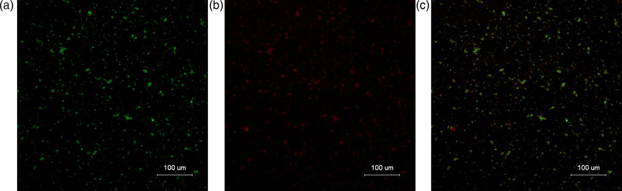
Monoclonal antibody conjugated to MPIO. (a) ICAM-1 mAb added to MPIO suspension and allowed to react for some time. Green fluorescence points = MPIO. (b) Cy3 secondary antibody added to the targeted microparticle suspension. Red fluorescence points = secondary antibody. (c) Merged image (a) and (b). It is discovered that punctiform red fluorescence points of Cy3 secondary antibody almost coincided with the green fluorescence points of ICAM-MPIO
ICAM-MPIO bound to TNF-α stimulated endothelial cells
As assessed by light microscopy, ICAM-MPIO bound to the TNF-α-stimulated endothelial cells, and the number of cell-bound ICAM-MPIO increased in response to increasing doses of TNF-α and the concentration of Fe in cell culture (Fig. 2). However, it bound sparsely to non-stimulated endothelial cells. The number of adherent microspheres on TNF-α-stimulated endothelial cells was found to be 5 ± 0.5-fold greater than that of the control group. The negative control constructs free-MPIO did not bind to TNF-α-stimulated endothelial cells. As a demonstration of specificity, pre-incubation of ICAM-MPIO with free-ICAM-1 abolished the targeted MPIO retention almost entirely. Assessed by Prussian blue staining experiments, it showed a much stronger blue appearance surrounding the stimulated cells incubated with the ICAM-MPIO than non-stimulated ones. In the contrast experiment, no obvious blue spots could be observed around the stimulated cells incubated with free-MPIO (Fig. 3).

ICAM-MPIO bound to EA.hy 926 cells stimulated with TNF-α (50 ng/mL). (a) Microscopy of activated EA.hy 926 cells incubated with ICAM-MPIO. Green fluorescence points reflect ICAM-MPIO bound to the activated endothelial cells. (b) EA.hy 926 cells nuclei stained blue with Hoechst 33342. (c) Merged image a. and b. The green fluorescence points were mostly located at the cell surface. (d) ICAM-MPIO binding to activated endothelial cells increased in response to increasing TNF-α in the culture medium
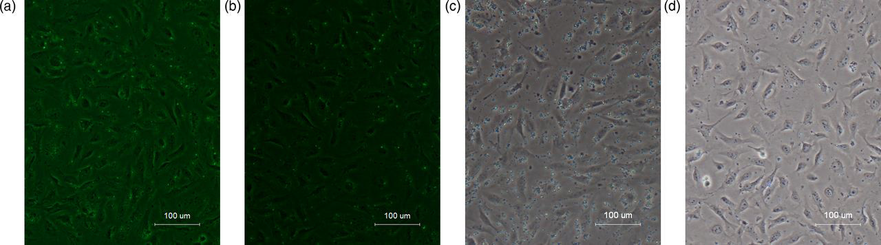
MPIO bound to EA.hy 926 cells stimulated TNF-α (50 ng/mL). (a) Microscopy of activated EA.hy 926 cells exposed to free-MPIO. There was little green fluorescence on the cell surface. (b) Prior incubation of ICAM-MPIO with monoclonal antibody of ICAM-1 almost completely eliminated ICAM-MPIO retention. Prussian blue staining images of stimulated EA.hy 926 cells (5 × 10⁵) incubated with ICAM-MPIO (c) and free-MPIO (d), respectively. Staining showed a much stronger blue appearance surrounding the stimulated cells incubated with the ICAM-MPIO than with free-MPIO
In vitro MRI
The signal intensity for ICAM-MPIO incubated with stimulated endothelial cells decreased significantly. This low signal intensity decreased in response to increasing concentrations of Fe in cell culture on T2-weighted imaging. No similar decrease was observed in the signal intensity for ICAM-MPIO incubated with non-stimulated cells, and negative control free-MPIO incubated with stimulated cells showed no contrast effect. In a competitive inhibition experiment, decreased signal intensity was not observed in the MRI (Fig. 4).

T2 signal intensity of the endothelial cells. (a) T2-weighted imaging of stimulated EA.hy 926 cells after 1 h incubation with ICAM-MPIO and free-MPIO, respectively, at final Fe concentrations of 0, 0.01, 0.1, 1, 5, 10, and 15 µg/mL in culture medium. The T2 signal intensity of the stimulated endothelial cell incubation with ICAM-MPIO decreased significantly as compared to free-MPIO. (b) T2 signal intensity of stimulated EA.hy 926 cells incubation with ICAM-MPIO and free-MPIO, which decreased in response to increasing Fe concentration
Increase in ICAM-1 expression following irradiation
There was a marked increase of ICAM-1 immunoreactivity in the endothelial cells in the irradiated brain tissues at 24 h, which strongly up-regulated at 48 h, and remained increased at 7 days after irradiation. However, control rats showed no evidence of ICAM-1 immunoreactivity in the normal brain tissues (Fig. 5).

ICAM-1 expression in endothelial cells. (a) There was a marked increase of ICAM-1 immunoreactivity in the endothelial cells in the irradiated brain tissues following irradiation. (b) Control rats showed no evidence of ICAM-1 immunoreactivity in normal brain tissues
In vivo MRI detects ICAM-MPIO accumulating in irradiated brain tissues
Our results showed that ICAM-MPIO was distributed in the irradiated hemisphere, and that it caused a marked MRI contrast effect manifested as intensely low signal areas in irradiated brains; this contrast effect was found 24 h following irradiation, remained conspicuous 48 h after irradiation, and lasted for 1 week. By comparison, control free-MPIO did not accumulate in the irradiated brains, as few low-signal areas could be found in the irradiated hemisphere.
The T2 signal intensity of the irradiated brains following ICAM-MPIO injection decreased 28% as compared to brains prior to ICAM-MPIO administration. As compared with brains without irradiation, the specific contrast effect increased more than seven-fold after administration of ICAM-MPIO (F = 751.495, P < 0.05). The T2 values of the irradiated brains following ICAM-MPIO injection decreased 15% as compared to brains prior to ICAM-MPIO administration. As compared to brains without irradiation, the specific contrast effect increased more than five-fold after administration of ICAM-MPIO (F = 122.359, P < 0.05). Pre-injection of the antibody to rat ICAM-1 prior to administration of ICAM-MPIO almost eliminated the retention of ICAM-MPIO in the irradiated brains (Figs. 6 and 7).
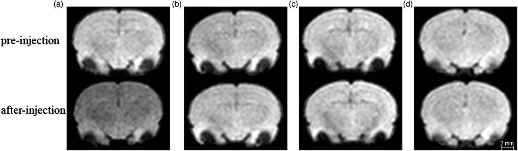
In vivo T2 *-weighted coronal images from FFE data at 48 h after irradiation. (a) Irradiated rat brains showed intense low-signal areas after being injected with ICAM-MPIO via tail vein. (b) T2 signal intensity did not decrease in the irradiated rats after being injected with free-MPIO. (c) Absence of MPIO effects in an irradiated rat injected with ICAM-MPIO intravenously after pretreatment with ICAM-1 antibody. (d) Blank control rat injected with ICAM-MPIO, which showed no negative contrast effects

T2 signal intensity (a) and T2 value (b) of the irradiated rats injected with ICAM-MPIO decreased significantly as compared to those injected with control free-MPIO at 24 h, 48 h, and 5 days. These differences peaked at 48 h, and vanished at 7 days. Pre-injection of free-ICAM-1 prior to administration of ICAM-MPIO almost eliminated the retention of ICAM-MPIO in the irradiated brain tissues. Blank control rats that underwent injection of ICAM-MPIO showed no contrast effect. ** P < 0.05, data are means ± SD
Safety and tolerability
Aside from a decrease in appetite, none of the 21 rats that underwent irradiation showed signs of ill effects during 1 week of close observation following the experiment. Injection of antibody-conjugated MPIO was well-tolerated in all rats.
Discussion
Radiation-induced up-regulation of ICAM-1 in the early period of RBI provides a potential marker for functional molecular imaging and targeted therapeutics, and MPIO that produced a larger r2*-relaxivity than SPIO can be used for molecular imaging and detected by clinical MRI (12, 13, 15, 16). Our study examined the consequences of radiation therapy by combining intense MRI contrast effects attainable by MPIO with a targeting strategy modeled on activated endothelium cells in irradiated brains in vitro and in vivo.
Radiation-induced brain injury of the central nervous system can occur within hours after exposure to a radiation dose higher than 15 Gy, and death may occur within approximately 2 days of exposure (22). However, more attention is generally paid to late radiation injury than to the early phase of RBI. Furthermore, the underlying molecular mechanism of the early stage of RBI remains unclear.
Molecular imaging techniques that use targeted agents that bind specifically to radiation-induced encephalitis are increasingly allowing earlier diagnosis and assessment of a treatment response in RBI. One pioneering study demonstrated the feasibility of targeted optical imaging of ICAM-1 on the endothelium to diagnose the early phase of RBI (11). MRI provides three-dimensional non-invasive imaging at near-cellular resolution and tissue contrast, without ionizing radiation, which makes it the most effective imaging modality of choice for molecular imaging as compared with optical and traditional imaging techniques such as computed tomography (CT), ultrasound, and single-photon emission CT (SPECT) (23). At present, no targeted MR contrast-enhancing materials specific for changes in endothelial receptor expression to evaluate the early period of RBI have been described. In contrast, this study selected ICAM-1 as a molecular target for combined MR imaging, and represented the first study to use molecular MR imaging to evaluate the early phase of RBI.
Neuroinflammation is a prominent feature of radiation injury response, and may play a causative role in the development of damage to brain tissues (3, 4). It is well established that activated vascular endothelial cells play an important role in the development of acute RBI, and that the extent of radiation damage is dependent on the number of infiltrating leucocytes.
ICAM-1on the endothelial cells have been shown to be up-regulated at the mRNA level following single and fractionated radiation (5, 9). Injection with the anti-ICAM-1 monoclonal antibody significantly reduces leukocyte adhesion and permeability. However, ICAM-1 does not have a causative role in the histopathological injury and behavioral dysfunction that develop after moderate single doses of cranial irradiation. Despite these limitations, we demonstrated that ICAM-1 has great potential diagnostic value and may be able to image an inflammatory process by detecting an up-regulation of this protein in the early phase of RBI.
Although late RBI also induces the up-regulation of endothelial ICAM-1 expression, imaging ICAM-1 may not be of incremental diagnostic value for late RBI because Gd-DTPA-enhanced MRI and several emerging MRI methods are sufficient to confirm the diagnosis. Thus, combined with conventional MRI and several emerging MR methods, molecular MR imaging with MPIO targeted to ICAM-1 may be useful for differentiation of the various stages of RBI.
In vivo molecular MR imaging can demonstrate the neuro-inflammatory process of the early phase of RBI, in concordance with the up-regulated ICAM-1 expression observed with immunohistochemical staining. It should be noted that in our study, small numbers of ICAM-MPIO also adhered to the non-stimulated endothelial cells in vitro, which may be attributed to low levels of ICAM-1 expression on normal endothelium. In addition, immunohistochemical examination of non-irradiated brain tissues did indeed show the baseline expression of ICAM-1 on endothelial cells. Our study also found that in vitro few particles of free-MPIO bound to the stimulated endothelial cells, which may be non-specific and attributable to the phagocytosis of the endothelial cells.
It should be noted that there are several limitations to this study. It is known that radiation caused a molecular response characterized by an increase in the expression of adhesion molecules. These adhesion molecules included ICAM-1, vascular cell adhesion molecule-1 (VCAM-1), E-selectin, and P-selectin. Among them, the up-regulation of ICAM-1 was greater than that of the other adhesion molecules, so for this study we selected ICAM-1 as a molecular target to evaluate the early phase of RBI. We did not evaluate the diagnostic capabilities of other adhesion molecules in this study. A previous study has shown that dual-ligand (P-selectin and VACM-1) MPIO binding to the endothelium in animal models of atherosclerosis was five- to seven-fold greater than that of P-selectin-MPIO or VACM-MPIO alone (13). This work provided the basis for future evaluation of the diagnostic value of molecular MR imaging targeted to other single- or two-adhesion molecules.
The MPIO reported here are non-biodegradable and are not suitable for human use. However, the basic iron contrast mechanism is potentially transferable to humans with suitable adaptation of the carriage particle (13, 24). We realize that there is still a long way to go to transfer the contrast molecular imaging technique studied here to use in clinical work.
In conclusion, molecular MR imaging with ICAM-MPIO displayed specific images targeted to the ICAM-1 protein that is expressed in the early inflammatory phase of RBI. This novel imaging tool may prove valuable for the early diagnosis and differentiation of the various stages of RBI. Such targeted contrast agents could be exploited to deliver targeted drugs and/or genes to lesions in the future, and to provide early intervention.
Footnotes
ACKNOWLEDGEMENTS
This work was partially supported by the Science and Technology Planning Project of Guangdong Province, China (2011B031800145 and 2009B308001120).