
Abstract
Background
CT enterography (CTE) is a valuable tool in the management of patients with inflammatory bowel disease. Reducing imaging time, reduced motion artifacts, and decreased radiation exposure are important goals for optimizing CTE examinations.
Purpose
To assess the potential impact of new CT technology (ultra-high pitch CTE) for the ability to reduce scan time and also potentially reduce radiation exposure while maintaining image quality.
Material and Methods
This retrospective study compared 13 patients who underwent ultra-high pitch CTE with 25 patients who underwent routine CTE on the same CT scanner with identical radiation emission settings. Total scan time and radiation exposure were recorded for each patient. Image quality was assessed by measurement of image noise and also qualitatively by two independent observers.
Results
Total scan time was significantly lower for patients who underwent ultra-high pitch CTE (2.1 s±0.2) than by routine CTE (18.6 s±0.9) (P < 0.0001). The mean radiation exposure for ultra-high pitch CTE was also significantly lower (10.1 mGy±1.0) than routine CTE (15.8 mGy±4.5) (P < 0.0001). No significant difference in image noise was found between ultra-high pitch CTE (16.0 HU±2.5) and routine CTE (15.5 HU±3.7) (P > 0.74). There was also no significant difference in image quality noted by either of the two readers.
Conclusion
Ultra-high pitch CTE can be performed more rapidly than standard CTE and offers the potential for radiation exposure reduction while maintaining image quality.
Computed tomography enterography (CTE) employs multidetector CT (MDCT) along with low density oral contrast and intravenous contrast to provide a rapid and highly accurate assessment of patients with inflammatory bowel disease (IBD) (1,2). CT enterography is increasingly preferred over fluoroscopic small bowel follow-through examinations as it provides additional clinical information regarding the extent of disease; however, often has a higher radiation exposure level (3). The potential long-term risks from radiation exposure are particularly relevant in the management of IBD given the young age of many of the patients and the potential need for serial imaging (4,5).
New imaging technologies offer the potential for significant reductions in the radiation exposure. One such advancement is the development of ultra-high pitch CT imaging. As background, early CT technology only permitted CT images being performed as a “single slice”, whereby each axial image was obtained following a single revolution of the CT gantry. With the advent of MDCT, it became possible to obtain an image slice with less than a complete revolution. In essence, with MDCT, the patient is imaged as a volume instead of as a number of individual slices. “Pitch” is a number which quantifies the revolutions of the CT gantry over the length of the scan, with higher numbers representing fewer revolutions per distance. Higher pitch imaging leads to faster scanning and also offers the potential for reduced radiation exposure in proportion to the degree of pitch.
Until recently, MDCT has been performed nearly exclusively with single source CT. However, studies with single source CT have shown a marked reduction in image quality at higher pitch levels (6). This fact can be explained by the under-sampling of data as the gantry does not complete an entire revolution to form each image. With dual source CT, which incorporates the use of two separate CT radiation emitters (sources), there is increased data sampling per rotation as the two sources are set 90° apart. This technology allows for higher pitch examinations due to fewer data under-sampling.
The most direct benefit of increased pitch is reduced scan time, which typically leads to improved image quality due to reduced motion (breathing, bowel peristalsis). Increased pitch also theoretically allows for reduced radiation. To date, no studies have directly evaluated ultra-high pitch CTE for the ability to reduce image time or its effect on radiation exposure. This study seeks to compare the differences in scan time and radiation exposure of an ultra-high pitch CTE performed with dual source technique compared to routine single source CTE with similar radiation emission settings.
Material and Methods
This Health Insurance Portability and Accountability Act compliant, retrospective study was approved by our institution's review board with a waiver of informed consent.
Study population
Following a retrospective review, 38 consecutive patients were identified that had undergone CTE on a single CT scanner (Somatom Definition Flash; Siemens Healthcare, Forchheim, Germany) in our department. Thirteen studies had been performed at ultra-high pitch using dual source CTE technique and 25 studies performed using routine single source CTE technique. All examinations were included in the study. The choice of performing ultra-high pitch CT or routine CT was left solely to the technologist's discretion at the time of the examination. As only two technologists had been trained in the use of the ultra-high pitch technique, they often employed it while others performed conventional CTE.
CTE technique
All CTE examinations were performed using intravenous contrast (100 mL of Omnipaque 350 mg/mL; GE Healthcare, Milwaukee, WI, USA) at a rate of 4 mL/s followed by a 50-mL saline flush injected with a mechanical power injector. Scan timing was optimized for the enteric mucosal enhancement phase at 45 s following contrast injection. Prior to the examination, patients ingested 1350 mL of a neutral oral contrast agent (VoLumen, Bracco Diagnostics, Princeton, NJ, USA). No antispasmodic agents were used for any of the CTE examinations, per routine at our institution.
CT parameters
All CTE studies were performed using the following settings: attenuation-based anatomical tube current modulation (CARE Dose 4D, Siemens); quality reference mAs 258; tube potential 120 kV; effective slice collimation 128 × 0.6 mm (employing a z-flying focal spot technique and two physical 64 × 0.6 mm channel detector arrays). Also, a dynamic beam collimator (Adaptive Dose Shield, Siemens) was used.
Routine CTE had a gantry rotation time of 0.5 s. Ultra-high pitch CTE was set at a gantry rotation time of 0.28 s. For all studies, pitch, total scan time, and scan length (z-axis) were recorded and differences in the means between the two groups were assessed using Student's t test.
Radiation dose analysis
Radiation exposure was recorded for each CT study from the radiation dose sheet output by the scanner. Computed tomography dose index of the scanned patient volume (CTDIvol) was chosen to represent the radiation dose of each CT examination. For the two groups (routine CTE and ultra-high pitch CTE) mean CTDIvol was calculated and the difference in the means was assessed using Student's t test.
Image quality assessments
Image noise was recorded based on the standard deviation (SD) of a measurement within a region of interest (ROI) (7) placed within the subcutaneous fat of the bilateral flanks several centimeters above the iliac bone for each patient. The ROI had a minimum volume of 4 cm3 and the two ROI for each patient were averaged to record a single number for each patient. Mean noise was calculated for each group and the difference in the means was assessed by Student's t test.
Assessment of image quality was performed by two separate, independent readers, who were blinded to the type of study. The readers were both board certified radiologists and had 9 and 6 years of experience in the interpretation of abdominal CT images, respectively. Each reader was presented with all CTE studies in random order. They were directed to score each examination on a five-point scale for image quality specifically related to two diagnostic tasks: (i) imaging the small bowel for assessment of IBD; and (ii) imaging of extra-luminal structures for assessment of potential complications of IBD (fistula, abscess). The scale was as follows: 1, non-diagnostic; 2, severely limited diagnostically; 3, limited diagnostically but able to evaluate gross abnormalities; 4, reduced image quality but not diagnostically limited; 5, highest diagnostic quality.
Results
Patient demographics
The 13 patients who underwent ultra-high pitch CTE had a mean age of 36.8 years, with nine women, while the 25 patients who underwent routine CTE had a mean age of 51.7 years (17 women). All patients presented as outpatients and were referred with clinically known or suspected Crohn's disease. All patients were also symptomatic at the time of examination. On the final interpretations of the studies, 5/13 (38.5%) of patients who underwent ultra-high pitch CTE had evidence of bowel inflammation typical of Crohn's disease. Eleven of 25 (44%) of patients who underwent routine CTE had evidence of bowel inflammation.
CT parameters
The mean image pitch of ultra-high pitch CTE 2.15±0.25 was greater than routine CTE (0.79±0.03) (P < 0.001). Also, mean total scan time was significantly lower for ultra-high pitch CTE (2.1 s ±0.2) than for routine CTE (18.6 s ± 0.9) (P < 0.0001). The scan length of the two imaging protocols were not significantly different (ultra-high pitch CTE = 50.4±1.7; routine CTE = 48.7±4.2 (P > 0.15).
Radiation dose
The mean radiation exposure, as measured by CTDIvol, was 10.1 mGy ± 1.0 for ultra-high pitch CTE, which was significantly less than the mean dose of routine CTE 15.8 mGy ± 4.5 (P < 0.0001). The mean dose reduction was 36% for ultra-high pitch CTE compared with routine CTE.
Image quality assessment
Image noise demonstrated no significant difference in image noise between ultra-high pitch CTE (16.0 HU±2.5) and routine CTE (15.5 HU±3.7) (P > 0.74).
For the qualitative assessment of image quality, reader 1 rated all 13 ultra-high pitch CTE as being of the highest diagnostic quality (“5”) for both small bowel findings and extra-luminal findings. Example cases are presented in Figs. 1–3. This reader did rate a single routine CTE study as “4” (reduced image quality but not diagnostically limited) for the evaluation of small bowel (due to motion artifact), which is shown in Fig. 4. The remaining 24 were recorded as “5.” All routine CTE were recorded as “5” for extra-luminal disease assessment for reader 1. Reader 2 scored all ultra-high pitch CTE and all routine CTE at the highest diagnostic quality (“5”) for luminal and extra-luminal assessments.
Axial ultra-high pitch CTE in a patient without findings of bowel inflammation. This case was scored by both reviewers as “5” – highest diagnostic quality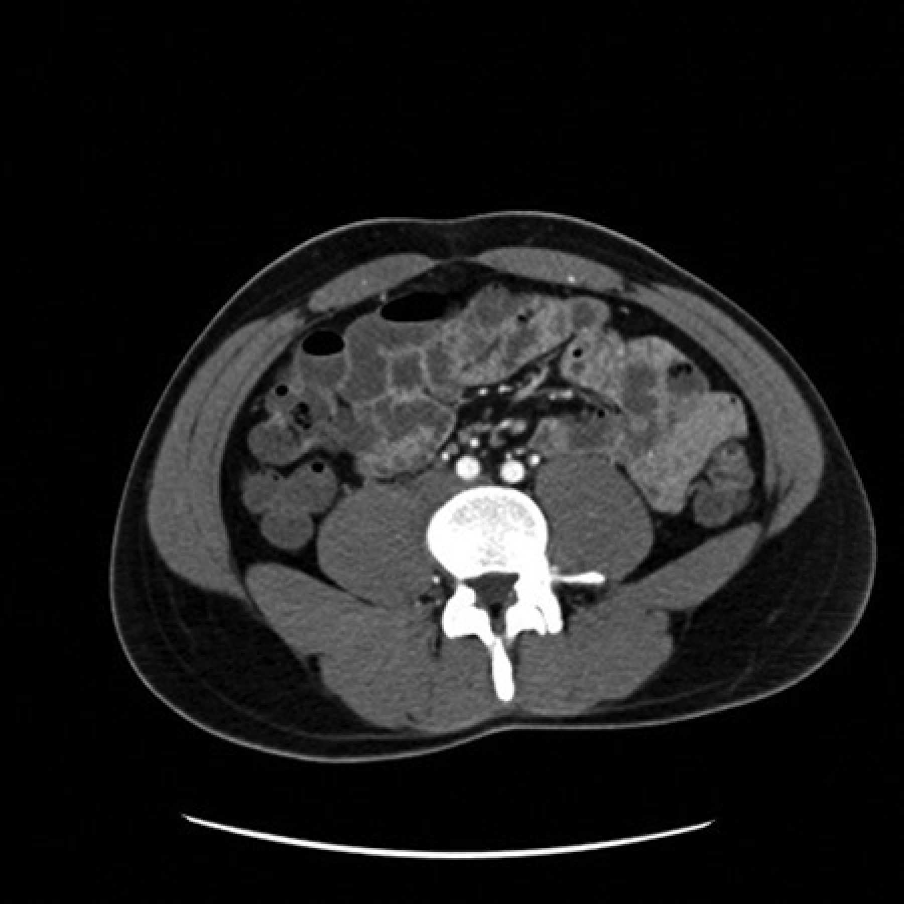
Reconstructed coronal image from same ultra-high pitch CTE study in Fig. 1
Axial ultra-high pitch CTE in a patient with findings of bowel inflammation in the distal ileum. This case was scored by both reviewers as “5” – highest diagnostic quality
Example case of conventional CTE which was interpreted by reader 1 as having slight motion artifact present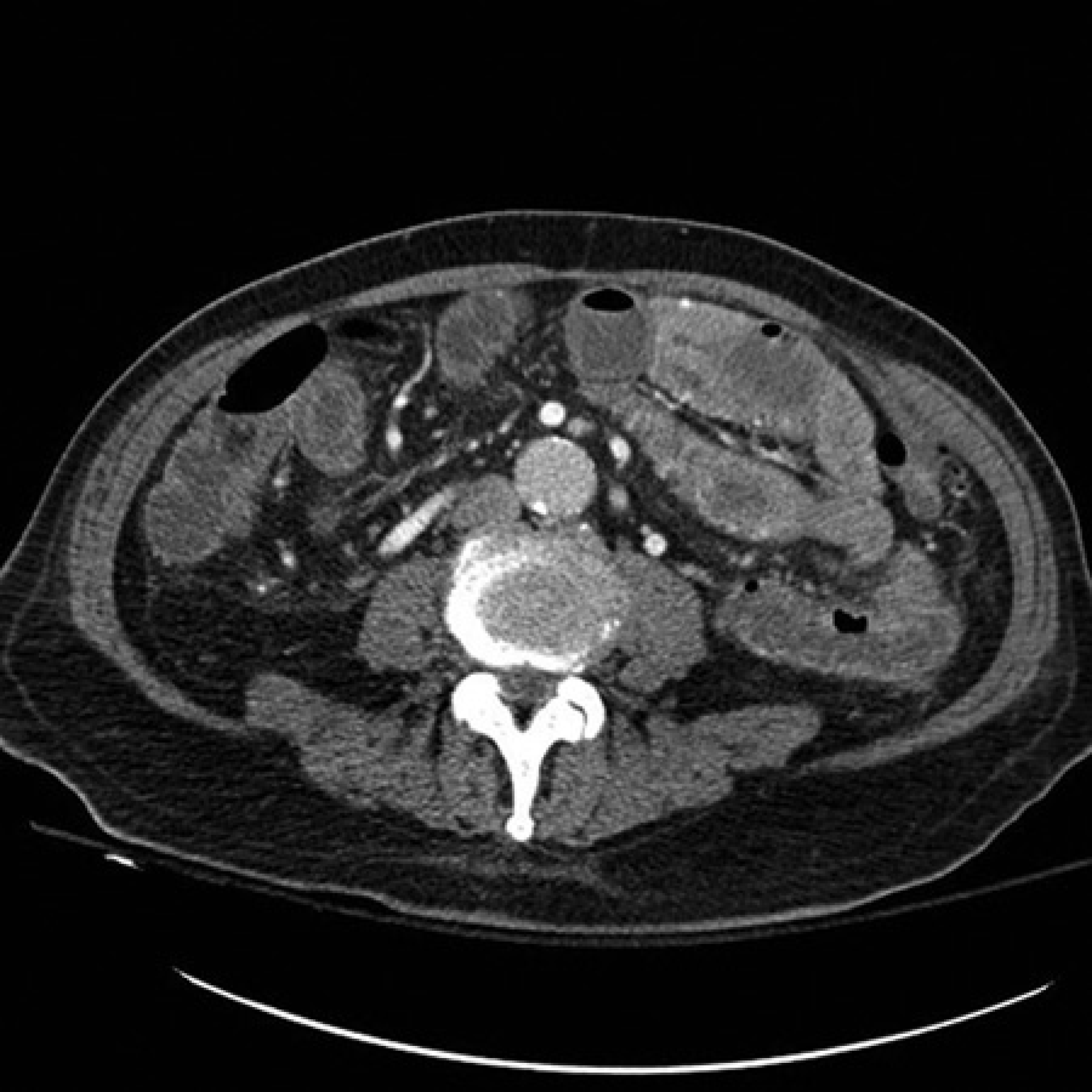
Discussion
This study has demonstrated the feasibility of ultra-high pitch CTE. Most directly, there was a marked reduction in total scan time realized with ultra-high pitch CTE compared with routine CTE. Importantly, image quality was maintained, as demonstrated by a lack of significant difference in both quantitative (noise) and qualitative image quality evaluations. Further, there was a markedly lower radiation exposure in the ultra-high pitch CTE group vs. routine CTE. Although additional studies are indicated in order to more fully evaluate the effect of ultra-high pitch CTE on radiation exposure, in this study mean radiation dose was lower by approximately one-third. Given the young age of many patients with IBD and the likelihood of needing multiple examinations in a lifetime, cumulative radiation exposure is highly relevant. Ultra-high pitch CTE may be useful as a means to reduce radiation exposure.
To our knowledge, no reports have assessed dual source CT for the ability to perform ultra-high pitch CT enterography. Although ultra-high pitch CTE imaging is a new technology, it is currently available for clinical use and is likely to increasingly become widely available. The authors have previously evaluated the feasibility of dual source ultra-high pitch CT in the abdomen and pelvis (8). However, in the previous study the diagnostic task was simpler, to exclude acute intra-abdominal infection, and utilized only images at 5-mm thick section. In this study, the robustness of ultra-high pitch CT was evaluated further by assessing a significantly more demanding diagnostic task, to evaluate for the inflammatory changes of Crohn's disease. Also typical CTE technique is performed at thinner CT sections, as it was in this study at 2 mm. Also, this study also looked at the effect of dual source, ultra-high pitch CTE on radiation dose, which has also not been previously reported.
Although numerous reports exist of reduced radiation exposure CT examinations, most of the previously reported techniques depend on reducing radiation emission settings, such as kVp or mAs. However, reducing these factors often produces more image noise, which reduces image quality. In this study, image noise was not significantly higher in the ultra-high pitch CTE group than routine CTE with otherwise identical radiation emission settings. Further, many prior reports of reduced dose CT were designed specifically for limited indications such as for visualizing the appendix or renal stones (9,10). Given that these approaches fail to provide a comprehensive abdominal-pelvic evaluation, many of these techniques have failed to gain widespread support. In this study, qualitative image quality assessment was not found to be reduced for both small bowel and extra-luminal findings. In fact, a significantly reduced time required for imaging may lead to reduced motion artifacts and improved quality for ultra-high pitch CTE. In this study, one routine CTE study was found to have lower image quality due to motion, which was not found on any ultra-high pitch CTE study, although this would need to be evaluated in larger study.
Limitations of this study include the small sample size and the lack of the ability to have a single cohort imaged using both techniques. However, given the large differences in pitch, total scan time, and radiation reduction between the two CTE protocols, a large sample size is likely not necessary to appreciate the potential advantages in terms of radiation exposure.
In conclusion, ultra-high pitch CTE is feasible and provides a more rapid examination without significantly compromising image quality. Although further studies are needed to fully assess the effect of ultra-high pitch CTE on radiation exposure, the technique may have advantages in imaging of patients with inflammatory bowel disease.