
Abstract
Optimal function of the serotonin system is essential for mental health and its role in psychopathologies is undisputed. Enhancing the ability to study primate serotonin neurons in culture would facilitate understanding of intracellular signaling pathways that mediate the action of drugs and other epigenetic or developmental factors impacting human mental health. We were the first group to report differentiation of the non-human primate rhesus monkey embryonic stem cell (ESC) line 366.4 into cultures of serotonin neurons. In this study, we optimized yield and obtained functional characteristics of the derived serotonin neurons. Sequential treatments of ESC 366.4 during expansion stage with fibroblast growth factor 4 and sonic hedgehog markedly increased the yield of serotonin neurons. These serotonin neurons propagated action potentials and expressed GABA receptors. Also, for the first time we demonstrate that these ESC-derived serotonin neurons exhibit functional high-affinity transporter sites, as well as high-affinity 5HT1A binding sites, which are essential targets of common psychoactive drugs. Finally, to test the generality of this method, we utilized another rhesus ESC line, ORMES-22, which efficiently differentiated into serotonin neurons. Together, these findings demonstrate the feasibility of our protocol to direct different primate ESC lines to serotonin neurons with physiological characteristics, which makes them a useful in vitro model system.
Introduction
The serotonin neural system plays a pivotal role in mood, stress sensitivity, integrative cognition and other autonomic functions. Serotonin functions through many different serotonin receptors, and it is important during development for guidance of neurogenesis, ultimate structure of the central nervous system (CNS) and neuronal plasticity. 1,2 Dysfunction of the serotonin system has been implicated in many psychopathologies such as depression, anxiety, schizophrenia, bipolar disorder, obsessive–compulsive disorder and eating disorders. Various drugs such as serotonin reuptake inhibitors, monoamine oxidase inhibitors and receptor antagonists have been designed for treatment; however, these medications only partially alleviate the symptoms and have numerous undesirable side-effects. 3,4 Intracellular signaling cascades, neurotransmitter synthesis, trafficking and release in primate serotonin neurons, as well as the effects of drugs on these events, have been unavailable for detailed study in vitro. The availability of a higher primate serotonin cell line would greatly facilitate study of the serotonin neural system at the molecular level in a way that would be relevant to human mental health.
Embryonic stem cells (ESCs) are unique pluripotent cells that can proliferate indefinitely in an undifferentiated state and are theoretically capable of differentiating into any cell type. 5 Our laboratory was the first group to report the utilization of non-human primate rhesus monkey ESC 366.4 to differentiate into predominantly serotonin neurons 6 in the hope of providing a cell culture model for cellular and molecular studies, as well as generating neurons for experimental transplantation into monkeys. The ability to experimentally put rhesus stem cells into the brain of rhesus monkeys provides a unique preclinical approach that cannot be currently attempted with humans.
Our ESC-derived serotonin cultures were previously shown to express serotonergic markers such as tryptophan hydroxylase (TPH), serotonin, serotonin reuptake transporters (SERT) and 5HT1A autoreceptors, in addition to synthesizing serotonin, which was measured by enzyme-linked immunosorbent assays. 6 However, the attractive pluripotent characteristics of stem cells also present significant challenges since they do not necessarily generate a single type of neuron (i.e. a pure culture). Other studies using mouse ESCs were able to derive cultures containing 25–60% serotonergic neurons. 7,8 Recently, Kumar et al. 9 showed differentiation of serotonin neurons from human ESCs with ∼50% differentiation rate, highlighting the difficulty of obtaining a pure culture. The goal of this study was to improve our previous protocol in order to optimize yield and to obtain physiological characterization of the ESC-derived serotonin neurons. Different combinations of mitogenic signaling molecules, specifically fibroblast growth factor 4 (FGF4) with sonic hedgehog (SHH), improved the yield of the serotonin neurons. These ESC-derived serotonin neurons have neuronal membrane properties and express specific functional SERT and 5HT1A autoreceptors, thus making them a useful in vitro model. The feasibility of our new optimized protocol was shown by using another rhesus ESC line ORMES-22, which efficiently differentiated to serotonin neurons.
Materials and methods
ESC culture and in vitro differentiation
The rhesus monkey ESC line 366.4 was obtained from Dr James Thomson (Wisconsin National Primate Research Center). This line was derived from an in vivo flushed preimplantation embryo and has been characterized for its pluripotency, including its potential to differentiate into cells of the neural lineage. 10 The rhesus monkey ESC line ORMES-22 was derived by Dr Shoukhrat Mitalipov (ART Core, Oregon National Primate Research Center). 11
ESCs were propagated and maintained as previously described. 6 Briefly, ESCs were co-cultured with mitotically inactive (mitomycin C-treated; 1 mg/mL at 37°C for 30 min; Sigma-Aldrich, St Louis, MO, USA) mouse embryonic fibroblasts (MEFs). ESC culture medium consisted of 85% Dulbecco's modified Eagle's medium (DMEM/F12) supplemented with 1% non-essential amino acids, 2 mmol/L glutamine, 0.1 mmol/L β-mercaptoethanol (Sigma-Aldrich) and 15% fetal bovine serum (Thermo Fisher Scientific Inc, Waltham, MA, USA). ESCs and their colonies were observed daily and passaged every 6–8 d when colonies reached 1–1.5 mm in diameter. The pluripotency of ESCs was evaluated periodically by immunocytochemistry (ICC) with antibodies to Oct-4, stage-specific embryonic antigens (SSEA-3 and 4) and embryonic proteoglycans (TRA-1–60 and TRA-1–81) as described previously. 6
A protocol consisting of multiple sequential steps was used to induce differentiation as follows:
Isolation of ESC colonies and formation of embryoid bodies (EBs): After ESC colonies attained a 1–1.5 mm diameter, they were isolated mechanically, triturated into intermediate-sized clumps (>200 cells/clump), transferred into 60 mm dishes (BD Biosciences, San Jose, CA, USA) and cultured in ESC medium at 37°C for 7 d. EBs were defined as Oct-4 negative, three-dimensional structures that could potentially give rise to endo-, ecto- and mesodermal cell lineages;
Selection (N1 stage): After formation of EBs, ESC medium was replaced with a selection medium composed of DMEM/Nutrient Mixture F12 (1:1) containing
Expansion (N2 stage): At the end of the selection period, neurospheres were cultured in expansion medium composed of DMEM/Nutrient Mixture F12 (1:1), 1% N2 supplement (500 μg/mL insulin; 10,000 μg/mL transferrin; 0.63 μg/mL progesterone; 1611 μg/mL putrescine and 0.52 μg/mL selenite) and FGF4 (10 ng/mL) for 2 d supplemented with SHH (50 ng/mL) for an additional 5 d changing the medium daily;
Maturation of differentiated neural cells (N3 stage): Expanded neurospheres were gently dispersed into single-cell suspension using TrypLE (Invitrogen, Carlsbad, CA, USA) and then plated at 90% confluency on growth factor reduced (GFR)-matrigel-coated coverslips or wells depending on the application. Cells were cultured in Neurobasal medium with N2 and B27 supplement (Invitrogen) up to two months.
Immunocytochemistry
Cells grown on coverslips were fixed with 4% paraformaldehyde for 15 min at room temperature. The cells were permeabilized with 0.2% Triton-X and 0.1% Tween-20 in phosphate-buffered saline (PBS) for 40 min, followed by incubation with blocking solution (10% normal goat serum in PBS) for one hour. Cells were then incubated overnight at 4°C with primary rabbit polyclonal anti-TPH2 antibody (catalog#: NB100-74555; immunizing peptide corresponds to residues 15–30 of human TPH2, Novus Biologicals Inc, Littleton, CO, USA) and antibody–antigen complexes were detected with Alexa Fluor 488 secondary antibody (1:1000, Invitrogen) by incubation for one hour at room temperature. The samples were washed five times with PBS after each incubation, and then counterstained with 4′,6-diamidino-2-phenylindole (DAPI).
Treatment with different mitogenic factors and SHH
To improve the viability and yield of serotonin neurons, the ESCs were treated with N2 medium (see above) during the expansion stage (N2 stage) with the following:
N2 media with 10 ng/mL FGF2 (Sigma-Aldrich) for 7 d; N2 media with 10 ng/mL FGF2 for 2 d then addition of 50 ng/mL SHH (R&D Systems, Minneapolis, MN, USA) for 5 d; N2 media with 10 ng/mL FGF4 (Sigma-Aldrich) for 7 d; N2 media with 10 ng/mL FGF4 for 2 d then addition of 50 ng/mL SHH for 5 d.
The cells were immunostained for TPH2 with a rabbit antibody generated against human TPH2 followed by development with an anti-rabbit mouse monoclonal antibody conjugated to Alexa Fluor-488. The cultures were photographed at a low magnification (10×) using the Marianas stereological workstation (Intelligent Imaging Innovations, Denver, CO, USA). The Marianas stereological workstation with Slidebook 4.2 was used to obtain a montage and for analysis. The green-labeled, TPH2-positive pixel area and the DAPI-positive pixel area, indicating the total number of cells, were both reported in square microns (μ
2 positive area). Two montages from two independent experiments for each treatment group (total of four montages) were analyzed. The TPH2-positive pixel area was divided by the DAPI-positive pixel area in each montage to obtain a ratio of TPH2 labeled neurons to total cells in the differentiated culture (n = 4/treatment). Differences between the treatment groups were determined by ANOVA followed by Newman–Keuls post-test using Prism Statistical software (Graph-Pad Software, Inc, San Diego, CA, USA). P < 0.05 was considered statistically significant.
Electrophysiology
Whole-cell patch-clamp recordings were made with electrodes pulled to 2–4 MΩ resistance filled with an internal solution (138 mmol/L potassium methylsulfate, 10 mmol/L 4-(2-hydroxyethyl)-1-piperazineethanesulfonic acid) [HEPES]; 10 mmol/L KCl, 1 mmol/L MgCl2, 1 mmol/L ethylene gylcol tetraacetic acid, 0.3 mmol/L CaCl2, 4 mmol/L MgATP, 3 mmol/L NaGTP, pH 7.4). Cells were superfused with an external solution (146 mmol/L NaCl, 30 mmol/L dextrose, 5 mmol/L KCl, 5 mmol/L HEPES, 2.5 mmol/L CaCl2, 1.2 mmol/L MgCl2, pH 7.35). Junction potentials were calculated (JPCalc, Molecular Devices, Sunnyvale, CA, USA) and corrected at the beginning of the experiments. Capacitance and series resistance compensation (>80%) were corrected and data were collected with a Multiclamp amplifier (Molecular Devices) at 10 kHz and filtered with a low-pass Bessel filter at 2 kHz. Currents were digitized with Digidata1322 (Molecular Devices), collected and analyzed using Axograph (Axograph Scientific, Sydney, Australia). Action currents were elicited with a depolarizing step from −70 to −30 mV. Current–voltage plots were generated from a series of 10 mV steps from −100 to +20 mV. Drugs were diluted in appropriate buffers in concentrated stock solutions, then diluted further in external solution to final concentration. Drugs were applied by bath superfusion.
SERT-binding assay
To determine whether the ESC-derived serotonin neurons manifested a specific serotonin reuptake-binding site (SERT), a binding assay was conducted. The SERT-binding assay was adapted from Lu et al. 12 Briefly, N3 stage serotonin neurons were preincubated in binding assay buffer (50 mmol/L Tris-HCl, 120 mmol/L NaCl, 5 mmol/L KCl, pH 7.4) for 15 min at room temperature. A volume of 2 μmol/L [3H]-paroxetine (15 Ci/mmol, Perkin-Elmer Life Sciences, Waltham, MA, USA) alone or in combination with 1 μmol/L Fluoxetine (Eli Lilly, Indianapolis, IN, USA) was added for 1.5 h at room temperature. To further verify the specificity, 1 μmol/L mazindol (Sigma-Aldrich), a noradrenaline reuptake inhibitor, was also added along with [3H]-paroxetine. Cells were collected in 100 μL of binding assay buffer and radioactivity was measured using a liquid scintillation counter (LS6000IC, Beckman, Brea, CA, USA). Differences between the treatment groups were determined by ANOVA followed by Newman–Keuls post-test using Prism Statistical software (Graph-Pad Software Inc) (n = 6/treatment). P < 0.05 was considered statistically significant.
5HT1A-binding assay
To determine whether the ESC-derived serotonin neurons manifested a specific 5HT1A autoreceptor, a binding assay was conducted. The 5HT1A autoreceptor-binding assay was adapted from Lu and Bethea.
13
Briefly, N3 stage serotonin neurons were incubated in preincubation buffer (170 mmol/L Tris-HCl, 4 mmol/L CaCl2, pH 7.6) for 15 min at room temperature. Then, the cultures were incubated in binding assay buffer (preincubation buffer plus 0.01%
Results
Improvement in viability and yield of serotonin neurons
Previously, we found that exposure of neurospheres to FGF2 helped differentiate the rhesus 366.4 ESC into serotonin neurons. These cultures were extensively examined by ICC and found to express markers that typify serotonergic neurons of the dorsal raphe of the monkey brain including TPH, serotonin and SERT. 6 In order to improve the yield of monkey serotonin neurons, we cultured neurospheres in expansion (N2) medium with different combinations of FGF2 or FGF4 plus or minus SHH after three days for a total of seven days (Figure 1A). Figure 1C shows low-power montages of cultures that were immunolabeled for tryptophan hydroxylase 2 (TPH2), the rate-limiting enzyme for serotonin synthesis, 14 and illustrate the colonies of TPH2-positive serotonin neurons. In higher magnification, these TPH2-positive neurons showed a typical serotonergic morphology as previously observed with extensive neurite outgrowth. 6,8,15 TPH2-positive pixel area was reported in square microns (μ 2 positive area) along with DAPI-positive pixel area in square microns (μ 2 positive area). TPH2-positive neurons were expressed as a percentage of DAPI-positive area (Figure 1B). Compared with FGF2 treatment, the addition of FGF4 followed by addition of SHH gave a higher yield of serotonin-positive neurons resulting in ∼75% of serotonin neurons.

Improvement in differentiation of ESC-derived serotonin neurons by mitogenic factors and SHH. (A) Schematic diagram of differentiation protocol. (B) Percentage of TPH2-positive cells in the differentiated cultures with different combination treatments with mitogenic factors (FGF2 or FGF4) and SHH (n = 4, *P < 0.05). (C) Montage of serotonin neurons immunolabeled for TPH2 (green). (a) FGF2 alone, (b) FGF2 with SHH, (c) FGF4 alone, (d) FGF4 with SHH. Scale bar 200 μm. ESC, embryonic stem cell; SHH, sonic hedgehog; TPH, tryptophan hydroxylase; FGF, fibroblast growth factor; EB, embryoid body; DAPI, 4′,6-diamidino-2-phenylindole
ESC-derived serotonin neurons have basic membrane properties
We examined whether these ESC-derived serotonin neurons have neuronal membrane properties. ESC-derived serotonin neurons had resting membrane potentials between −60 and −70 mV (n = 12). Figure 2A illustrates the striking neuronal morphology attained by a neuron that has been immunolabeled for TPH2. The electrophysiological recordings showed that ESC-derived serotonin neurons exhibit action potentials that are blocked by tetrodotoxin (TTX) (Figure 2B). Figure 2C illustrates that these cells express the GABAA receptor/channel as is common in cultured raphe cells. 16 Currents elicited by a series of depolarizing voltage steps are increased in the presence of GABA (10 μmol/L; n = 4) (Figure 2D) and reversed at hyperpolarized potentials as would be expected for the GABAA-induced chloride current (Figure 2E). Together these data indicate that ESC-derived serotonin neurons have membrane properties that are consistent with a neuronal phenotype.
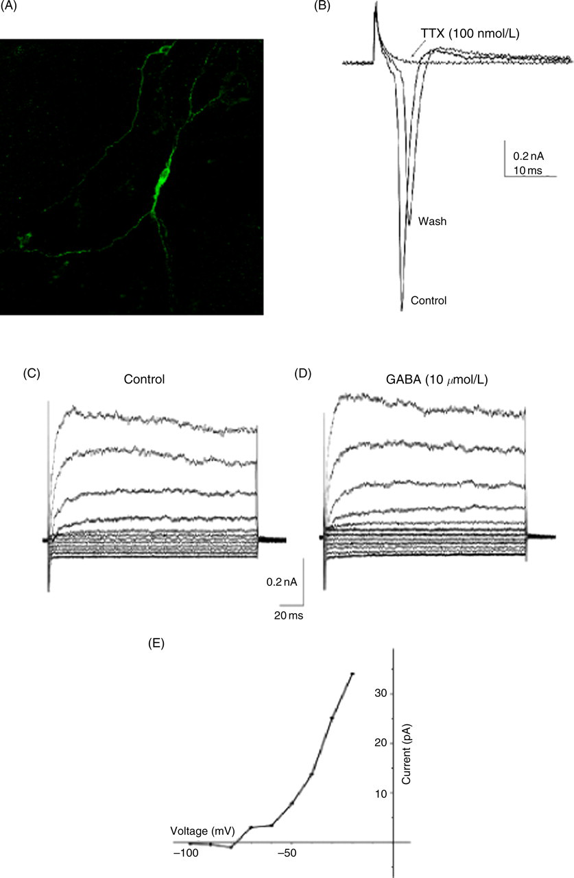
ESC-derived serotonin neurons elicit action currents. (A) Representative serotonin neuron. (B) Action currents in depolarizing voltage step −70 to −30 mV (n = 12). Voltage-dependent sodium channel implicated by absent action potential during TTX superfusion, recovers after washout. (C) Currents evoked by depolarizing 10 mV voltage steps. (D) Presence of GABA (n = 4). (E) Current–voltage plot of the subtracted (GABA-control). TTX, tetrodotoxin (A color version of this figure is available in the online journal)
ESC-derived serotonin neurons have high-affinity transporter and autoreceptor sites
CNS serotonin neurons have a high-affinity uptake mechanism that utilizes SERT, which is one of the major targets of the class of psychoactive drugs known as selective serotonin reuptake inhibitors (SSRIs). 17 To examine whether ESC-derived serotonin neurons express SERT, [3H]-paroxetine, a common SSRI, was used in a radioligand-binding assay. ESC-derived serotonin neurons exhibited an average [3H]-paroxetine-specific binding of 87.36 fmol at a substrate concentration of 2 μmol/L. To determine the specificity of [3H]-paroxetine binding, 1 μmol/L fluoxetine, another common SSRI, was used for competition. Fluoxetine significantly displaced [3H]-paroxetine binding (Figure 3A, *P = 0.03). To further verify the specificity of [3H]-paroxetine binding, mazindol, which has a high affinity for norepinephrine transporters, was incubated along with [3H]-paroxetine. Mazindol did not displace [3H]-paroxetine binding.
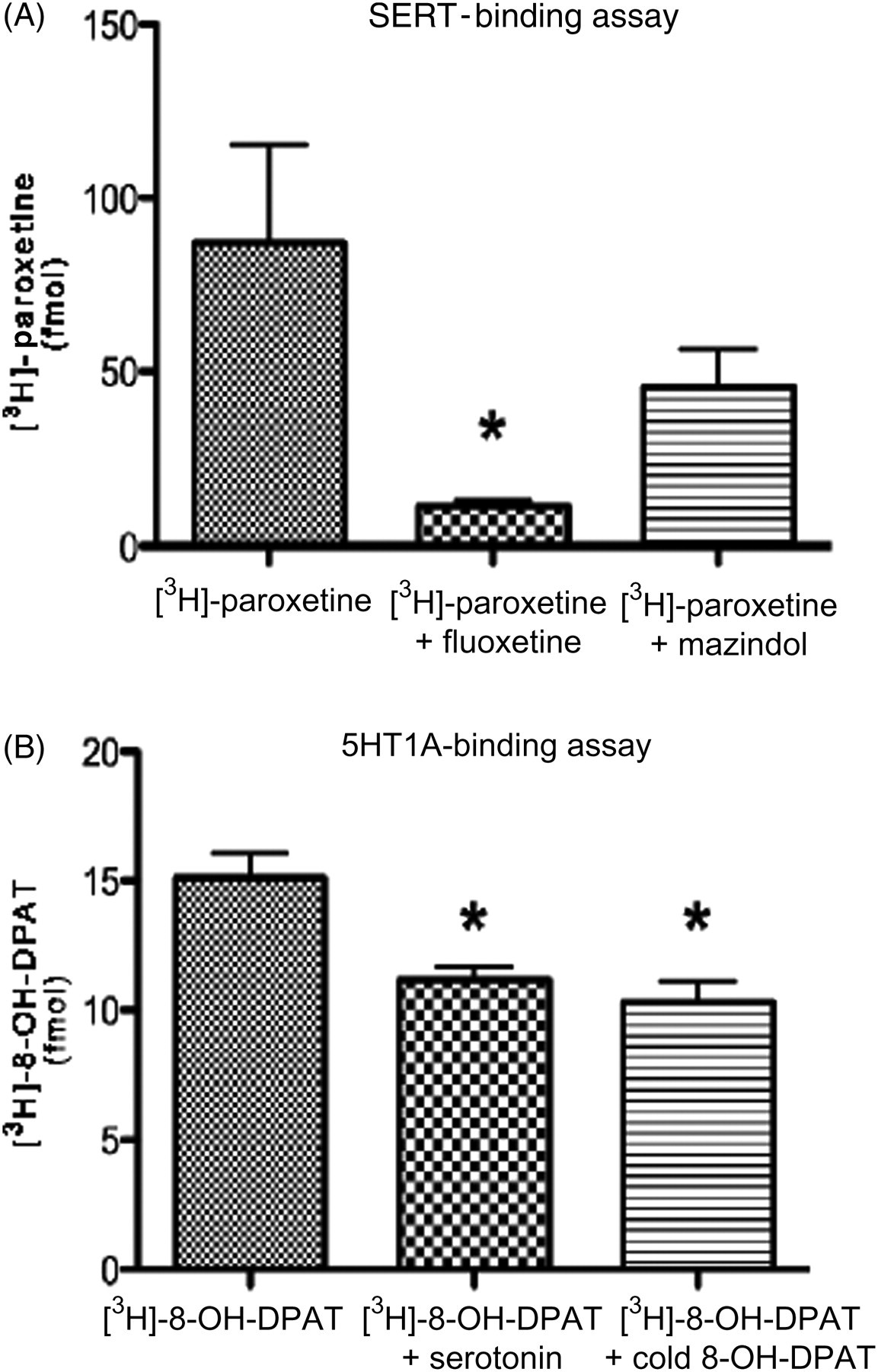
ESC-derived serotonin neurons express functional transporter and autoreceptor sites. (A) SERT binding using [3H]-paroxetine radioligand assays. A significant reduction in [3H]-paroxetine binding in the presence of fluoxetine (n = 6, *P < 0.03) but not mazindol. (B) 5HT1A binding using [3H]-8-OH-DPAT radioligand assays. A significant reduction in [3H]-8-OH-DPAT binding in the presence of serotonin and cold 8-OH-DPAT (n = 6, *P < 0.05). ESC, embryonic stem cell; SERT, serotonin reuptake transporter
The 5HT1A autoreceptor is one of the major serotonin receptors that binds serotonin in the extracellular space and decreases neuronal firing and serotonin release. 18–20 Thus, it is essential that our ESC-derived serotonin neurons express functional 5HT1A autoreceptors in order for us to proceed with psychopharmaceutical studies. ESC-derived serotonin neurons were incubated with [3H]8-OH-DPAT radioligand, 5HT1A autoreceptor agonist, to measure the affinity of 5HT1A autoreceptor binding. [3H]8-OH-DPAT had an average specific binding of 15.09 fmol at a substrate concentration of 2 nmol/L. The binding was specific to 5HT1A autoreceptor as [3H]8-OH-DPAT radioligand binding was significantly displaced when incubated with either serotonin or cold 8-OH-DPAT (Figure 3B, *P < 0.05). Moreover, the Bmax was similar to that previously obtained in macaque brain. 13
Altogether, the binding studies confirm that the ESC-derived serotonin neurons express functional proteins that define a neuron as serotonergic, and thus will be a useful in vitro primate model system.
Rhesus ESC line, ORMES-22, differentiates to serotonergic neurons with high efficiency
We used another rhesus ESC line, ORMES-22, to validate our optimized protocol's efficiency to differentiate ESC into serotonin neurons. ORMES-22 is a recently established in vitro fertilized-derived rhesus ESC line, 11 which was subjected to our modified protocol. Similar to rhesus ESC line 366.4, ORMES-22 differentiated into serotonin neurons as determined by TPH2 ICC (Figure 4). This suggests that our modified protocol can efficiently differentiate another rhesus ESC line, ORMES-22, into serotonin neurons.
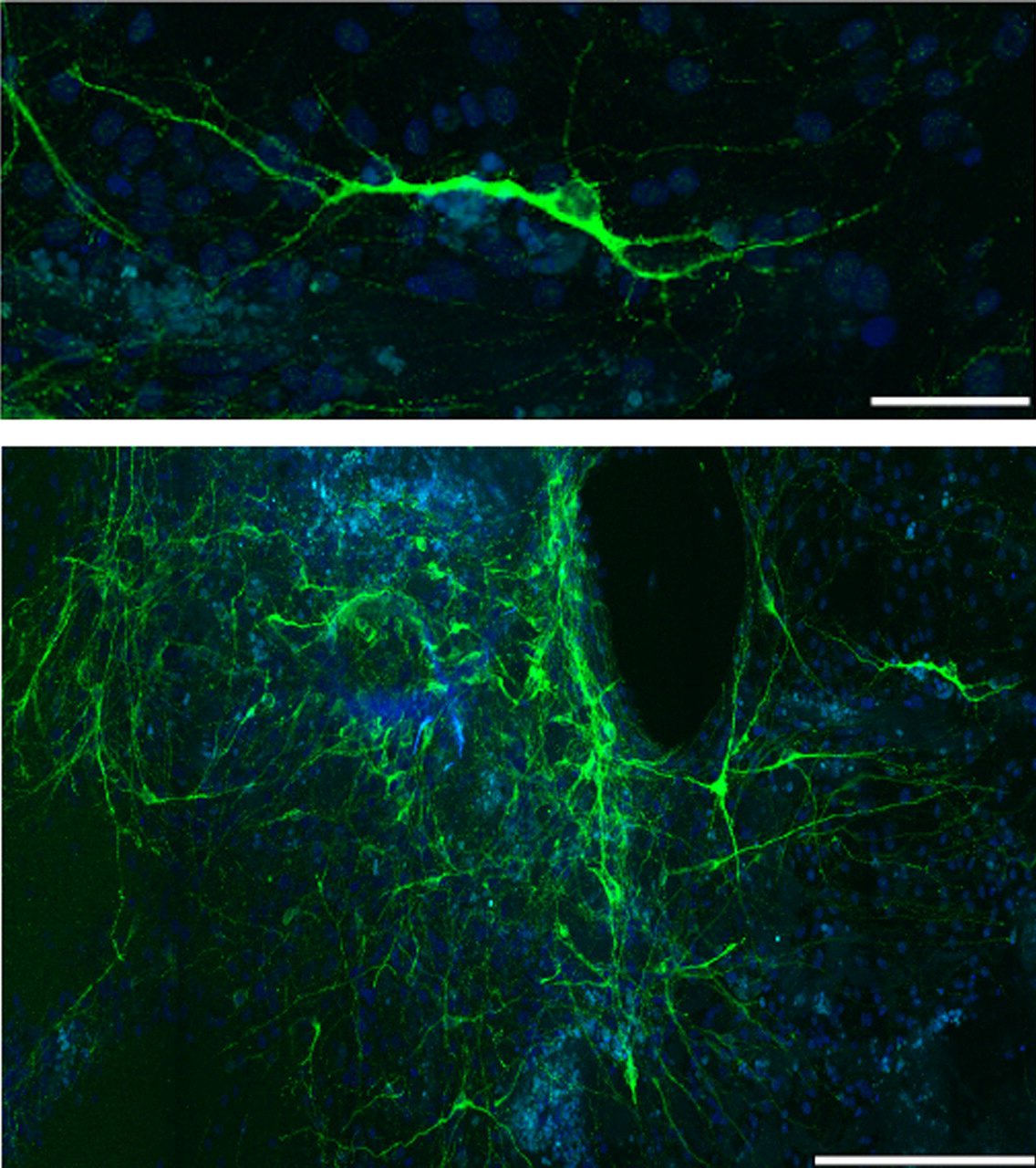
Rhesus ESC ORMES-22 differentiates into serotonin neurons. Confocal photomicrograph of representative differentiated serotonin neurons immunolabeled for TPH2 (green) and DAPI (blue). Scale bars, 50 μm (top), 250 μm (bottom). TPH, tryptophan hydroxylase; DAPI, 4′,6-diamidino-2-phenylindole; ESC, embryonic stem cell (A color version of this figure is available in the online journal)
Discussion
The development of a physiologically relevant primate serotonin neural culture system that allows direct examination of cellular and molecular signaling mechanisms is essential to our understanding and development of new psychopharmacotherapies. Human and monkey ESCs differ substantially from mouse ESCs in terms of morphology and surface marker expression. The differences between human and mouse ESCs may result from fundamental differences in embryonic development between the species, or they may reflect a difference in the embryonic stage of origin of ESCs in mouse and primate. Recently, we obtained information on the genetics of the developmental cascade throughout the differentiation stages from undifferentiated ESCs to serotonin neurons. 21 Most of our understanding in the developmental pathway leading to serotonin neurons comes from rodent models 22,23 ; however, fetal development in rodents and primates differs significantly. We observed similarities and differences between in vivo rodent studies and rhesus ESC-derived serotonin neurons in our microarray study. Several genes thought to be critical signals for a neural and serotonin phenotype in mice such as SHH, Pet1 (Fev), TGFβ, VEGF, and Wnt pathways were similarly expressed during in vitro differentiation of ESC-derived serotonin neurons. However, we also observed some differences in the ontogeny of Phox2b, Mash1 and Lmx1b gene expressions between our microarray study and in vivo mice studies. 24 In the ESC-derived serotonin neurons, Fev appeared to be the key gene for expression of other serotonin markers. Nonetheless, the differences between in vivo rodent studies and our in vitro ESC study may point to genes that are important for the organization of the serotonin system rather than major differences between rodents and primates in the expression of the serotonin phenotype.
The attractive pluripotent characteristic of stem cells also presents a significant challenge to obtain pure cultures of only one cell type. The goals of this study were to enhance our ability to differentiate rhesus ESCs into serotonergic neurons at a high yield in a predictable manner and to use these cultures to obtain physiological characterization of the ESC-derived serotonin neurons. Previously, we found that exposure of neurospheres to FGF2 helped differentiate the rhesus 366.4 ESC into serotonin neurons. 6 Other studies using mouse ESCs showed sequential exposure of FGF4 then FGF8 and SHH led to a high yield of differentiated serotonin neurons. 7,8,25,26
Our results corroborate the mouse studies in that the sequential combination of FGF4 and SHH improved the yield of serotonin-positive neurons to ∼75% as determined by TPH2 ICC and stereology analysis. Overall, the yield of serotonin neurons and the architecture of the cultures were significantly improved. We have also confirmed that our modified protocol is efficient in differentiation of serotonergic neurons from another rhesus ESC line, ORMES-22. It may be noted that TPH2-positive ESC 366.4-derived serotonergic neurons in Figure 1C does not illustrate neurite extension as seen in Figure 4 of ESC ORMES-22-derived serotonin neurons; however, this is due to the difference in the low magnification used to obtain the montages (see scale bars in figure legends). In high magnification, both ESC 366.4 and ORMES-22-derived serotonergic neurons showed similar striking neuronal morphology with extensive neurite extensions.
Other attempts to obtain a pure monolayer ESC-derived serotonin culture included: (1) treatment with cytosine arabinoside (Ara-C), a well-known antimitotic agent, to eliminate unwanted rapidly proliferating cells while preserving non-proliferating serotonergic neurons 27 ; (2) priming with heparin and laminin to encourage migration and spreading of the neurons 28 ; (3) using different substrata such as an astrocyte cell line, extracellular matrix from astrocytes and neural cell adhesion molecule. 29,30 Although these methods showed initial promise by enhancing neuronal outgrowth and attainment of monolayers (data not shown), they also led to overgrowth of unwanted cell types and Ara-C treatment was toxic for the ESC-derived serotonin neurons.
Nonetheless, application of our modified protocol provided monolayers of ESC-derived serotonin neurons with consistent well-to-well cell numbers. This allowed us to perform functional studies and determine whether these neurons have neuronal membrane properties, as well as SERT and autoreceptor expression in a manner similar to primate CNS serotonin neurons. The electrophysiological recordings showed that ESC-derived serotonin neurons exhibit action potentials that are blocked by TTX, and that they express GABA receptors in a fashion similar to dissociated postnatal rat serotonin neurons. 16 Together these data indicate that ESC-derived serotonin neurons have membrane properties that are consistent with a neuronal phenotype.
SERT is responsible for the recycle and clearance of serotonin in the brain and is the major target of SSRIs. Therefore, in order for the ESC-derived serotonin neurons to be a good in vitro model, it is essential that they express a functional transporter. Indeed, the ESC-derived serotonin neurons exhibited a high-affinity-binding site for [3H]-paroxetine that was effectively blocked with fluoxetine. In rats, [3H]-paroxetine exhibits an average Bmax of 120 fmol/mg protein, which is comparable to our SERT-binding assay. 31 Addition of mazindol, a high-affinity norepinephrine transporter ligand, had a minor, non-significant effect in blocking [3H]-paroxetine binding. Although paroxetine has been shown to be a potent and selective inhibitor of SERT, it has recently been demonstrated to also have moderate affinity for the norepinephrine transporter (NET). 32 Thus, it is possible that mazindol may exhibit low-affinity binding to serotonin transporters or that [3H]-paroxetine bound to a low number of norepinephrine transporters that were in the cultures and that were displaced by mazindol.
The 5HT1A autoreceptor is located on the soma and dendrites of the serotonin neurons and it provides an ultra-short loop feedback mechanism thought to cause a delay in the onset of efficacy of antidepressant drugs. 33 In depressed patients, 5HT1A autoreceptor levels are elevated and supplementation of antidepressant therapy with 5HT1A antagonists outperforms SSRIs alone. 34,35 Thus, it is essential that our ESC-derived serotonin neurons express the 5HT1A autoreceptor in order for us to proceed with psychopharmaceutical studies. The ESC-derived serotonin neurons exhibited a high-affinity-binding site for [3H]-8-OH-DPAT that was blocked to a significant degree with cold 8-OH-DPAT or serotonin. Moreover, the Bmax was similar to that previously obtained in macaque brain. 13 The commonly used 5HT1A autoreceptor agonist 8-OH-DPAT has been shown to have affinity for non-5HT1A autoreceptors, such as serotonin uptake sites, 5HT1D and 5HT1B receptors, adrenoceptors and dopamine D2 receptors, 36–39 which may explain some of the [3H]-8-OH-DPAT binding that was not blocked by serotonin or cold 8-OH-DPAT in Figure 3B. Thus, it will be interesting to further examine the specificity of 5HT1A autoreceptor binding in the presence of non-5HT1A autoreceptor drugs such as SSRIs, ketanserin (non-5HT1A receptor antagonist), prazosin (adrenoceptor antagonist) and raclopride (dopamine receptor antagonist). Also, there are seven distinct serotonin receptors families with at least 15 subpopulations to date; thus further studies will be needed to examine other possible receptors expressed in these ESC-derived serotonin neurons.
In conclusion, we are able to produce ESC-derived serotonin neurons with high efficiency in a predictable manner. Altogether, the electrophysiological and binding studies establish that the ESC-derived serotonin neurons express several functional proteins that define a neuron as serotonergic, and suggest that this culture system will be a useful in vitro primate model system. This will open the door for use of these cells in high throughput pharmaceutical screening for novel drugs, and for studies on the intracellular mechanism of action of antidepressants and antipsychotics. In addition, it will further the goal of transplantation into a macaque model of mental illness for restoration of serotonin function or for transport of gene vectors that promote regeneration of in situ systems.
Footnotes
ACKNOWLEDGEMENTS
Supported by NIH grants: MH73564 and MH62677 to CLB, U54 contraceptive Center Grant HD 18185, S10RR024585, and RR000163 for the operation of Oregon National Primate Research Center (ONPRC). We are deeply grateful to the Assisted Reproductive Technology Core (Dr Shoukhrat Mitalipov) for the generation of the MEFs and embryonic stem cell cultures and to Dr Anda Cornea for her assistance in confocal microscopy.