
Abstract
Brain death (BD) induces acute lung injury and makes donor lungs unfit for transplantation. Carbon monoxide (CO) inhalation at 50–500 ppm exerts anti-inflammatory and anti-apoptosis effects in several lung injury models. We examined whether CO inhalation would show favorable effects on lung injury in BD rats. BD rats inhaled 250 ppm CO for two hours. Inhalation decreased the severity of lung injury, as checked by histological examination. CO treatment reversed aggravation in PaO2/FiO2, base excess and pH of BD rats. CO inhalation downregulated the pro-inflammatory cytokines (tumor necrosis factor-α, interleukin-6), and inhibited activity of myeloperoxidase in lung tissue. Inhalation significantly decreased cell apoptosis of lungs, and inhibited mRNA expression of intercellular adhesion molecule-1 and caspase-3 in the lungs. Further, the inhalation activated phosphorylation of p38 expression and inhibited phosphorylation of extracellular signal-regulated kinase expression in the lungs. In conclusion, CO exerts potent protective effects on lungs from BD rats, exhibiting anti-inflammatory and anti-apoptosis functions by modulating the mitogen-activated protein kinase signal transduction.
Introduction
Lung transplantation is an established therapeutic option for end-stage lung disease, but the deficiency of suitable donors has limited the quantity and quality of transplantation. 1 Brain death (BD) donor is still a major source of lung for transplantation. Nonetheless, the BD process induces significant sympathetic discharge, producing a hypertensive crisis and endocrine change, 2,3 resulting in acute lung injury and aggravating chronic rejection after transplantation. 4
Elimination of the hypertensive crisis during the BD process and correction of the neurogenic hypotension following BD could mitigate the systemic inflammatory response and ameliorate oxygenation. 2 In addition, optimizing ventilatory support and hemodynamics management, 5 and high-dose steroid administration 6 before organ recovery could improve oxygenation. However, measures to improve or to maintain the lung function between confirmation of BD and procurement of the donor lung are still scarce.
Carbon monoxide (CO) inhalation at a low concentration (50–500 parts per million, ppm) exerts anti-inflammatory and anti-apoptosis effects on lung injury induced by hyperoxia, 7 sepsis, 8 ischemia–reperfusion, 9 inadequate mechanical ventilation 10 and transplantation. 11 Furthermore, Kohmoto et al. 12 found that exogenous low-dose CO (250 ppm) treatment of donors and recipients improved gas exchange, diminished intragraft and systemic inflammation, and retention of graft vascular endothelial cell ultrastructure. The same team also found that 5% CO as an additive to the cold flush/preservation solution could exert potent anti-inflammatory and cytoprotective effects after cold preservation and transplantation of lung grafts. 13 In our previous study also, 14 CO inhalation in BD rats exerts potent protective effects on lung grafts from BD donor, exhibiting anti-inflammatory and anti-apoptosis functions, but the direct effects of CO inhalation on lung injury induced by BD was unknown. Therefore, we hypothesized that CO inhalation at 250 ppm during the phase from confirmation of BD to lung procurement could ameliorate lung function.
Methods and materials
Animals
Male pathogen-free Wistar rats (250–300 g) were supplied by the animal experiment center of Harbin Medical University and allowed to acclimate on arrival for seven days before experiment. The animals were fed rodent chow and water ad libitum. All procedures were approved by the Institutional Animal Care and Use Committee of Harbin Medical University.
Model of BD
BD was produced as previously described. 15 Briefly, rats were anesthetized with sodium pentobarbital 60 mg/kg intraperitoneally and intubated through a tracheostomy. BD was produced by injecting saline (20 μL/min) into a balloon catheter (Fogarty 4-Fr, Baxter Health Care Corporation, Irvine, CA, USA) introduced into the intracranial cavity through an occipital burr hole. BD was confirmed by apnea, sudden decrease and increase in mean arterial blood pressure (MAP), the disappearance of electroencephalogram (EEG) activity, and maximally dilated and fixed pupils. 2 The MAP was maintained between 80 and 120 mmHg by infusing a mixture of norepinephrine (0.01 μg/kg/min) and saline. Rats with MAP <80 mmHg for over five minutes were excluded to avoid ischemic effects. When respiration ceased, the animals were ventilated (Model 683, Harvard Apparatus, South Natick, MA, USA) with 40% oxygen, a tidal volume of 10 mL/kg, a positive end expiratory pressure of 2 cm H2O and the breath rate of 30–50 beats per minutes, which was adjusted to maintain PaCO2 within 35–45 mmHg. Rectal temperature was maintained at 38 ± 0.5°C with a heating pad. Normal saline was infused at a rate of 10 mL/kg/h through the left femoral vein.
Experimental design
Rats were randomly divided into three groups. In the sham group (n = 9), a balloon catheter was inserted into the cranial cavity of rats, but the balloon was not inflated. Rats were ventilated with 40% oxygen and anesthesia was maintained by sodium pentobarbital and pipecuronium bromide (0.4 mg/kg/h) intraperitoneally. In the BD group (n = 11), rats were ventilated with 40% oxygen after BD confirmation for two hours. In the BDCO group (n = 10), rats inhaled, after BD confirmation, 250 ppm CO in 40% oxygen and 60% nitrogen (XueLong Gas Corporation, DaQing, China) for two hours. The concentration of CO was continuously monitored (T40 Rattler, New York, NY, USA), and the arterial blood gases were measured every 30 min (Rapidlab 248, Bayer, Medfield, MA, USA). All rats were sacrificed by exsanguinations after two hours of observations, and their arterial blood samples and lungs were collected for further detection.
Assay of inflammatory cytokines in serum and myeloperoxidase activity in lungs
The upper section of left lungs was excised and desiccated at 70°C for one week to measure the wet- to dry-weight ratio (W/D). The levels of interleukin (IL)-6 and tumor necrosis factor (TNF)-α in serum were measured by enzyme-linked immunosorbent assay (R&D Systems, Minneapolis, MN, USA). The lungs stored at −80°C was ground into homogenate to measure myeloperoxidase (MPO) activity using a special Regent-Kit (Jiancheng Bio-Technology, Nanjing, China) and a spectrophotometer. One unit of MPO was defined as the quantity that degraded 1.0 μmol of peroxide per minute at 37°C. The results were expressed as units per gram of wet lung tissue (U/g).
Histological examination and grading of lung sections
Formalin-fixed lungs were embedded in paraffin, sectioned into 6 μm thicknesses, and then stained with hematoxylin and eosin. All of the sections were evaluated microscopically by a pathologist blinded to this study. The evaluation was based on the following criteria: (1) neutrophil infiltration; (2) airway epithelial cell damage; (3) interstitial edema; (4) hyaline membrane formation; and (5) pulmonary hemorrhage. Each criterion was scored on a semi-quantitative scale of 0–4, where 0 = normal, 1 = minimal change, 2 = mild change, 3 = moderate change and 4 = severe change. An overall histological score was calculated by totaling the scores for criteria 1 through 5. 16
Immunohistochemistry of lung sections
Apoptosis of cells was examined using terminal deoxynucleotidyl transferase dUTP nick end-labeling (TUNEL) assays (Zhongshan Golden Bridge Biotechnology, Beijing, China). Sections were stained with brown 3,3'-diaminobenzidine (DAB) color-developing agent. Cell apoptosis was considered when cells had a brown–yellow staining in the nuclei. The sections were examined by a pathologist with a single blind method. The number of positive cells per section was counted in five random high-power (×40) fields from every specimen, and evaluated by the apoptotic index (AI). AI is a measure of the number of positive cells in each 100 cells counted in five different fields in the same section. 17
Reverse transcriptase-polymerase chain reaction
Levels of mRNA for intercellular adhesion molecule (ICAM)-1 and caspase-3 in lungs were assessed by reverse transcriptase-polymerase chain reaction (RT-PCR) according to a previous study. 11 Total RNA was extracted from lung tissues applied by Trizol reagent (Invitrogen, Carlbad, CA, USA). Total RNA of 3 μg from each sample was used for RT with the Access RT-PCR system (Promega, Mannheim, Germany). The PCR mixture was performed as described in the RT-PCR kit (TaKaRa, Tokyo, Japan). β-Actin mRNA was used as an internal control to assess the quality and quantity of the DNA extraction and the efficiency of the RT-PCR. Data were expressed in a ratio of targeted mRNA to β-actin mRNA by integrated density values (band area times relative intensity).
Western blot
Western blot analysis was performed with the use of 25 μg of tissue protein from each lung tissue as described previously. 18 Protein samples were boiled for five minutes and resolved by a 12% sodium dodecyl sulfate-polyacrylamide gel electrophoresis, then electroblotted onto nitrocellulose membranes. After blocking of non-specific binding with non-fat milk overnight, the membranes were incubated with primary antibodies including anti-p38, anti-phosphorylated p38 (p-p38), anti-extracellular signal-regulated kinase (ERK) and anti-phosphorylated ERK (p-ERK) (Santa Cruz Biotechnology, Santa Cruz, CA, USA) for two hours, followed by incubation with horseradish peroxidase-conjugated anti-mouse antibody (1:500) for 1.5 h. The membranes were developed with DAB staining and exposed to film. The level of measured materials was normalized to the level of β-actin. Data were expressed in a ratio of targeted protein to β-actin protein by integrated density values (band area times relative intensity).
Statistical analysis
Results were expressed as mean ± SD. Differences between groups were analyzed by one-way analysis of variance (ANOVA) followed by SNK test. Continuous data (e.g. MAP, PaO2/FiO2, pH and base excess [BE] values) were assessed by repeated measures ANOVA. Differences were considered significant at the level P < 0.05.
Results
The disturbance of hemodynamics induced by BD
Eight rats were included in each group; the rest of the rats were excluded either by bleeding from the hole of cranium or by hemodynamics instability. The hemodynamics induced by the BD process and management is shown in Figure 1. In the sham group, the MAP remained constant at 102 ± 9 mmHg during the 150-min period. In the BD group, inflation of the intracranial balloon caused a hypertensive response showing a peak MAP of 175 ± 14 mmHg at 9.2 ± 2.1 min, and then declined within five minutes to 68 ± 11 mmHg. With the infusion of noradrenalin, MAP in BD rats was maintained within 80–120 mmHg until removal of the lung. The infusion volume was not statistically different among groups. The onset of apnea, fixation of pupils at the maximal size and flatting of EEG were noted at 9 ± 3, 10 ± 3 and 11 ± 3 min after the start of the balloon inflation, respectively.
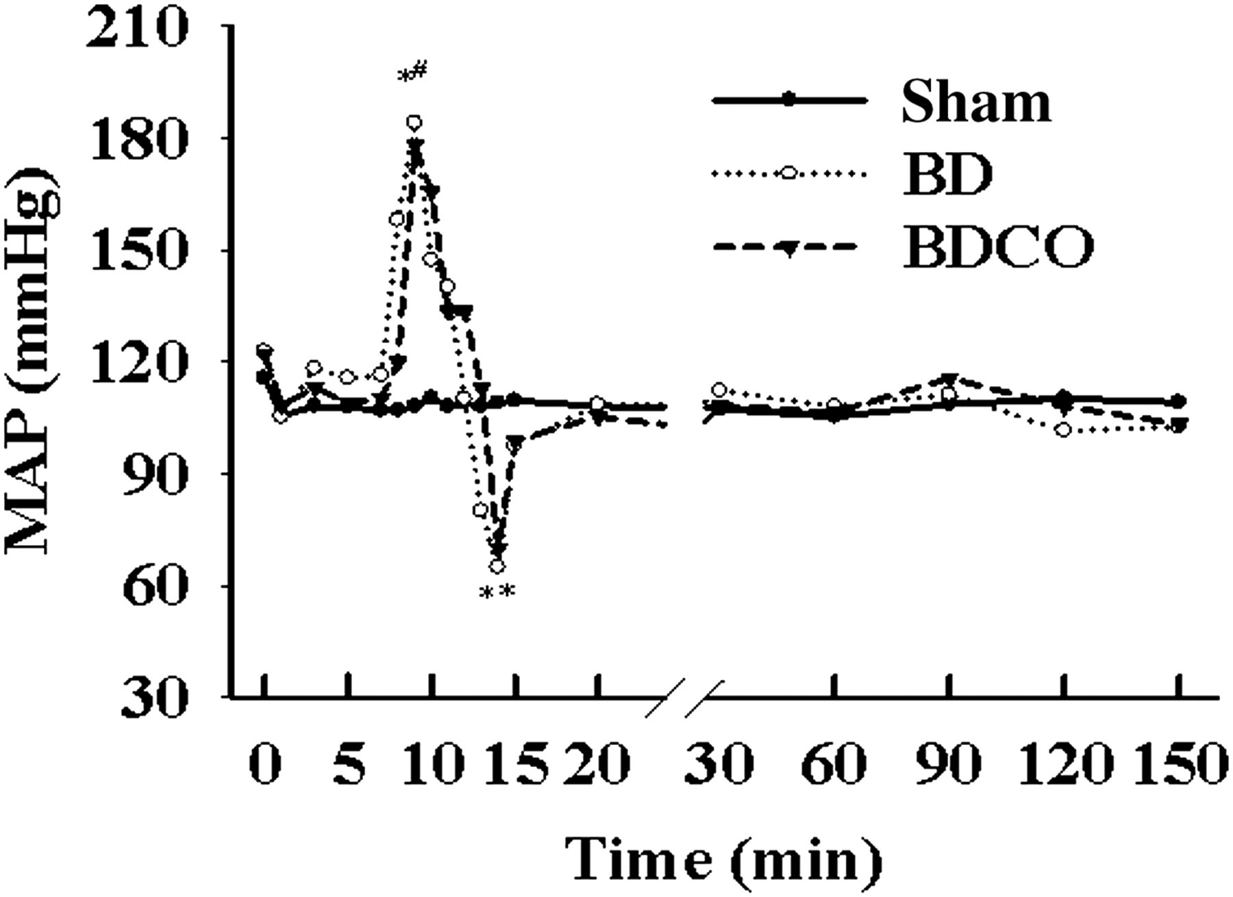
Changes of MAP after induction of BD in rats. BD process values are means; SD bars were omitted for clarity. 0 min means the start of BD induction. BD caused an immediate increase and subsequent decline of the MAP to hypotension. After infusion of noradrenalin when MAP <80 mmHg, the MAP returned to normotension. MAP of the sham group rats remained normotensive throughout the period of 150 min. *P < 0.05 versus sham group. BD, brain death; MAP, myeloperoxidase
CO inhalation improves the indexes of blood gas analysis in BD rats
The results of blood gas analysis are shown in Figure 2. In the sham group, the values, including ratio of arterial oxygen tension to fraction of the inspired oxygen (PaO2/FiO2) (Figure 2a), BE (Figure 2b) and pH values (Figure 2c) were relatively stable. BD resulted in a decrease of PaO2/FiO2 from 453 ± 22 to 386 ± 21 mmHg (P < 0.01), BE from 0.22 ± 0.25 to −1.74 ± 0.38 mmol/L (P < 0.05) and pH from 7.37 ± 0.02 to 7.31 ± 0.05 (P < 0.05) at 30 min after the induction. The values of PaO2/FiO2, BE and pH in the BD group were aggravated gradually, and were improved significantly in the BDCO group (P < 0.05), but the indexes were lower than those from the sham group (P < 0.05).

Blood gas analysis of arterial blood. 0 min means the start of BD induction. Brain death caused a decrease in PaO2/FiO2 (a), BE (b) and pH (c). The indexes were aggravated progressively in the BD group. CO inhalation in the BDCO group improved the condition. *P < 0.05 versus sham group; # P < 0.05 versus BD group. BD, brain death; BE, base excess
CO inhalation exerted anti-inflammatory effects in BD rats
As shown in Table 1, the W/D in the BD group (6.0 ± 0.9) was higher than that of the sham group (5.1 ± 0.4) (P < 0.05). Although alleviated by CO inhalation, W/D in the BDCO group (5.4 ± 0.4) was higher than that of the sham group (P < 0.05). The MPO activity in lungs was higher in the BD group (1.12 ± 0.38) compared with the sham group (0.55 ± 0.14) (P < 0.01). MPO activity in the BDCO group (0.81 ± 0.21) was lower than the BD group (P < 0.05), but was higher than the sham group (P < 0.05). The level of IL-6 in serum was higher in the BD group (130 ± 34) compared with the sham group (43 ± 14), and were lower in the BDCO group (100 ± 26) compared with the BD group (P < 0.05). The level of TNF-α in serum had a similar tendency as IL-6.
Comparison of W/D, level of TNF-α and IL-6 in serum; MPO activity in lungs and lung injury score among groups (mean ± SD, n = 8)
W/D, wet- to dry-weight ratio; TNF, tumor necrosis factor; IL, interleukin; MPO, myeloperoxidase; BD, brain death
*P < 0.05 versus sham group; # P < 0.05 versus BD group
CO inhalation alleviated lung injury induced by BD
Microscopic findings in the lungs revealed normal lung parenchyma in the sham group (Figure 3a) but severe lung injury in the BD group. Lungs from BD rats (Figure 3b) showed extensive alterations, i.e., moderate to severe edema in the alveolar septum and spaces, hyaline membrane formation and intraalveolar hemorrhage occasionally. Much less severe changes were present in lungs from the BDCO group (Figure 3c): rare polymorph nuclear leukocyte infiltration, moderate interstitial edema and less hyaline membrane formation. Lung injury score in the BD group (14.7 ± 2.7) was increased compared with the sham group (0.6 ± 0.5) (P < 0.05). Lung injury score in the BDCO group (8.8 ± 4.3) was lower than the BD group (P < 0.05), but was higher than the sham group (P < 0.05) (Figure 3d).

Histological analysis of lungs: (a) sham group; (b) BD group; (c) BDCO group; and (d) lung injury score in the three groups. Formalin-fixed lung sections were stained with hemotoxylin and eosin. All panels represent ×10 original magnification. The data are representative of five rats. *P < 0.05 versus sham group; #, P < 0.05 versus BD group. BD, brain death (A color version of this figure is available in the online journal)
CO inhalation suppressed the expression of ICAM-1 mRNA and cell apoptosis in lungs from BD rats
The expression of ICAM-1 mRNA in the BD group was higher than the sham group (P < 0.01). In the BDCO group, the expression was decreased compared with the BD group (P < 0.05), but was increased compared with the sham group (P < 0.05) (Figure 4a). The expression of caspase-3 mRNA was increased in the BD group compared with the sham group (P < 0.01), and was decreased in the BDCO group significantly, but was higher in the BDCO group than the sham group (Figure 4b). TUNEL staining demonstrated the ratio of cell apoptosis in lungs: the BD group exhibited a highly significant induction in the AI (24 ± 5) compared with the sham group (4 ± 2) (P < 0.05). In contrast, the BDCO group (17 ± 3) demonstrated a significant reduction compared with the BD group (P < 0.01) (Figure 5).

Expression of ICAM-1 mRNA and caspase-3 mRNA in lungs. (a) Expression of ICAM-1 mRNA in lungs. (b) Expression of caspase-3 mRNA in lungs. The BD group activated the expression of ICAM-1 mRNA and caspase-3 mRNA, and CO inhalation in the BDCO group reversed the condition. Each lane represented mRNA extracted from one rat (n = 5). Normalization for RNA loading was shown by labeling β-actin of the same gel. *P < 0.05 versus sham group; # P < 0.05 versus BD group. ICAM, intercellular adhesion molecule; BD, brain death

Cell apoptosis of lungs by TUNEL staining: (a) and (d) sham group; (b) and (e) BD group; (c) and (f) BDCO group. (g) Compared with the sham group, the AI was increased in lungs from BD rats. CO inhalation in the BDCO group reduced the AI. The upper panels (a, b and c) represent ×10 original magnification. The lower panels (d, e and f) represent ×40 original magnification. The data were representative of five rats. *P < 0.05 versus sham group; # P < 0.05 versus BD group. BD, brain death; AI, apoptotic index (A color version of this figure is available in the online journal)
CO inhalation activated p-p38 protein expression and inhibited p-ERK protein expression in lungs from BD rats
The expressions of p38 protein (Figure 6a) and ERK protein (Figure 6b) were not statistically different among groups, but the expressions of p-p38 protein and the p-ERK protein showed activation in the BD group compared with the sham group (P < 0.05). The expression of p-p38 protein in the BDCO group was increased compared with the BD group (P < 0.05). The expression of p-ERK protein in the BDCO group was decreased compared with the BD group (P < 0.05), and was higher than the sham group (P < 0.05).
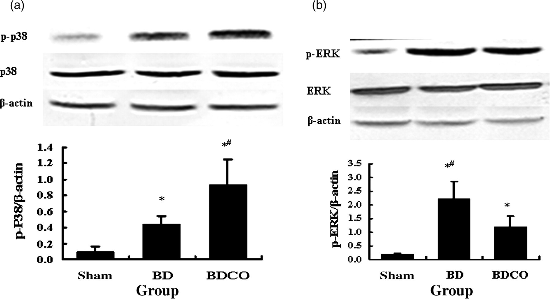
Expression of p38 and ERK proteins in lungs. (a) Expression of p38 proteins in lungs. (b) Expression of ERK proteins in lungs. The BD group activated p-p38 and p-ERK protein expression, and CO inhalation in BDCO group increased the p-p38 protein expression, but inhibited p-ERK protein expression. The data were representative of five independent experiments. The same loading volume was probed with an antibody against β-actin to assure equal loading of the gel. *P < 0.05 versus sham group; # P < 0.05 versus BD group. ERK, extracellular signal-regulated kinase; BD, brain death
Discussion
Our data showed that BD induced a significantly higher grade of deteriorations in the W/D, lung injury scores, inflammatory cytokine contents, progressive metabolic acidosis and cell apoptosis than those from normal rats. However, inhalation of CO at a concentration of 250 ppm in the BD rats greatly reversed these deteriorations. Furthermore, the inhalation of CO also increased the p-p38 protein expression and inhibited the p-ERK protein expression.
BD can cause severe changes in the lungs, presenting massive interstitial edema, alveolar hemorrhage, protein-rich intra-alveolar deposits and ultra structural changes in pneumocyte type II cells. 19,20 In the present study also, the BD process in rats induced an acute lung inflammatory response, substantiated by the increased W/D, the higher level of inflammatory cytokines (IL-6, TNF-α), increased MPO activity and histology changes of lungs. Simultaneously, early hemodynamic damage during the BD process of the donor could increase reperfusion injury after lung transplantation, 21 and progressive systemic inflammatory response induced by the BD donor aggravated chronic rejection after transplantation. 4 Thus, the lung injury caused by BD must be minimized to improve the function of the donor lung.
In the present study, the CO inhalation decreased the level of pro-inflammatory cytokines in serum from the BD rats. Although the mechanism has not been entirely elucidated, the anti-inflammatory effect through which CO provided cytoprotective effects may play a pivotal role. Ryter et al. 8 reported that CO confers protection against endotoxin shock in vitro and in vivo by inhibiting the production of pro-inflammatory cytokines. Miyazaki et al. 22 also found that CO inhalation in rats might modulate the inflammatory response to lipopolysaccharide. In addition, neutrophils are generally believed to be the primary cellular mediator triggering lung injury 23–25 and ICAM-1 is a key factor that adjusts the adhesion and activation of neutrophils in lung tissue. 26,27 Determining the activity of MPO, found principally in neutrophils, yields an estimate of neutrophil content. 28 Our data showed that CO inhalation inhibited the expression of ICAM-1 mRNA, decreased the MPO activity in lungs from BD rats and decreased the quantity of infiltrated neutrophil in lungs. From these results and considerations, it could be supposed that suppression of the lung injury and the inflammatory response, which were induced by BD, might play an important role in the favorable effects of CO inhalation.
Furthermore, the inhibition of apoptosis by CO may represent an additional mechanism by which CO provided protection against BD-induced injury. Although the precise physiological function of apoptosis in the lung has yet to be established, several studies showed that apoptosis was an important contributor to lung injury and severe organ dysfunction. 29–31 Modulation of apoptosis may provide an important avenue of therapeutic intervention in lung donors. Wang et al. 7 reported that CO confers lung protection by modulating apoptosis during hyperoxic stress. Zhang et al. 32 also found that CO could attenuate lung ischemia reperfusion injury by modulation of apoptosis and could modulate some key proteins during the apoptosis process, including Fas/Fas ligand, caspases and Bcl-2 family proteins. Our data indicated that CO inhalation decreased the ratio of apoptosis, and the expression of caspase-3 mRNA was also inhibited by CO inhalation in lungs from BD rats. Therefore, we inferred that the protective effects of CO in lungs from BD rats might be, at least partly, due to anti-apoptosis effects.
The cytoprotection of CO is also regulated and activated through the phosphorylation of mitogen-activated protein kinases (MAPKs), which occupy a central position in mediating cell survival and cell death. 33 Three major subgroups of the MAPK family were identified, including ERK, c-Jun N-terminal kinase and p38. In vitro studies using mouse endothelial cells and a warm lung ischemia reperfusion injury model in mouse had demonstrated that CO exerts antiapoptosis effects through p38. 32,34 Recently, Kohmoto et al. 35 reported that the protective effects of CO against ischemia reperfusion injury induced by lung transplantation from a living donor were associated with p38 MAPK phosphorylation. In the present study, the CO inhalation of BD rats increased the p-p38 protein expression and inhibited the p-ERK protein expression. Accordingly, we inferred that these two signal transduction pathways may be related to the anti-inflammatory and anti-apoptosis effects of CO.
The limitation of the present study was that we did not measure the concentration of carboxyhemoglobin in blood, but the CO concentration we applied might be safe. 11,36 In the preliminary experiment, we found that the lung function of BD rats was aggravated profoundly if the observation time was more than two hours, and therefore the donor lung may be unfit to be accepted as a donor, 37 so the observation time in this study was two hours. In conclusion, the present study demonstrated that CO exerted anti-apoptosis and anti-inflammatory effects on the lung injury induced by BD, and the protective effects were related to modulation of the MAPK signal transducer pathway.
Footnotes
Acknowledgements
This work was supported by Nature Science Foundation of China (Grant No. 30571784) and the Heilongjiang Province postdoctoral Foundation (No. LRB-68254).