




Abstract
Previous studies have suggested that zinc exerts anticarcinogenic and antiproliferative effects against prostate cancer both in vitro and in rat ventral prostate. Zinc accumulation diminishes early in the course of prostate malignancy and it inhibits the growth of several carcinoma cells through induction of cell cycle arrest and apoptosis. In this study, we have investigated the influence of zinc on N-methyl-N-nitrosourea (MNU) and testosterone (T)-induced prostatic intraepithelial neoplasia in the dorsolateral prostate of Sprague Dawley (SD) rats. The results indicate that zinc plays an important role in prostate carcinogenesis. Increased tumor incidence was accompanied by a decrease in prostatic acid phosphatase activity, citrate, zinc, glutathione-S-transferase, reduced glutathione, p53, B-cell lymphoma protein (Bcl-2)-associated X protein and caspase-3 levels in MNU + T-treated rats. On the contrary, significantly increased phase I drug metabolizing enzyme activities, lipid peroxide, hydrogen peroxide, proliferating cell nuclear antigen, Bcl-2 and Bcl-XL protein levels were observed in the dorsolateral prostate of MNU + T-treated rats. Simultaneous zinc supplementation significantly reversed these effects in MNU + T-treated rats. Signs of dysplasia, a characteristic of prostatic intraepithelial neoplasia, were evident in the dorsolateral prostatic tissue sections by MNU + T administration. However, zinc supplementation has reversed these effects in the dorsolateral prostatic histoarchitecture. These results suggest that zinc may act as an essential trace element against MNU and testosterone-induced prostatic preneoplastic progression in SD rats.
Introduction
Prostate cancer is the most common malignancy in men and the second leading cause for male cancer-related death worldwide. 1 The etiology of the disease is complex and it has a multitude of potential contributing factors including genetic and environmental factors. 2 Research on prostatic carcinogenesis has been impeded by a lack of adequate and reliable animal models. Growth of prostatic carcinomas beyond the phase of progression to invasively growing carcinoma can be studied in a few transplantable systems. 3 Prostate cancer development is a multistep process, which is thought to progress from premalignant lesions or prostatic intraepithelial neoplasia (PIN) through several stages of locally invasive, metastatic and hormone refractory disease. 4
Previous reports from our and other laboratories have described 5–7 the Sprague Dawley (SD) rat as an experimental model for hormonally induced prostatic dysplasia and cancer. Shorter-term (16 weeks) treatment with the combined hormone and carcinogen regimen consistently generated prostatic dysplasia, a putative preneoplastic lesion, exclusively within the enlarged dorsolateral prostates. 6 Mitotic activity was increased in these glands, with mitoses restricted almost entirely to epithelium of the dysplastic foci. 6 Close histological similarities exist between this proliferative lesion in the rat gland and PIN, a presumed precursor of prostate adenocarcinoma in the human prostate. 5,6 Spontaneous prostatic adenocarcinomas have been reported in different rat strains. 3,8 In most strains, these lesions develop in the ventral prostate lobe 8 for which there is no homologue. The adenocarcinomas should originate exclusively from the dorsolateral prostate, because that part of the rodent prostate is most likely a homologue to the region of the prostate from which prostatic cancer originates in man. 9
Zinc is one of the essential markers for prostate function. Low zinc status has been observed in cancer patients, suggesting a strong association between zinc and cancer development. 10 It is currently unknown as to how prostate cells could lose the ability to accumulate high levels of intracellular zinc, and whether this occurs as a consequence of acquiring cancerous properties. Zinc treatment causes decreased incidence of the spontaneous lung tumors arising in A/J mice, and also effectively inhibits chemically induced rat colonic preneoplasia. 11 Zinc replenishment has been shown to induce apoptosis in esophageal epithelial cells, thereby providing growth inhibition for the development of esophageal cancer. 12 Zinc has significant potency to inhibit prostate cancer cell proliferation and cell cycle arrest, and also inhibits prostate cancer cell survival by regulation of insulin-like growth factor-I receptor signaling. 13–16 Based on the clinical and experimental observations, zinc also acts as an anti-inflammatory agent. 17 It has been suggested that zinc supplementation may be a useful agent in the prevention and treatment of prostate cancer. 18,19
With the speculated literature limitations, the present study was designed to explore the possibility of zinc being used as a measure for long-term prophylactic therapy in delaying the cascade of events, leading to the development of prostate tumor. Our recent study demonstrated that zinc inhibits PIN, an early stage of prostate cancer, and also evidences the ability of zinc to restore the PIN changes particularly in the rat ventral prostate induced by carcinogen and testosterone, thereby indicating its anticarcinogenic potential. 7
As the dorsolateral prostate of rodents is most likely homologous to the human prostate, this study was interested in observing the role of zinc on the regulation of key enzymes of prostate function, oxidative stress markers, phase I and phase II drug metabolizing enzymes, p53, proliferating cell nuclear antigen (PCNA) and apoptotic and antiapoptotic proteins, as well as investigating the dorsolateral prostate histological alterations of rats subjected to N-methyl-N-nitrosourea and testosterone (MNU + T) treatment.
Materials and methods
Animals
Healthy male SD rats (eight weeks of age) were obtained from the Institutional Central Animal House Facility. The animals were housed in clean polypropylene cages (three rats/cage) and maintained in an air-conditioned animal house with constant 12-h light/dark cycle. Rats were permitted free access to drinking water throughout the experimental period. The animals were fed with a standard rat pellet diet (Lipton India Ltd, Mumbai, India). The experiment was approved by the Institutional Animal Ethical Committee (IAEC No. 03/027/07).
Chemicals
Testosterone propionate, MNU and zinc chloride were purchased from Sigma-Aldrich Chemicals Private Ltd (St Louis, MO, USA). Primary antibodies such as mouse monoclonal p53, mouse monoclonal PCNA, mouse monoclonal B-cell lymphoma protein (Bcl-2), mouse monoclonal Bcl-2-associated X protein (Bax), goat polyclonal caspase-3 and mouse monoclonal Bcl-XL antibodies were obtained from Santa Cruz Biotechnology Inc (Santa Cruz, CA, USA). Secondary antibodies were purchased from GeneI, Bangalore, India. Other chemicals were obtained from SISCO Research Laboratories Pvt Ltd, Mumbai, India. All the chemicals were extra pure and of analytical grade.
Induction of prostate tumor
Rats were injected carcinogen and hormone by a modified protocol derived by McCormick and Rao 20 and Liao et al. 5 First, each rat received daily intraperitoneal injections of testosterone propionate (50 mg/kg body weight) for 21 consecutive days. After the last dose of testosterone injection, rats received daily intraperitoneal injections of 100 mg testosterone propionate/kg body weight in 0.3 mL propylene glycol for three days. After the last dose of testosterone injection, all the rats received a single intraveneous dose (50 mg/kg body weight) of MNU (dissolved in saline at 10 mg/mL) via the tail vein. One week after MNU administration, rats received intraperitoneal injection of 4 mg testosterone propionate/kg body weight alternatively for 120 d.
Experimental protocol
The rats were divided into four groups, and each group consisted of 10 animals. Group I animals received vehicle (propylene glycol) by intraperitoneal injection, considered as control. Group II animals were induced prostate cancer by using carcinogen and hormone. Group III animals were induced prostate cancer and simultaneous supplementation of zinc chloride (100 ppm) weekly thrice in drinking water for 20 weeks. Zinc supplementation begun a week before administration of initial dose of testosterone propionate administration and throughout the studies. Group IV animals received zinc chloride (100 ppm).
We estimated that the daily zinc intake by a 292 g rat (average body weight at study termination) from 100 ppm zinc-supplemented water was ∼ 0.75 mg based on a water intake of ∼16 mL per day. This zinc dose translates to the human supplementation of ∼ 70 mg zinc per day for a 65-kg individual.
The zinc dose was selected based on the previous studies by Arriazu et al. 21 After the treatment period, rats were killed, the prostatic fluid was removed and the dorsolateral prostate was dissected from the adhering connective tissue, washed several times with physiological saline, weighed accurately and separated. The organ/body weight ratio was calculated.
Histopathological examination
Fresh tissue pieces of the dorsolateral prostate were fixed in Bouin's fluid for the histopathological observations. Following an overnight fixation, the specimens were dehydrated in ascending grades of alcohol, cleared in benzene and embedded in paraffin wax. Blocks were made and 4-μm-thick sections were double-stained with hematoxylin and eosin. The sections were permanently mounted with dibutyl phthalate xylene and observed under a light microscope (Nikon Optiphot, Tokyo, Japan).
Biochemical assays
Sample preparation
Prostate tissue was homogenized in 0.1 mol/L of Tris-hydrochloric acid buffer, pH 7.4 and centrifuged at 3000
Dorsolateral prostatic acid phosphatase (PAcP), citrate, phase I drug metabolizing enzymes such as cytochrome P450 (cyt P450), cytochrome b5 (cyt b5), NADH-cyt b5 reductase (cyt b5 red), NADPH-cyt C reductase (cyt C red) activities, phase II enzyme, glutathione-S-transferase (GST), lipid peroxide (LPO), hydrogen peroxide (H2O2) and reduced glutathione (GSH) were estimated. 22–30
Zinc analysis
Zinc concentration was determined using an atomic absorption spectrophotometer (AAS), according to the method of O'Halloran et al. 31 To obtain a clear dissolved solution, the prostatic tissue was digested repeatedly with concentrated nitric acid and 60% perchloric acid. Finally, the solution was diluted to 10 mL with deionized water and analyzed by AAS. The zinc level was expressed as micrograms per gram of tissue.
Western blot analysis
After the sacrifice, the dorsolateral prostate tissue was homogenized with 1 mL of radioimmunoprecipitation assay buffer (150 mmol/L sodium chloride, 50 mol/L Tris, 1 mmol/L ethylenediamine tetraacetic acid, 1% non-idet protein-40, 0.5% sodium deoxycholate and 0.1% sodium dodecyl sulphate [SDS], pH 7.4) with protease inhibitor cocktail. Tissue homogenate was then centrifuged at 12,000
Statistical evaluation
Data were expressed as mean ± standard error mean. Software Package for Social Studies (SPSS Inc, Chicago, IL, USA) was used for the statistical analysis. Statistical differences between means were evaluated using one-way analysis of variance followed by post hoc Duncan's new multiple range test. P < 0.05 was considered statistically significant.
Results
Body and dorsolateral prostate weights
Body weight was significantly decreased in MNU + T-treated rats compared with controls, whereas simultaneous zinc supplementation significantly increased the body weight. Zinc-alone-supplemented rats showed significant gain compared with controls (Table 1). It was observed that MNU + T-treated rats showed significant increase in both absolute and relative dorsolateral prostate weights when compared with control and drug-control rats. The increase in absolute and relative dorsolateral prostate weight was decreased following zinc supplementation to MNU + T-treated rats. This decrease in body weight clearly denotes that the tumor incidence was depressed by zinc in MNU + T-treated rats comparable with that of both control and drug control groups.
Effect of zinc on body weight and dorsolateral prostate weights treated with MNU + T, Zn and Vehicle
MNU, N-methyl-N-nitrosourea; T, testosterone; SEM, standard error of mean
Data presented are mean ± SEM and N = 10 animals. The statistical significance was considered at the level of P < 0.05 following post hoc Duncan's new multiple range test
†Control versus MNU + T, MNU + T + Zn
‡MNU + T versus MNU + T plus Zn-treated rats
¶Control versus Zn
Tumor incidence
The percentage of tumor incidence is summarized in Figure 1. Survival was limited to 20 weeks from the start of the treatment. Increased tumor incidence was observed in the dorsolateral prostate of MNU + T-treated rats. Four (40%) of 10 animals showed PIN in the dorsolateral lobe of the prostate. PIN is a pattern of epithelial cell proliferation, preneoplastic lesion containing epithelial cells protruding into the lumen. Two to six PIN regions were observed in each MNU + T-treated rat. MNU + T-treated rats showed hyperplasia and dysplasia of about 70% (7) and 50% (5), respectively. About 10% (1/10) of animals developed PIN in the dorsolateral prostate of zinc-supplemented rats. Occurrence of hyperplasia, dysplasia and PIN lesions were less in the dorsolateral lobe (20%, 10% and 10%, respectively) in simultaneous zinc-supplemented rats. Moreover, the zinc-alone group did not show any evidence of tumor.

Effect of zinc on the incidence of prostate carcinogenesis in Sprague Dawley rats. Number of animals in each group was 10. Hyp, hyperplasia; Dys, dysplasia; PIN, prostatic intraepithelial neoplasia (A color version of this figure is available in the online journal)
Histopathology
Control animals displayed normal dorsolateral prostate architecture; the tubules are lined by a single layer of cuboidal cells with secretion in the lumen (Figure 2a). MNU + T-induced animals showed hyperplastic and dysplastic sites within the same glandular epithelium. An architectural pattern of high-grade prostatic intraepithelial neoplasia (HGPIN) such as tufting (Figure 2b1), cribriform (Figure 2b2) and micropapillary (Figure 2b3) structures were observed. MNU + T + zinc-treated animals illustrated an absence of dysplastic and hyperplastic nodules, and it also showed less number of mitotic figures and the stroma was very loosely organized (Figure 2c). Zinc-treated animals showed the normal appearance of epithelial tubules lining the lumen secretions (Figure 2d).
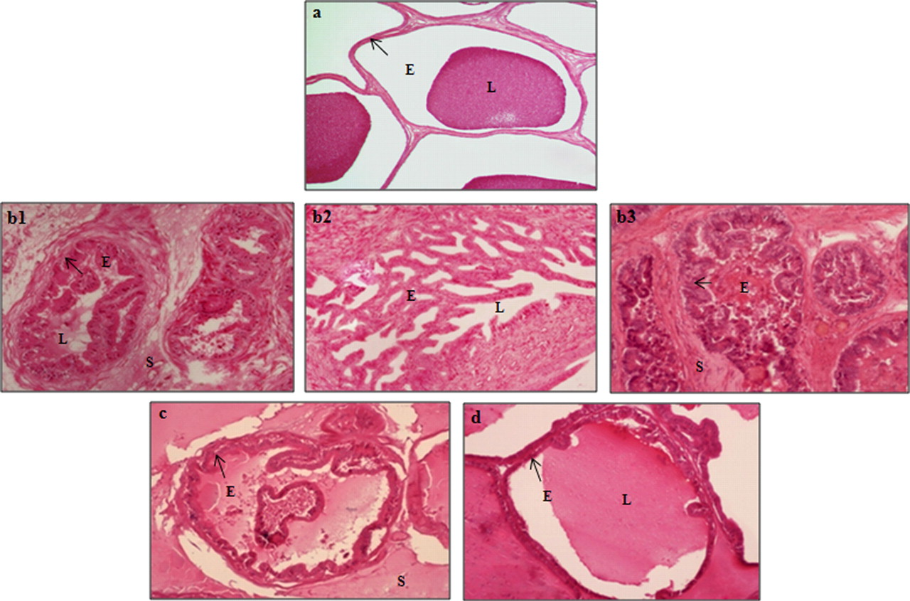
Histopathology of dorsolateral prostate tissues from MNU + T-induced Sprague Dawley rats treated with zinc. (a) Normal dorsolateral prostate epithelium. A single layer of cuboidal cell and the tubules with secretion in the lumen. Epithelial tubules, surrounded by a thin layer of smooth muscle cells are distributed (×200 magnification; hematoxylin and eosin [H & E]). (b1) MNU + T treated rats. Hyperplastic and dysplastic sites were seen within the same glandular epithelium. Architectural pattern of high-grade prostatic intraepithelial neoplasia (PIN) such as tufting (b1), cribriform (b2) and micropapillary (b3) structures were observed (×200 magnification; H & E). (c) MNU + T + Zn-treated group. Hyperplasia and dysplasia are less common and loosely organized stoma was observed. It also showed less number of mitotic figures (×200 magnification; H & E). (d) Zn group showing the normal appearance of epithelial tubules lining the lumen secretions (×200 magnification; H & E). E, epithelium; L, lumen; S, stroma; MNU, N-methyl-N-nitrosourea; T, testosterone (A color version of this figure is available in the online journal)
Biochemical analysis
There was a significant decrease in PAcP activity (51%), and zinc (60%) and citrate (52%) levels in the dorsolateral prostate of MNU + T-treated rats, which was only partially reversed to control levels by simultaneous zinc supplementation. Further, a significant increase in prostatic zinc (83%) and citrate (77%) levels were observed in the zinc-alone group in comparison with controls (Figure 3).
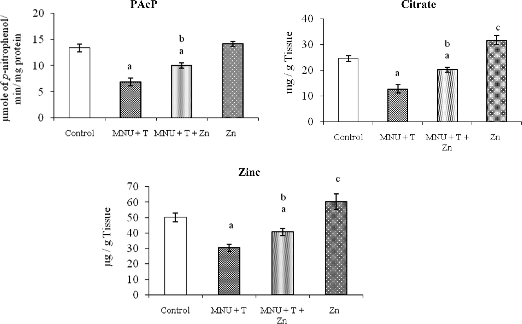
Effect of zinc on PAcP activity, citrate and zinc levels in the dorsolateral prostate of Sprague Dawley rats treated with MNU and testosterone. Data presented are mean ± SEM and N = 10 animals. The statistical significance was considered at the level of P < 0.05 following post hoc Duncan's new multiple range test. aControl versus MNU + T, MNU + T + Zn, bMNU + T versus MNU + T plus Zn-treated rats, cControl versus Zn. MNU, N-methyl-N-nitrosourea; T, testosterone; SEM, standard error of mean; PAcP, prostatic acid phosphatase
Statistically significant increases in cyt P450 (53%), cyt b5 (48%), cyt b5 red (26%) and cyt C red (67%) enzyme activities were obtained in the dorsolateral prostate of the MNU + T-treated group compared with controls, whereas simultaneous zinc supplementation significantly decreased these enzyme activities compared with MNU + T-treated rats (Figures 4a–4d). The activity of GST (38%) was decreased significantly in the dorsolateral prostate of MNU + T-treated rats, whereas it was increased significantly by simultaneous zinc supplementation (Figure 4e). However, the increases in cyt P450 (68%), cyt b5 (70%), cyt b5 red (36%) and cyt C red (82%) enzyme activities and the reduction in GST (71%) observed in the simultaneous zinc supplementation were significantly higher or lower, respectively, than the controls. Moreover, zinc supplementation alone exerted no changes in these parameters as compared with controls.
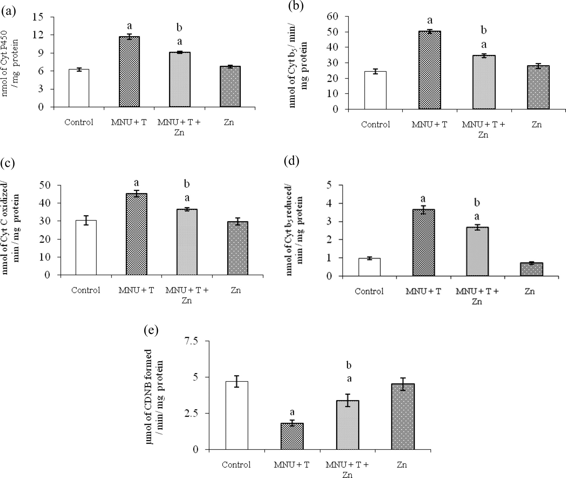
Effect of zinc on phase I and phase II drug metabolizing enzymes activities in dorsolateral prostate of Sprague Dawley rats treated with MNU + T, Zn and Vehicle. Assay of phase I and II drug metabolizing enzyme activities such as cytochrome P450 (a), cytochrome b5 (b), cytochrome C reductase (c), cytochrome b5 reductase (d) and glutathione-S-transferase (e). Data presented are mean ± SEM and N = 10 animals. The statistical significance was considered at the level of P < 0.05 following post hoc Duncan's new multiple range test. aControl versus MNU + T, MNU + T + Zn, bMNU + T versus MNU + T plus Zn-treated rats. MNU, N-methyl-N-nitrosourea; T, testosterone; SEM, standard error of mean; CDNB, 1-chloro,2,4-dinitrobenzene
A statistically significant increase in the levels of lipid peroxides (75%) and H2O2 (44%) levels were observed in the dorsolateral prostate of MNU + T-treated rats compared with controls. The MNU + T-treated group showed a significant decrease in GSH level (30%) compared with controls. Simultaneous zinc supplementation of MNU + T treatment shows reduced lipid peroxidation and H2O2 level than MNU + T treatment alone, but the reduction did not reach the level of controls. Simultaneous zinc supplementation has increased GSH level compared to MNU + T alone, but the GSH level was still lower than controls (Table 2).
Effect of zinc on lipid peroxidation, H2O2 and GSH levels in dorsolateral prostate of Sprague Dawley rats treated with MNU + T, Zn and Vehicle
MNU, N-methyl-N-nitrosourea; T, testosterone; SEM, standard error of mean; GSH, glutathione; LPO, lipid peroxide; H2O2, hydrogen peroxide, TBARS, thiobarbituric acid reactive substances
Data presented are mean ± SEM and N = 10 animals. The statistical significance was considered at the level of P < 0.05 following post hoc Duncan's new multiple range test
†Control versus MNU + T, MNU + T + Zn
‡MNU + T versus MNU + T plus Zn-treated rats
Western blot analysis
A significant increase in PCNA (>100%) and decrease in p53 (75%) were observed in the dorsolateral prostate of MNU + T-treated rats compared with controls. However, these protein levels were reversed to control levels by simultaneous zinc supplementation compared with MNU + T-treated rats (Figures 5a and 5b). No significant PCNA and p53 levels were observed in the zinc-alone group as compared with controls.
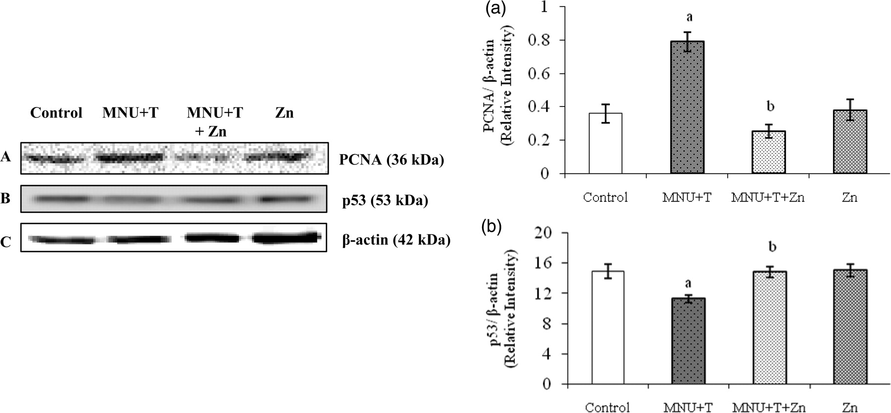
Western blot analysis of PCNA and p53 proteins in rat dorsolateral prostate treated with MNU + testosterone. Quantitative representation of protein levels of PCNA (a) and p53 (b). Data presented are mean ± SEM and N = 10 animals. The PCNA and p53 protein levels were assessed using anti-PCNA (A) and anti-p53 (B) antibodies and β-actin (C) was used as internal control. Relative intensities of PCNA and p53 with β-actin levels are shown in the right panel, respectively. The statistical significance was considered at the level of P < 0.05 following post hoc Duncan's new multiple range test. aControl versus MNU + T, MNU + T + Zn, bMNU + T versus MNU + T plus Zn-treated rats. MNU, N-methyl-N-nitrosourea; T, testosterone; SEM, standard error of mean; PCNA, proliferating cell nuclear antigen
Protein levels of antiapoptotic proteins, Bcl-2 (71%) and Bcl-XL (70%) were significantly increased, whereas apoptotic protein, Bax (59%) and caspase-3 (57 %) protein levels were significantly decreased in MNU + T-treated rats compared with control rats. However, these changes in protein levels were reversed toward respective levels in controls by simultaneous zinc supplementation (Figures 6a–6d).
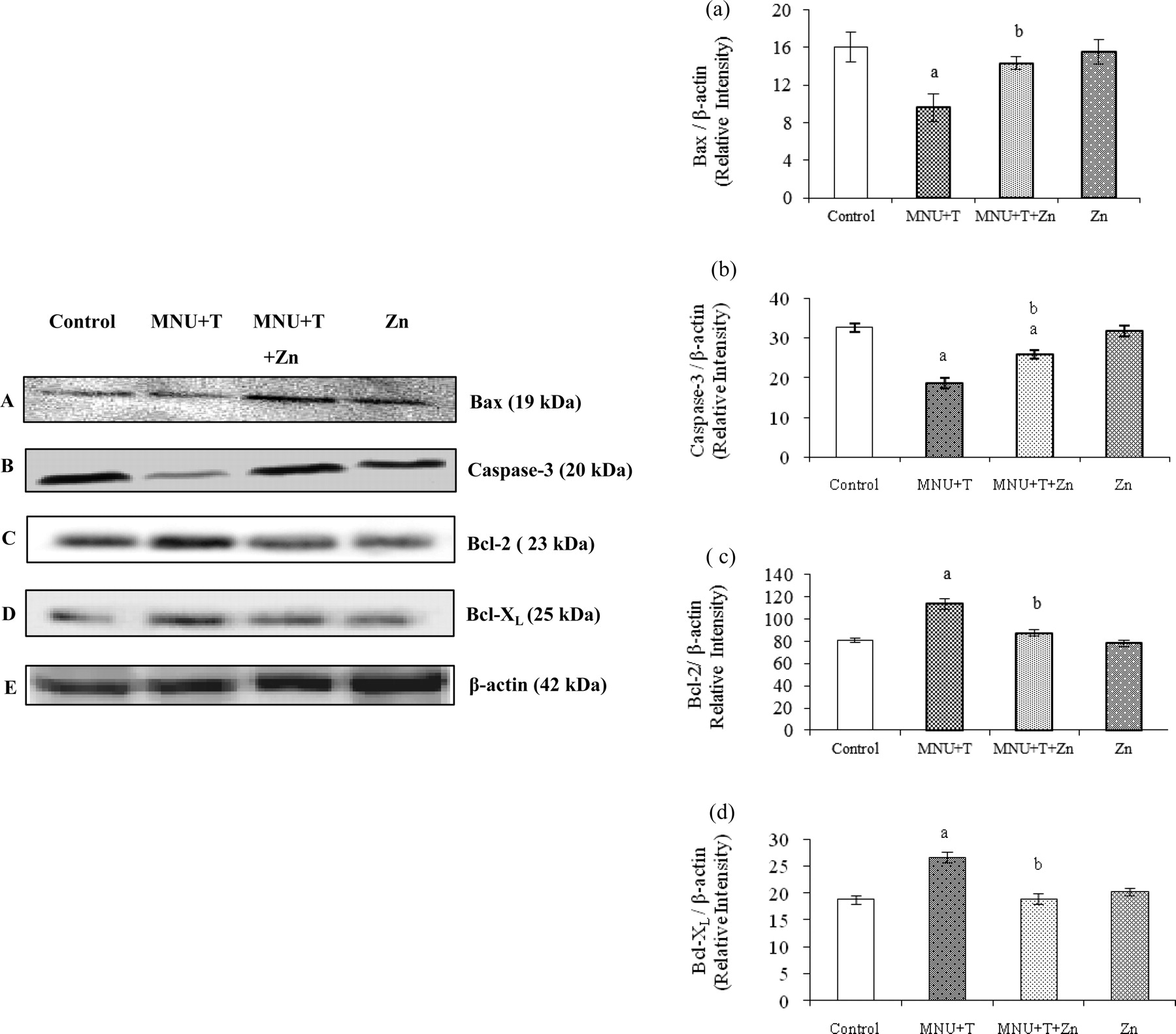
Western blot analysis of apoptotic and antiapoptotic proteins in rat dorsolateral prostate treated with MNU + T, Zn and Vehicle. Quantitative representation of protein levels of Bax (a), caspase (b), Bcl-2 (c) and Bcl-XL (d). Data presented are mean ± SEM and N = 10 animals. Bax and caspase-3 protein levels were assessed using anti-Bax (A), anticaspase-3 (B), anti-Bcl-2 (C) and anti-Bcl-XL (D) antibodies and β-actin (E) was used as internal control. Relative intensities of Bax, caspase-3, Bcl-2 and Bcl-XL with β-actin level are shown in the right panel, respectively. The statistical significance was considered at the level of P < 0.05 following post hoc Duncan's new multiple range test. aControl versus MNU + T, MNU + T + Zn, bMNU + T versus MNU + T plus Zn-treated rats. MNU, N-methyl-N-nitrosourea; T, testosterone; SEM, standard error of mean; Bcl-2, B-cell lymphoma protein; Bax, Bcl-2-associated X protein
Discussion
The importance of zinc in prostate health abide by the findings that the zinc concentration is highest in the prostate gland but tends to be very low in cancerous prostates. Analyses of malignant prostate tissues showed a 60–70% reduction in zinc levels in normal peripheral zone tissues. 32 The present study demonstrates the potential role of zinc on the detrimental effects caused by carcinogen and hormone in the dorsolateral prostate of SD rats.
Our data demonstrated that zinc reduced dorsolateral prostate weight and tumor incidence with concomitant increase in body weight following carcinogen and hormone treatment. The combined MNU and testosterone regimen induced a 40% incidence of prostatic preneoplastic lesion in the dorsolateral prostate of SD rats. A high incidence of early stage of prostate cancer was evidenced in the dorsolateral prostate when compared with the ventral prostate, whose incidence is 30%. 7 Furthermore, these putative preneoplastic lesions in the dorsolateral prostate appeared to have morphological similarity with premalignant lesions described in the human prostate gland. 33 Zinc supplementation caused a decrease in the tumor incidence, suggesting the capability of zinc in inhibiting or reducing tumorigenesis. In the present study, the daily zinc intake by a 292-g rat from zinc-supplemented water was ∼0.75 mg based on a water intake of ∼16 mL per day. This zinc dose translates to human supplementation of ∼70 mg zinc per day for a 65-kg individual. In general, the combined level of dietary and supplemental zinc in adults is 40 mg/d. The recommended daily allowance of zinc in human males is 15 mg/d. 34 Moreover, no tumor incidence was evident in the zinc-treated group, suggesting that zinc at this dose level causes no disruption of normal homeostasis and was determined to be non-toxic. 11
This model appears to provide a unique opportunity to investigate the spectrum of histological and molecular changes during the early progression of prostate cancer, i.e. PIN. In this model, MNU + T-induced prostate carcinogenesis showed the presence of hyperplasia, dysplasia and PIN. The characteristics of HGPIN such as micropapillary, tufting and cribrifom structures were observed in the dorsolateral prostate of MNU + T-treated rats. These are graded according to the conventional grading criteria as described by previous studies. 35,36 Zinc supplementation showed less PIN morphology compared with MNU + T-treated animals and reversed the above-mentioned criteria. Although the mechanisms involved in the protective effects against incidence of PIN formation are not clearly understood, the inhibitory action of zinc could be explained by its putative antioxidant activity. 11 Thus, zinc effectively reversed MNU and testerone-induced preneoplastic changes of prostate cancer that are described here and which have a large number of important characteristics with human prostatic cancer. However, the relationship between zinc and androgen sensitivity remains unclear.
Acid phosphatase is considered an important marker for the prostate function. The level of serum acid phosphatase of prostatic origin increased markedly in humans with extensive or metabolic carcinoma of the prostate. 37 The present study demonstrated a significant increase in serum PAcP (data not shown) and decrease in PAcP, which could be due to an increase in cell number of the tumor mass and increased leakage of the PAcP from plasma membranes of carcinoma cells. 37 Simultaneous zinc supplementation significantly increased the dorsolateral PAcP activity. However, the exact mechanism behind this remains unclear.
Loss of zinc and citrate is an important metabolic change associated with prostate cancer development, creating an optimal environment for uncontrolled proliferation, impaired apoptosis, cancer progression and metastasis. 38 In this study, zinc supplementation reverses prostatic zinc and citrate levels following carcinogen and hormone exposure, which is in line with the previous study. 11 Although the precise mechanistic role of zinc in human carcinogenesis is unclear, our observations together with the published literature for the diminished zinc levels in prostate cancer suggest that zinc directly or indirectly inhibits the growth of cancerous cells.
It was reported that MNU + T caused a significant increase in the total cyt P450 activity and also three regenerating enzymes activities such as cyt b5, NADH cyt b5 red and NADPH cyt C red. The observed reversal trend of these enzymes activities following zinc supplementation could possibly be due to the activation of pyrophosphatase II, a zinc-containing enzyme, which is capable of metabolizing both NADPH and NADH. 39 Zinc is known to form a complex with NADPH, and it is suggested that regulation of drug metabolism by zinc might be because this complex formation with reductase changes its oxidation − reduction potential, thus impacting on the regulation of cyt P450 level. 40–42
Considering that GST are detoxifying enzymes that catalyze the conjugation of a variety of electrophilic substrates to the thiol group of GSH, producing less toxic forms, 43 the significant decrease in GST activity in the rat dorsolateral prostate following carcinogen and hormone induction may indicate insufficient detoxification in rats. The reversal of GST activity following zinc supplementation could possibly be due to either induced metallothionein content or due to its indirect action in reducing the levels of oxygen reactive species. 44
Our results revealed that carcinogen and hormone caused a statistically significant increase in the levels of LPO and H2O2. Supplementation of zinc to the MNU + T-treated group of rats brought back the levels of LPO and H2O2 to controls. Decreased zinc level results in an increase in free iron and H2O2 in liver microsomes, 45 thus increasing a free-radical-mediated attack on DNA, proteins and membrane lipids. This competition in turn interrupts the oxidative inactivation of several enzymes involved in the binding site in the catalytic portion of enzymes; the Fe2+, by redox cycling, promotes the generation of hydroxyl radicals from H2O2 that oxidizes one or more amino acids. 45,46 The reversal of LPO following zinc supplementation is very likely due to its antiperoxidative properties, as had been shown previously. 46
GSH is the major cellular sulfhydryl compound that serves as an effective reductant and a nucleophile that interacts with numerous electrophilic and oxidizing compounds. Modulation of antioxidant protectors like GSH occurs during cellular apoptotic processes. 47 By the catalytic effects of metal ions, polymerization of aldehydes and hydrocarbons results in the formation of several byproducts, including malondialdehyde (in the terms of thiobarbituric acid reactive substances formed), which is an indicator of the rate at which oxidation is occurring. 48 MNU + T treatment caused a significant decrease in glutathione level in the dorsolateral prostate compared with controls. The observed reversal trend of GSH level followed by zinc supplementation may be due to its physiological role, as an antioxidant by protecting sulfhydryl groups against oxidation and by inhibiting the production of reactive oxygen species like H2O2 by transition metals. 49,50 The antioxidant role of zinc may be one of the key mechanisms to elucidate its protective effect.
PCNA emerged as an easily reproducible biomarker of proliferation of fixed tissue. 5 It is suggested that the progression of prostate carcinogenesis in the MNU model was associated with a profound increase in PCNA labeling from hyperplasia to adenocarcinomas of varying differentiation. 5 Zinc supplementation has downregulated PCNA level following carcinogen and hormone treatment, suggesting that it acts as an effective inhibitor of cell proliferation. However, the exact mechanism of this is still unclear. Zinc exerted its suppressive effect on PCNA not only in the dorsolateral prostate but also in the ventral prostate, suggesting that it act as an effective antiproliferative agent against prostate cancer.
A number of investigators have suggested that the development of new interventions for prostate cancer prevention and treatment should focus on enhancing apoptosis. 51 The p53-induced apoptosis can be blocked by gene-transfer-mediated elevations in the levels of the Bcl-2 protein, suggesting that p53 and Bcl-2 may participate in a common pathway for regulation of cell life and death. 21,52 The attenuated Bcl-2, Bcl-XL and increased level of p53 proteins by zinc supplementation is due to the conformational change in the protein structure and increases its affinity for specific sequences of p53 as it is tightly bound to zinc ions, 21 thereby stimulating the transcriptional activity of cell-cycle regulators (p21, GADD45, etc.) involved in the cell-cycle progression. 21 A decreased level of Bax protein was observed in MNU + T-treated rats; however, this protein level was reversed by simultaneous zinc supplementation. The exact mechanism for increased Bax level is not known; it may be due to the increase in zinc level. 13
Apoptosis is a molecular process driven by apoptotic machinery components termed as caspases. 21 The present study also evidenced a significant decrease in caspase-3 protein level in MNU + T treatment that was reversed by simultaneous zinc supplementation. Zinc has an important role in regulating the prostatic cell growth and induces mitochondrial apoptosis via activation of caspases-9 and -3, leading to the cleavage to nuclear poly (ADP)-ribose polymerase. 53 This may be the mechanism contributing to the regression of cancer cell growth of zinc-supplementation rats.
Conclusion
In conclusion, zinc was found to inhibit the growth and decrease prostatic PAcP, zinc, citrate levels, phase I drug metabolizing enzyme activities, lipid peroxide, H2O2 levels and PCNA, Bcl-2, Bcl-XL expressions, with a concomitant increase in phase II enzyme activities, GSH level, and p53, Bax and caspase-3 expressions in a MNU and testosterone-induced model of prostate carcinogenesis. Zinc may also be effective in inducing regression in PIN and its utility as a chemopreventive agent during the long latency period of prostate cancer should be further explored. We hope that our findings stimulate future progress towards understanding the complex relationship between zinc levels and prostate carcinogenesis.
Footnotes
Acknowledgements
This work was supported by the Council of Scientific and Industrial Research (CSIR), India, in the form of CSIR-SRF (Grant No. 09/115 (0685)/2008-EMR-I dt 23.10.2008). The authors thank Kavitha Kishore in proofreading the article.