




Abstract
Recent studies indicated that regular consumption of antioxidant-rich foods reduces cellular oxidative stress and protects against health-related problems. This study aimed to assess the in vitro antioxidant properties of the papaya epicarp extract against hydrogen peroxide (H2O2)-induced oxidative stress in human SH-SY5Y neuronal cells. Our study revealed that papaya epicarp extract acted as a potent free radical scavenger and provided neuroprotection against H2O2-induced oxidative stress. Papaya epicarp extract ameliorated glutathione depletion, restored total antioxidant capacity and augmented the inhibition of antioxidant enzymes (catalase, glutathione peroxidases and superoxide dismutase). In conclusion, papaya epicarp extract can be used as a functional dietary ingredient that might help in reducing the neurological health problems associated with various oxidative stress insults.
Introduction
Oxidative stress is a condition under which an imbalance exists between the factors that promote oxidation (such as reactive oxygen species, ROS) and antioxidant defenses, including intracellular glutathione (GSH), dietary antioxidants and antioxidant enzymes. Oxidative stress is involved in the cell degenerative changes during the aging process and in the pathogenesis of a wide variety of human chronic diseases, such as cancer, atherosclerosis, coronary heart disease, macular degeneration, Alzheimer's disease, inflammation and emphysema. 1–3
Hydrogen peroxide (H2O2) is a well-known ROS that is formed during normal metabolism, where it is produced in the brain during the catalytic degradation of neurotransmitters such as dopamine, and it is estimated that about 3% of the body's hemoglobin is autoxidized daily to produce H2O2. 4,5
GSH is the primary redox buffer in all human neuronal cells, and when two molecules of GSH combine to form oxidized glutathione, two reducing equivalents of H+ are made available for the quenching of ROS, thereby protecting neuronal cells from oxidative damage. 6 GSH can also combine with xenobiotics and heavy metals, resulting in an increase in their rate of excretion and provides an important mode of detoxification. 7 Depletion of GSH and low total antioxidant capacity (TAC) indicate that the production of ROS exceeds the capacity of cellular antioxidant protective mechanisms. 8
Antioxidant enzymes such as catalase (CAT), superoxide dismutase (SOD) and glutathione peroxidase (GPX) provide a primary defense against ROS, and activities of these enzymes are known to decrease under ROS-induced oxidative stress. 9 CAT catalyzes the detoxification of H2O2 to H2O. GPX activates the GSH-scavenging reactions of free radicals (.OH) and singlet oxygen species ( 1 O2), whereas SOD is responsible for the dismutation of two superoxide anion molecules to form H2O2 and O2. 8
Papaya (Carica papaya L.) is a tropical fruit that is rich in dietary antioxidants (vitamin C, tocopherols, total phenols and beta-carotene). 10 It also contains many bioactive phytochemicals that show antioxidant activities. 11 Benzyl isothiocyanate, a natural toxin, has been found to be present in papaya fruit and seeds, and it has potent diverse biological activities in inducing the phase 2 detoxifying enzymes and apoptosis. 12–15 However, the quantities in mature papaya fruit are low and are considered as safe for human health. 16
Papaya fruit and seeds are widely used in medicine, because of their high antioxidant contents, to prevent lipid peroxidation, and are considered as a preventive treatment against atherosclerosis and coronary heart diseases. 10,17 Papaya was reported to have an in vitro free radical scavenging property, 18 was effective in improving antioxidant defense and significantly decreased the risk of age-related macular degeneration in human clinical trials. 19–21
Papaya was reported to protect against H2O2-induced oxidative DNA damage in rat pheochromocytoma tumor cells. 22 Papaya fruit skin contains different bioactive compounds, such as ferulic acid, p-coumaric acid, caffeic acid, carotenoids (mostly lycopene, β-cryptoxanthin, and β-carotene) and vitamin C, which collectively can protect human cells from oxidative stress. 10 Papaya epicarp extracts were effective in promoting the wound healing process and cellular skin development and these remarkable effects of papaya were attributed to its photochemical and antioxidant activities. 23,24
There has been no previous research conducted to explore the protective role of papaya against oxidative stress insults in human cultured neuronal cells. Therefore, the aim of this study was to evaluate the effect of papaya epicarp extract against H2O2-induced oxidative stress in human SH-SY5Y neuronal cells. The findings of this study will shed light on the potential use of papaya, as a rich source of natural dietary antioxidants, against oxidative stress-related neurodegenerative disorders such as Alzheimer's disease.
Materials and methods
Chemicals
All chemicals, α-minimum essential media (α-MEM) cell culture medium, trypsin-EDTA solution, penicillin–streptomycin–fungizone (PSF), trypsin–EDTA solution and fetal bovine serum (FBS) were purchased from Sigma Chemical Co (St Louis, MI, USA).
Preparation of papaya epicarp extract
Fresh green papaya was washed and the epicarp separated by manual peeling using a knife. The water content of the epicarp was 77.4 g/100 g skin. The epicarp was dried in a air convection dryer at 40°C for 18 h and then placed in desiccators containing silica gel at 20°C for one week. Dried epicarp was ground into powder using a hammer mill with sieve size 1.0 mm (Model MF 10 Basic, IKA Works, Wilmington, NC, USA). The dried powder was equilibrated for four weeks in desiccators at 20°C containing a saturated lithium chloride solution to maintain the relative humidity of the environment at 11.3%. The dried powder was mixed with distilled water (10 g/150 g) and the mixture was stirred on a magnetic stirrer for four hours at room temperature. The mixture was then centrifuged at 2000 g at 4°C for 30 min and the supernatant was collected and then stored at −80°C for subsequent analysis. The solid content of the extract was 3 g/100 g extract.
Total phenolic assay
Total phenolics were determined with the Folin-Ciocalteu reagent, where different volumes (50, 100 and 150 μl) of papaya epicarp extract were added to 2.50 mL of Folin-Ciocalteu's reagent and 750 μL of Na2CO3 (7.5%, w/v). Distilled water was then added to bring the total volume to 5 mL and the mixture was incubated at room temperature (22°C) for two hours. The absorbance of all samples was measured at 765 nm using a UV–visible spectrophotometer. Results were expressed as milligrams of gallic acid equivalent (GAE) per gram of papaya dry weight (mg GAE/100 g dry powder). Four replicates were run for each concentration.
Evaluation of the free radical scavenging capacity of papaya epicarp extract by the DPPH photometric assay
The capacity of papaya epicarp extract to scavenge 1,1-diphenyl-2-picrylhydrazyl (DPPH) free radicals was measured by a spectrophotometric method as described by López et al.
25
Briefly, 50 μL of each papaya epicarp extract, at different concentrations (μg/mL), was mixed with 50 μL of a DPPH methanolic solution (0.04 mg/mL). Absorbance was measured at 517 nm after 30 min of reaction at room temperature. Controls contained all the reaction reagents except the papaya epicarp extract or 2,6-di-tert-butyl-4-hydroxytoluene (BHT) as a positive control. The free radical scavenging capacity of different samples was expressed as % DPPH inhibition; a higher % free radical scavenging activity value indicates a higher antioxidant activity and was calculated as follows:

Evaluation of the antioxidant activity of papaya epicarp extract by the ABTS antioxidant assay
A colorimetric method using ABTS Antioxidant Assay Kit (Cat#AOX-1; Zen-Bio, Research Triangle Park, NC, USA) was carried out. The assay is based on the incubation of papaya epicarp extract samples at different concentrations (μg/mL) with 2, 2′-azino-di-[3-ethylbenzthiazoline sulphonate (6)] diaammonium salt (ABTS) with a peroxidase (methmyoglobin) and hydrogen peroxide to produce the radical cation ABTS+, which has a relatively stable blue-green color that is measured at 405 nm. Antioxidants present in the assayed papaya epicarp extract samples inhibit the oxidation of ABTS to ABTS+ (cause suppression of the color production) to a degree that is proportional to their concentration. The total antioxidant capacity of the assayed papaya epicarp extract samples was compared with that of standard Trolox, a water-soluble tocopherol analogue.
Human SH-SY5Y neuronal cell culture
SH-SY5Y cells were purchased from the American Type Culture Collection (ATCC; Manassas, VA, USA), and were frozen in liquid nitrogen before culturing. Cells were grown as a monolayer in 10-cm Petri dishes that contained 10 mL of α-MEM supplemented with 1% PSF and 10% FBS. Cultured cells were placed in an incubator chamber containing humidified 95% air and 5% CO2 at 37°C. For passage, confluent cells were detached with 1.0 mL trypsin-EDTA solution. They were then re-suspended in 10 mL of cell culture medium and incubated for 24 h before conducting the desired experiment.
Human SH-SY5Y neuronal cell treatment
The study design was to grow the human cultured neuronal cells, SH-SY5Y, in cell culture media supplemented with papaya epicarp extract in the presence or absence of H2O2, as an oxidative stress-inducing agent, for 60 min. After treatments, cells were scraped, pelleted and re-suspended in 1 mL of 100 mmol/L phosphate buffer, pH 7.4. Cell membranes were disrupted by sonication on ice and the cell lysates centrifuged at 4°C, 10,000 g for 20 min. The supernatant was separated from cell debris and used for subsequent biochemical assay measurements. All measurements were normalized to the protein content of the cell lysates.
Biochemical assays
The following parameters were measured in the cell lysates according to the manufacturer's instructions: GSH concentration with a glutathione assay kit (K251, Biovision, Mountain View, CA, USA), TAC using a Randox assay kit (Randox Laboratories, Crumlin, UK), GPX activity with an assay kit (Oxis International, Inc, Foster City, CA, USA), CAT activity with an assay kit (Cayman Chemical Co, Ann Arbor, MI, USA) and SOD activity using an assay kit (Cell Technology Inc, Mountain view, CA, USA).
Analysis of protein content
The protein content of cell lysates was measured by the method of Lowry et al. 26 using bovine serum albumin as standard and the protein contents were expressed as mg/mL of sample.
Cell viability using MTS assay
MTS (3-(4,5-dimethylthiazol-2-yl)-5-(3-carboxymethoxyphenyl)-2-(4-sulfophenyl)-2H-tetrazolium), in the presence of phenazine methosulfate, is chemically reduced into formazan product which is soluble in tissue culture medium and its color absorbance can be measured at 490–500 nm. 27 A colorimetric method using an MTS assay kit (Cell Titer 96® Aqueous Non-Radioactive Cell Proliferation Assay; Promega Corporation, Madison, WI, USA) was carried out using 96-well microplates and the production of formazan was read at 490 nm using a standard 96-well plate reader. The intensity of the color produced was proportional to the number of living SH-SY5Y cells.
Measurement of cell lipid peroxidation
Malondialdehyde (MDA), a secondary end product of the oxidation of polyunsaturated fatty acids, is an indicator of cell membrane lipid peroxidation and cell damage. In this study, MDA was measured using a commercial kit (Thiobarbituric Acid Reactive Substances (TBARS) assay kit; Item # 10009055; Cayman Chemical Company, Ann Arbor, MI, USA). The SH-SY5Y cells (control and treated) were collected in 1 mL of phosphate-buffered saline solution (pH 7.4) and sonicated (3× for 5 seconds interval at 40 V setting on ice). The whole-cell lysates were used in the TBARS assay based on the manufacturer's instructions. MDA reacts with thiobarbituric acid forming a colored product, the absorbance of which is measured at 532 nm.
Measurement of lactate dehydrogenase activity
A colorimetric assay kit (CytoTox 96® from Promega) was used to evaluate the SH-SY5Y cell membrane damage by measuring the cytosolic lactate dehydrogenase (LDH) enzyme that was leaked into the cell culture medium upon cell lysis. On a predetermined day of the experiment, SH-SY5Y cells were incubated with papaya epicarp extract in the presence/absence of H2O2 for 60 min in FBS-free cell culture media. Thereafter, 50 μL of the culture media from each well was collected and added to 50 μL of reaction buffer in a 96-well plate and experiments were conducted according to the assay instructions.
The released LDH enzyme results in the conversion of a tetrazolium salt into a red formazan product. The amount of visible color formed is proportional to the level of cell death, and it was read at 490 nm using a standard 96-well plate reader. Data were expressed as % of LDH leakage, which was calculated as follows:

Statistical analysis
Statistical analysis was performed using GraphPad Prism (version 5.03; GraphPad Software Inc, San Diego, CA, USA). The results were expressed as means ± standard deviation (SD). Comparisons among groups were performed with one-way analysis of variance, followed by Tukey's test. The Student's unpaired t-test was used for pairwise comparisons and P < 0.05 was considered to be significant.
Results and discussion
In vitro free radical scavenging and antioxidant activities of papaya epicarp extract
As presented in Figure 1, papaya epicarp extract significantly (P < 0.05) scavenged DPPH free radicals in a dose-dependent manner (5–300 μg/mL). Both BHT and papaya epicarp extract inhibited the DPPH free radical formation and reached a plateau at a concentration of 30 μg/mL. The dose-dependent effects of both BHT and papaya epicarp extract were similar with no statistical significant difference (t = 0.720, P = 0.483).
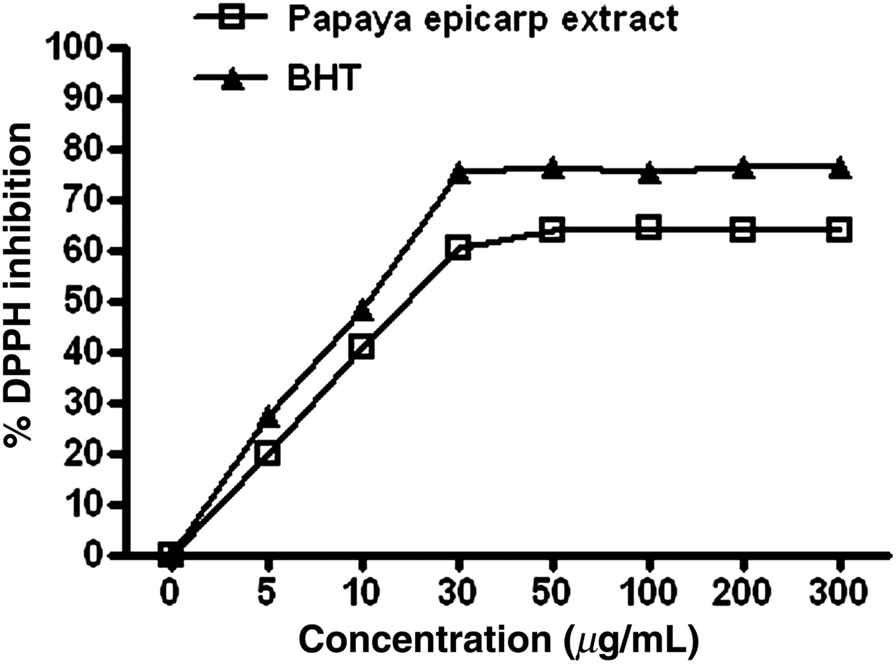
Scavenging effect of papaya epicarp extract and BHT against DPPH free radical formation. Papaya epicarp extract significantly scavenged DPPH free radicals in a dose-dependent manner. Both BHT and papaya epicarp extract inhibited the DPPH free-radical formation and reached a plateau at a concentration of 30 μg/mL. The dose-dependent effects of both BHT and papaya epicarp extract were similar with no statistical significant difference. Results are the means ± SD of six measurements. DPPH, 1,1-diphenyl-2-picrylhydrazyl
BHT is a synthetic additive that acts as an antioxidant and is used to preserve the food from oxidation and DPPH formation in a mechanism that is mainly attributed to its hydrogen-donating ability.
Both the papaya epicarp extract and Trolox standard (vitamin E analog) showed inhibition of ABTS radical formation in a dose-dependent manner. The IC50 (concentration of papaya epicarp extract or the Trolox standard in μg/mL required to scavenge 50% of ABTS+ formation) was comparable for papaya epicarp extract and Trolox standard (30 μg/mL) with no statistical significant difference (t = 0.866, P = 0.411) as shown in Figure 2.

Free radicals scavenging the ability of papaya epicarp extract and Trolox against ABTS+ radical formation. Both the papaya epicarp extract and Trolox standard (vitamin E analog) showed inhibition of ABTS radical formation in a dose-dependent manner. The IC50 was comparable for papaya epicarp extract and Trolox standard with no statistical significant difference. Results are the means ± SD of six measurements. ABTS, 2, 2′-azino-di-[3-ethylbenzthiazoline sulphonate (6)] diaammonium salt
In line with previous reports, our study suggests that papaya epicarp extract exhibits in vitro free-radical scavenging and antioxidant activities and we speculate that this may be related to its high total phenolic contents (396 ± 57 mg GAE/100 g dry skin powder) as measured in this study.
Effect of papaya epicarp extract and H2O2 on SH-SY5Y cells viability
In our study, we investigated the effect of H2O2 on SH-SY5Y cell viability in order to identify the concentration of H2O2 that would not be cytotoxic and would not induce cell death. Identification of such a dose was crucial for our study so that the observed H2O2-induced oxidative stress was not attributed to SH-SY5Y cell death.
SH-SY5Y cell viability decreased significantly after 60-min treatments with either 0.01% or 0.1% H2O2 as compared with controls (Figure 3, F = 5831, R 2 = 0.998, P < 0.001). However, treatment with 0.001% H2O2 did not significantly decrease SH-SY5Y cell viability. Therefore, we decided to adopt a concentration of 0.001% H2O2 for subsequent experiments that was intended to screen the papaya epicarp extract effect against H2O2-induced oxidative stress in SH-SY5Y cells.
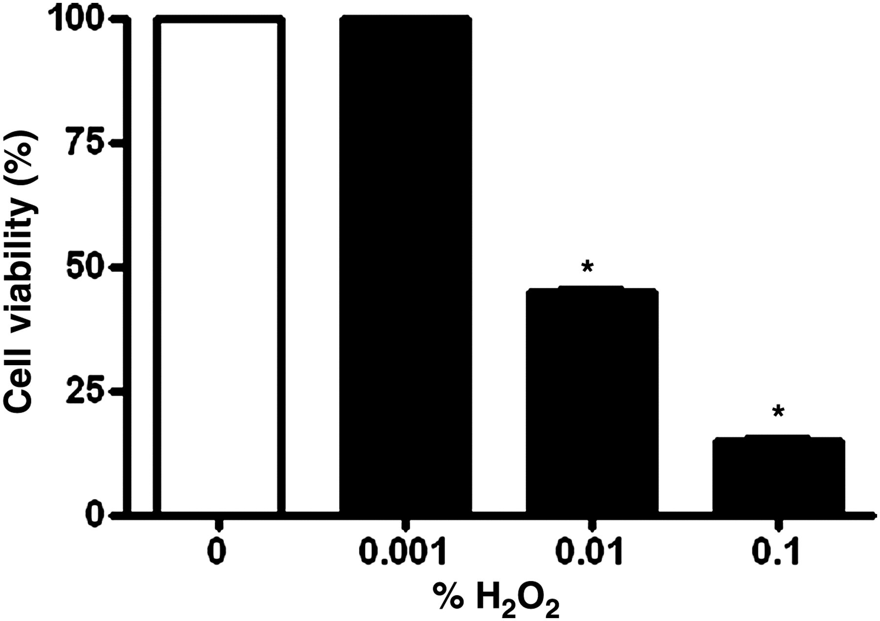
Effect of H2O2 on SH-SY5Y cell viability. Viability is expressed as a percent of control. Cells were treated for 60 min with 0.001%, 0.01% or 0.1% H2O2 and then subjected to a MTS assay. Treatment with either 0.01% or 0.1% H2O2 significantly decreased cell viability (*P < 0.001 as compared with control), whereas treatment with 0.001% did not affect cell viability. Results are the means ± SD of six measurements. MTS, (3-(4,5-dimethylthiazol-2-yl)-5-(3-carboxymethoxyphenyl)-2-(4-sulfopheny)-2H-tetrazolium)
H2O2 can induce SH-SY5Y cell death 28 and our study showed that the indicators of cell membrane damage (MDA level and % LDH leakage) were comparable in the control and 0.001% H2O2-treated cells (Figures 4 and 5).

SH-SY5Y neuronal cell membrane lipid peroxidation as expressed by MDA formation. The cells were incubated for 60 min with 30 μg/mL papaya epicarp extract in the presence of either 0.001% or 0.1% H2O2. MDA contents were measured in the control and treated cells as described in Materials and methods. *P < 0. 05 significantly higher as compared with control and other treated groups, ^P < 0.05 significantly lower as compared with 0.1% H2O2-treated groups. Based on one-way analysis of variance analysis followed by Tukey's test, F = 9.840, R 2 = 0.621. Results are the mean ± SD of six measurements. MDA, malondialdehyde

SH-SY5Y cells incubated with 0.1% H2O2 showed 90% increase in LDH leakage in the conditioned medium. Incubation of the cells with 30 μg/mL papaya epicarp showed significant reduction in the levels of % LDH leakage activity. *P < 0.05 significantly higher as compared with control and other treated groups, ^P < 0.05 significantly lower as compared with 0.1% H2O2-treated groups. Based on one-way analysis of variance analysis followed by Tukey's test, F = 450.6, R 2 = 0.984. Results are the means ± SD of six measurements. LDH, lactate dehydrogenase
H2O2 treatment at a dose of 0.1% induced a significant (P < 0.05) increase in both parameters, indicating SH-SY5Y cell death, and this effect was significantly (P < 0.05) reversed by concomitant incubation of SH-SY5Y cells with 30 μg/mL of papaya epicarp extract. We concluded that papaya epicarp extract intervention exhibited a protective effect against the 0.1% H2O2-induced cytotoxicity in SH-SY5Y cells.
Effect of papaya epicarp extract against H2O2-induced oxidative stress in SH-SY5Y cells
The measured oxidative stress indices included GSH and TAC markers as well as antioxidant enzymes (SOD, GPX and CAT) (Table 1). Acute incubation (60 min) of SH-SY5Y cells with 0.001% H2O2 significantly (P < 0.05) depleted GSH, decreased TAC and inhibited SOD, GPX and CAT enzymes. The observed effect is not attributed to SH-SY5Y cell death as evident by earlier cell viability assay. However, the observed 0.001% H2O2-induced oxidative stress was significantly (P < 0.05) ameliorated when the SH-SY5Y cells were incubated with 30 μg/mL papaya epicarp extract. It was observed that papaya epicarp extract alone did not significantly (P > 0.05) affect the measured oxidative stress indices GSH, TAC and antioxidant enzymes (SOD, GPX and CAT) as compared with the control group. Recently, it was reported that low activities of the antioxidant enzymes (CAT, GPX and SOD) indicate oxidative stress in SH-SY5Y cells, 29 and our results indicate that papaya epicarp extract restored the decreased CAT, GPX and SOD levels in SH-SY5Y cells that were treated with oxidative stress agent 0.001% H2O2. Further studies should be conducted to identify the presence of most active components in papaya epicarp extracts that may be responsible for the observed neuroprotective properties in human SH-SY5Y neuronal cells.
Protective effect of papaya epicarp extract against H2O2-induced inhibition of oxidative stress indices in human SH-SY5Y neuronal cells
GSH, glutathione; TAC, total antioxidant capacity; SOD, superoxide dismutase; GPX, glutathione peroxidase; CAT, catalase
SH-SY5Y neuronal cells were incubated with papaya epicarp extract (30 μg/mL) in the presence or absence of 0.001% H2O2 for 60 min. Values with a,b,c,d,e superscripts are significantly lower than the corresponding control group, based on Student's t-test, P < 0.05. Values not sharing a common superscript are not statistically significantly different, based on one-way analysis of variance, P > 0.05. Results are the means ± SD of six measurements
Conclusion
Papaya epicarp extracts augmented intracellular GSH and TAC levels in SH-SY5Y neuronal cells treated with H2O2 insults. Papaya epicarp extract can significantly ameliorate the oxidant inhibitory effect of H2O2 on the assayed antioxidant enzymes (SOD, CAT and GPX). Our study is the first report that specifically describes the amelioration of neurocytotoxicity of H2O2 by papaya epicarp extract. We hypothesize that papaya epicarp extract may act as a synergistic therapeutic dietary supplement in patients with neurological diseases related to oxidative stress. Further in vivo studies are required to explore the neuroprotective health aspects of papaya epicarp extract in animal models.
Footnotes
ACKNOWLEDGEMENTS
This study was supported by Sultan Qaboos University research funds, IG/AGR/FOOD/10/01 and IG/AGR/FOOD/10/02, and by the International Islamic University Malaysia Grant, IIUM/504/RES/G/14/3/RMGS/08-04.