
Abstract
Nanoscale carbon particles have emerged as versatile precursors for a new class of highly fluorescent nanomaterials that resemble semiconductor quantum dots. The surface-passivated fluorescent carbon nanoparticles, dubbed ‘carbon dots’, were already demonstrated for their potential optical bioimaging applications in vitro and in vivo. In this study, we conducted a systematic cytotoxicity evaluation on the carbon dots prepared by various combinations of precursor carbon nanoparticles and molecules for the particle surface functionalization. The results suggested that the cytotoxicity of carbon dots was dependent on the selection of surface passivation molecules. Those dots showing more significant cytotoxicity at higher concentrations were also evaluated for their effects on the fluorescence imaging of live cells. The implications of the results on the eventual use of carbon dots as cell imaging agents are discussed.
Introduction
Nanoscale carbon particles have emerged as versatile precursors for a new class of highly fluorescent nanomaterials 1–10 that resemble, at least phenomenologically, semiconductor quantum dots (QDs). 10 As originally reported by Sun and co-workers, 1,2 small carbon nanoparticles (less than 10 nm) could be treated in aqueous nitric acid to impart carboxylic acid moieties on the particle surface, followed by their functionalization with organic molecules to result in soluble particles of bright and colorful fluorescence emissions. These surface-passivated fluorescent carbon nanoparticles, dubbed ‘carbon dots’, have been demonstrated for their potential optical bioimaging applications in vitro and in vivo. 2,11,12 A variety of nanoscale carbon precursors and different surface passivation agents have been used for the synthesis of carbon dots or structurally and property-wise similar nanoscale configurations. 13 Mechanistically, the fluorescence emissions in carbon dots are attributed to radiative recombinations of the carbon particle surface-trapped electrons and holes, where the large surface (relative to the particle volume) and diverse surface energy trapping sites in the small carbon nanoparticles are stabilized by the surface passivation agents. According to more recent studies, carbon dots with fluorescence quantum yields more than 50% in the green could be prepared, with their optical performance competitive to that of the well-established semiconductor (CdSe/ZnS) QDs, 10 while for carbon dots emissive in the red, substantial improvements in fluorescence performance still present challenges.
An important advantage of carbon dots is that, unlike the semi-conductor QDs containing heavy metals such as cadmium, carbon is generally not considered as a toxic element. Carbon powders containing nanoparticles or their aggregates are presently used in many commercial products (automobile tires, for example). In the laboratory, carbon dots with oligomeric poly(ethylene glycol) diamine (PEG1500N) as the surface passivation agent in an aqueous solution were injected into mice for toxicity evaluation over a period of up to 28 d, and the results suggested no significant toxic effects in vivo. 14 There have also been several cytotoxicity studies on carbon dots of specific configurations. 14–17 For example, luminescent carbon nanoparticles synthesized by the electrochemical treatment of graphite were evaluated in terms of established cytotoxicity assay with a human kidney cell line, in which the cell viability was not negatively affected by the particles. 15 The PEG1500N-functionalized carbon dots were also found to be non-toxic to several human cancer cell lines, as measured by the cell proliferation, mortality and viability. 14
Carbon dots have been shown to internalize into various cells, amenable to being used as fluorescence probes in cellular imaging applications.
2,12,18
While carbon dots in terms of their nanoscale configuration are not intrinsically cytotoxic, nor are the precursor bare carbon nanoparticles,
14–17,19,20
and there are some indications on the surface passivation molecules being somewhat different in their cytotoxicity profiles when free in solution versus being attached to the carbon nanoparticle surface.
17
In the study reported here, we conducted more systematic cytotoxicity evaluations on the carbon dots prepared by various combinations of precursor carbon nanoparticles and molecules for the particle surface functionalization (Scheme 1). The results suggested that the cytotoxicity of carbon dots was dependent on the selection of surface passivation molecules. Those dots showing more significant cytotoxicity at higher concentrations were also evaluated for their effects on the fluorescence imaging of live cells.
Structure of carbon dots with various surface passivation agents used. PEG1500N, O,O′-bis(3-aminopropyl) polyethylene glycol; PPEI-EI, poly(propionylethyleneimine-co-ethyleneimine); PEI, polyethyleneimine; PAA, polyallyl amine (A color version of this scheme is available in the online journal)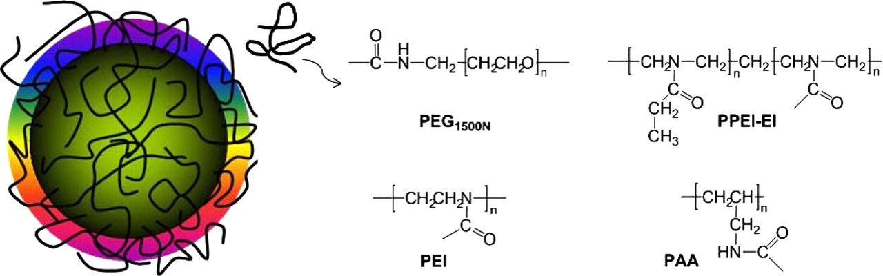
Materials and methods
Materials
Carbon nanopowders (produced by laser ablation), pyrrole and thionyl chloride were purchased from Sigma-Aldrich (Atlanta, GA, USA), O,O’-bis(3-aminopropyl) polyethylene glycol (PEG1500N; average molecular weight ∼1500) from Frontier Chemicals (Oak Creek, WI, USA), ferric chloride hexahydrate, decyltrimethylammonium bromide (DeTAB) and poly(propionylethyleneimine) (PPEI; average molecular weight ∼5000, partially hydrolyzed to yield poly (propionylethyleneimine-co-ethyleneimine [PPEI-EI]) from Alfa Aesar (Ward Hill, MA, USA), and polyethyleneimine (PEI; average molecular weight ∼1200) and polyallyl amine (PAA; average molecular weight ∼1000) from Polysciences Inc (Washington, PA, USA). Membrane tubing for dialysis was supplied by Spectrum Laboratories Inc (Rancho Dominguez, CA, USA). Water was deionized and purified by being passed through a Labconco Water Pros water purification system.
Measurement
A Baxter centrifuge (Megafuge model 2630; Baxter, Deerfield, IL, USA) and a Beckman-Coulter ultracentrifuge (Optima L90K with a type 90 titanium fixed-angle rotor; Beckman Coulter, Brea, CA, USA) were used for centrifugation at various g values. UV/vis absorption spectra were recorded on a Shimadzu UV-2501 spectrophotometer (Shimadzu North America, Durham, NC, USA). Fluorescence spectra were measured on a Jobin Yvon luminescence emission spectrometer (HORIBA Jobin Yvon Inc, Edison, NJ, USA) equipped with a 450 W xenon source, a Gemini-180 excitation monochromator and a Triax-550 emission monochromator, and a single-photon counting detector (Hamamatsu R928P PMT at 950 V; Hamamatsu Corporation, Bridgewater, NJ, USA). Atomic force microscopy (AFM) images were obtained in the acoustic AC mode on a Molecular Imaging PicoPlus AFM system equipped with a multipurpose scanner and a NanoWorld point probe NCH sensor (Agilent Technologies Inc, Santa Clara, CA, USA). The height profile analysis was assisted by using the SPIP software distributed by Image Metrology A/S (Horsholm, Denmark). Optical cell imaging was performed on a Leica laser scanning confocal fluorescence microscope (DM IRE2, with Leica TCS SP2 SE scanning system; Leica Microsystems Inc, Buffalo Grove, IL, USA) equipped with an argon ion laser (JDS Uniphase Corporation, Milpitas, CA, USA).
Precursor carbon nanoparticles
For the synthesis of polymeric nanoparticles, 21,22 an aqueous solution of the surfactant DeTAB (0.4 mol/L, 40 mL) was stirred at 3°C, and to the solution was added pyrrole (1 g) drop-wise to result in a microemulsion. An aqueous ferric chloride solution (6.9 mol/L, 5 mL) was added, and the mixture was stirred at 3°C for three hours. The polymerization reaction was quenched by excess methanol, followed by harvesting the polypyrrole nanoparticles via centrifugation. Upon drying in a vacuum oven at room temperature for 12 h, the polymeric nanoparticles in a crucible were carbonized in a furnace under inert atmosphere (heated to 800°C at a rate of 5°C/min and then held at 800°C for 5 h).
The commercially supplied carbon nanopowder sample and the sample obtained from carbonizing polypyrrole nanoparticles were both refluxed in aqueous nitric acid (2.6 mol/L) for 12 h, followed by dialysis (membrane pore size equivalent to molecular weight ∼1000) against fresh water, and then the removal of water to obtain the precursor carbon nanoparticle samples.
Carbon dots
The functionalization of carbon nanoparticles with PEG1500N for PEGylated carbon dots was reported previously, 1,14 and so was the gel-column fractionation of the as-synthesized sample to harvest the more fluorescent dots. 10 A similar procedure to the one for PPEI-EI-functionalized carbon dots 2,17 was used for the PEI- and PAA-functionalized carbon dots. Briefly, the precursor carbon nanoparticles from the carbonization experiment were refluxed in neat thionyl chloride for 12 h. Upon the removal of excess thionyl chloride, the sample (100 mg) was mixed well with carefully dried PEI (1 g) in a flask. The mixture was heated to 110°C, and the melt thus formed was vigorously stirred under nitrogen protection for 72 h. The reaction mixture was brought back to room temperature and dispersed in water, followed by high-speed centrifugation to retain the supernatant (a colored aqueous solution). It was dialyzed against fresh water to eliminate any residual impurities (such as reagents used in the synthesis, including thionyl chloride, for example), and then water was removed to obtain the carbon dots. The same procedure was applied to the synthesis of the PAA-functionalized carbon dots.
Cytotoxicity assays
The human breast cancer cell line MCF-7 (American Type Culture Collection [ATCC]; Manassas, VA, USA) and human colon adenocarcinoma grade II cell line HT-29 (ATCC) were grown at 37°C with 5% CO2 in Eagle's minimum essential medium (ATCC, with non-essential amino-acids, 1 mmol/L sodium pyruvate, 2 mmol/L
Cell imaging
HT-29 and MCF-7 cells at an initial density of 1 × 10⁴ per well were seeded in each well of a four-chambered Lab-Tek coverglass (Thermo Scientific, Rockford, IL, USA) and cultured at 37°C until reaching approximately 80% confluence. The separately prepared aqueous solution of carbon dots was filtered (0.2-μm Acrodisc syringe filter), and then diluted with fresh culture medium to the specific concentrations before being added to the glass slide chamber. Cells without exposing to carbon dots were taken as the control. Upon incubation for 24 h or another specific time period, the cells were washed three times with phosphate-buffered saline (PBS; 500 μL each time) and kept in PBS for fluorescence imaging (458 nm excitation and 470–820 nm emission collection). The images were processed and analyzed with the NIH ImageJ software (National Institutes of Health, Bethesda, MD, USA).
Results and discussion
The oligomeric PEG diamine, PEG1500N (Scheme 1), has been used as a surface passivation agent for carbon nanoparticles since the original finding of carbon dots; hence, the toxicity profile of the PEGylated carbon dots has received more attention. Free PEG1500N molecules are generally non-toxic to cells according to all available cytotoxicity evaluation results. In their corresponding carbon dots, different sources of precursor carbon nanoparticles have been used. For laser ablation-produced carbon nanoparticles (in house or acquired commercially),
14
the resulting carbon dots were found to be non-cytotoxic, as reported previously.
14
Subsequently, a gel-column fractionation method was developed to harvest more fluorescent carbon dots from the as-prepared mixture (Figure 1).
10
The much higher fluorescence quantum yields in the dots thus harvested were attributed structurally to improved passivation on the carbon particle surface by PEG1500N molecules.
10
Because of their bright fluorescence, which is competitive in performance to that observed in the well-established CdSe/ZnS QDs for the comparable spectral region,
10
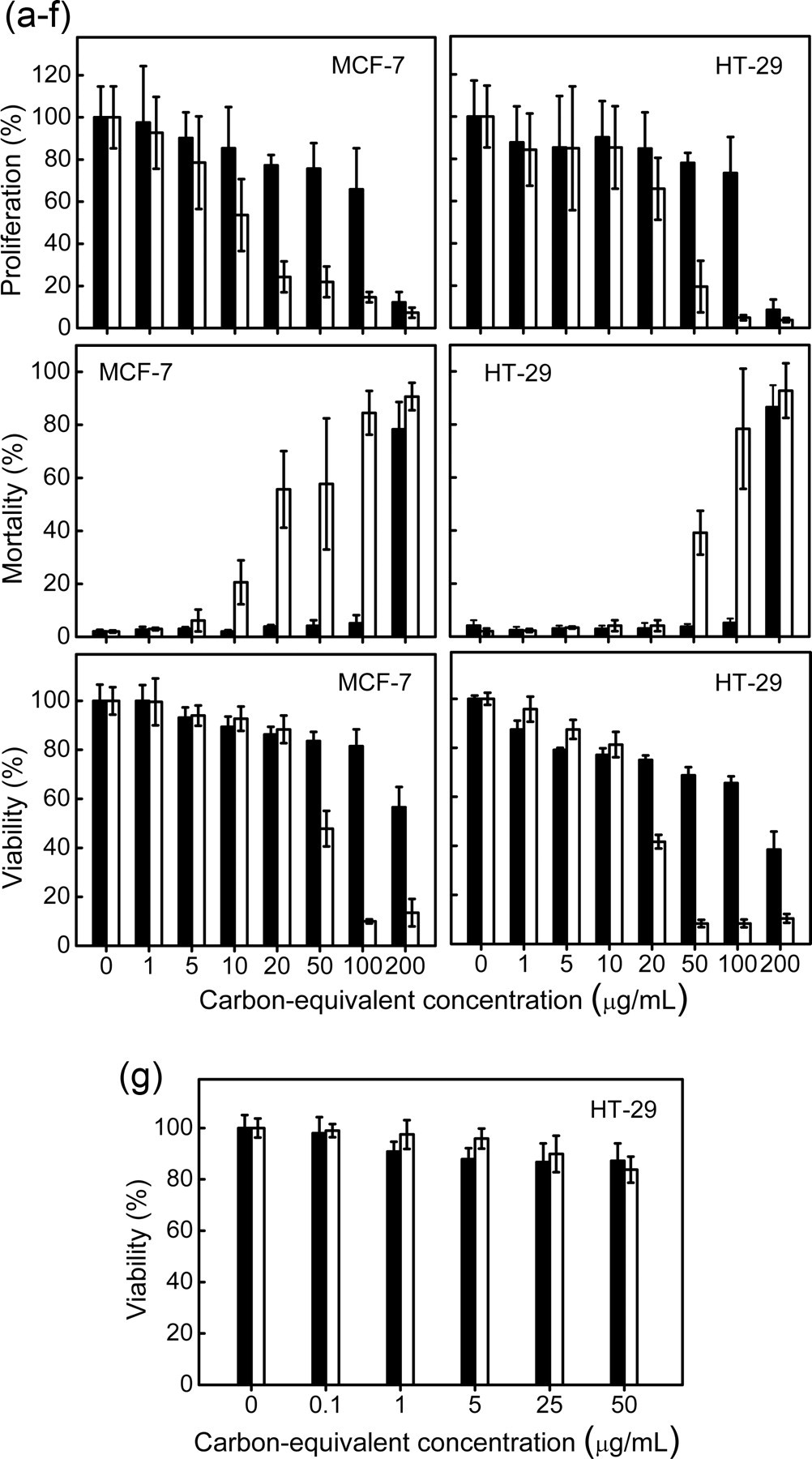
the fractionated carbon dots have been targeted for cell imaging applications, with an expectation that their cytotoxicity behavior should be similar to that of the as-prepared carbon dots sample. In this study, the expectation was confirmed experimentally, with results from the cytotoxicity assays on the fractionated highly fluorescent carbon dots (observed emission quantum yields all larger than 40%) exhibiting no significant cytotoxicity (Figure 2).
Absorption (ABS) and fluorescence (FLSC, 440 nm excitation) spectra of carbon dots. Upper: PEG1500N-functionalized dots with carbon nanoparticles from laser ablation (as-prepared: Results from cytotoxicity evaluations of PEG1500N-functionalized carbon dots (black) and free PEG1500N (white) with MCF-7 and HT-29 cell lines (concentrations of PEG1500N in terms of the carbon core-equivalent in the carbon dots). (a–f) With carbon nanoparticles from laser ablation. (g) With those from the carbonization of polypyrrole particles. Data presented as mean±SD (n = 4). PEG1500N, O,O′-bis(3-aminopropyl) polyethylene glycol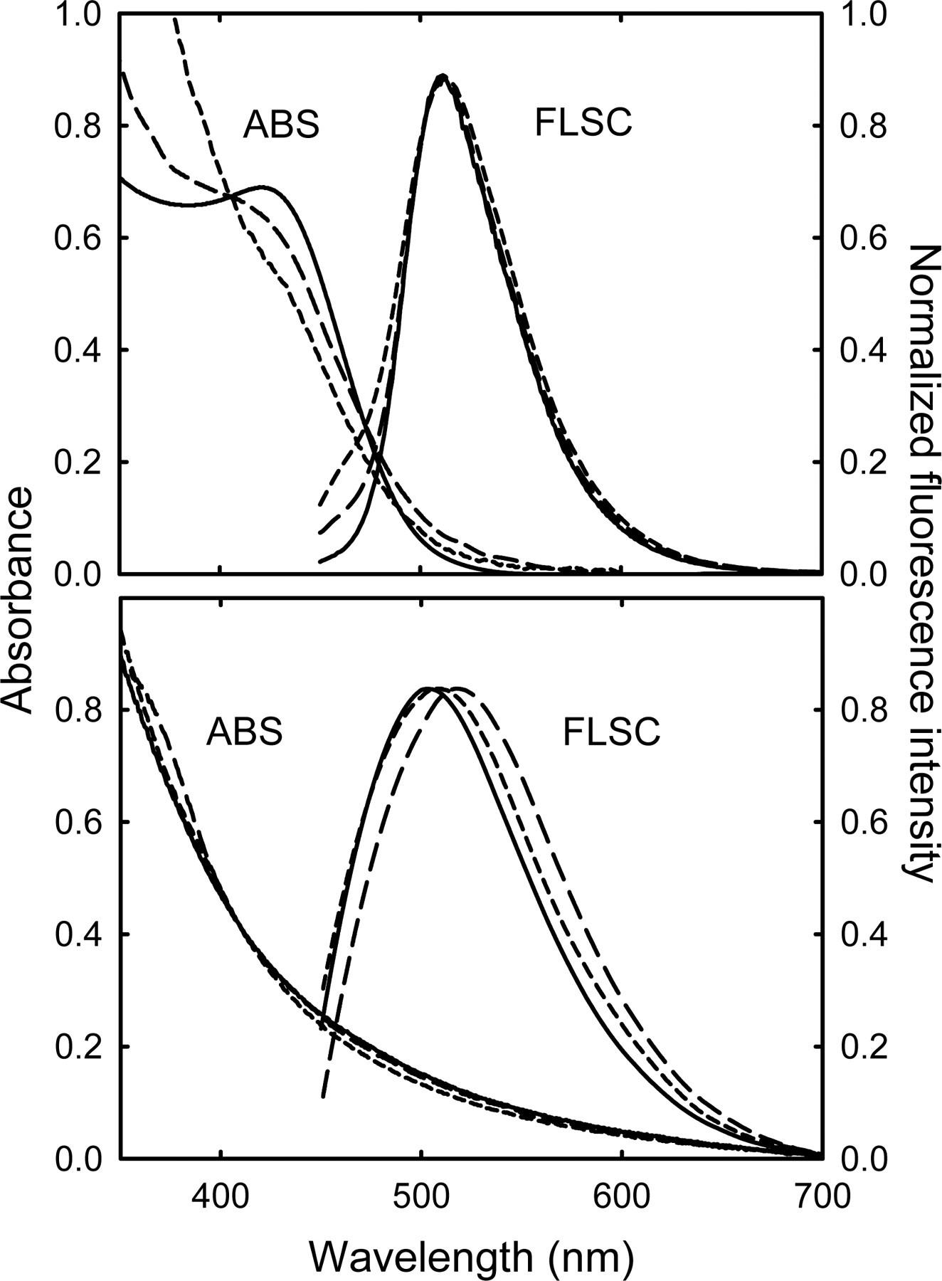
Precursor carbon nanoparticles for carbon dots were also produced by carbonizing polymeric nanoparticles, which were obtained from the emulsion polymerization of pyrrole.
21,22
The carbon nanoparticles thus produced were similarly processed with aqueous nitric acid, and then functionalized by PEG1500N for carbon dots (Figure 3). As compared in Figure 1, the absorption and fluorescence spectra of PEGylated carbon dots with different core carbon nanoparticles are similar. Since the synthesis of polymeric nanoparticles involved the use of metal catalysts and the subsequent carbonization might produce unwanted impurities, extra effort including dialysis with small pore-size membrane tubing was made to remove any residual catalysts and/or impurities from the as-prepared carbon dots. The resulting sample was evaluated in terms of the cell viability assay, from which the results again suggested no significant cytotoxicity in comparison with that of free PEG1500N (Figure 2).
A representative atomic force microscopy topography image of PEG1500N-functionalized carbon dots (carbon nanoparticles from the carbonization of polypyrrole particles) on mica, with the height profile analysis along the line. PEG1500N, O,O′-bis(3-aminopropyl) polyethylene glycol (A color version of this figure is available in the online journal)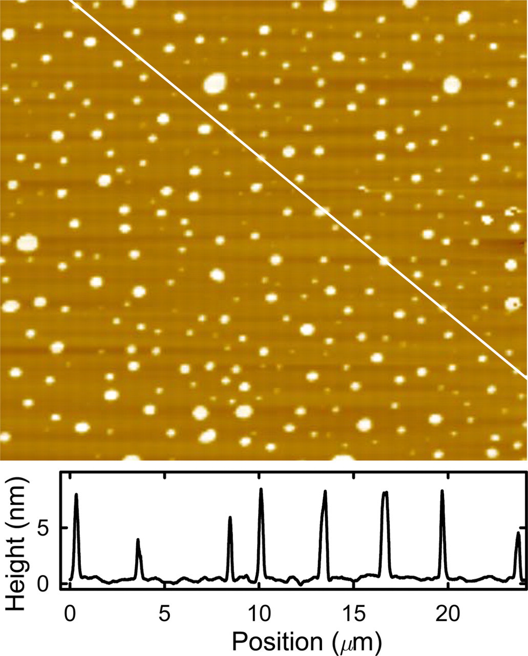
The PEGylated carbon dots in all available configurations are apparently non-cytotoxic up to concentrations much higher than what is necessary for optical cell imaging and related applications. The highly fluorescent dots harvested from gel-column fractionation, equally benign to cells, are particularly promising for potentially fluorescence imaging and/or labeling at a very high sensitivity.
Oligomeric aminopolymers (Scheme 1) represent another class of surface passivation agents for carbon nanoparticles in the preparation of carbon dots, with the fact that PPEI-EI (Scheme 1) was used in the original finding of carbon dots.
1,2
The PPEI-EI-functionalized carbon dots were also found to be readily internalized into cells.
2
As reported previously,
17
the cytotoxicity of PPEI-EI-functionalized carbon dots was meaningfully higher than that of free PPEI-EI beyond a threshold concentration, and the increased cytotoxicity was apparently associated with a larger ethyleneimine (EI) fraction in the PPEI-EI copolymer. In this study, PPEI-EI was used with precursor carbon nanoparticles from the route of carbonizing polymeric nanoparticles (Figure 1). The results from the cytotoxicity evaluation of the resulting carbon dots further confirmed that below a relatively high threshold of carbon core-equivalent PPEI-EI concentration, the carbon dots were mostly non-toxic to the cells (Figure 4).
Results from cytotoxicity evaluations of carbon dots (black) and corresponding surface functionalization agents (white) with MCF-7 and HT-29 cell lines (concentrations of the polymers in terms of their carbon core-equivalent in the carbon dots): PPEI-EI with EI mole fraction in the copolymer of ∼20% (upper), and PEI (lower). Data presented as mean±SD (n = 4). PEI, polyethyleneimine; PPEI-EI, poly (propionylethyleneimine-co-ethyleneimine); EI, ethyleneimine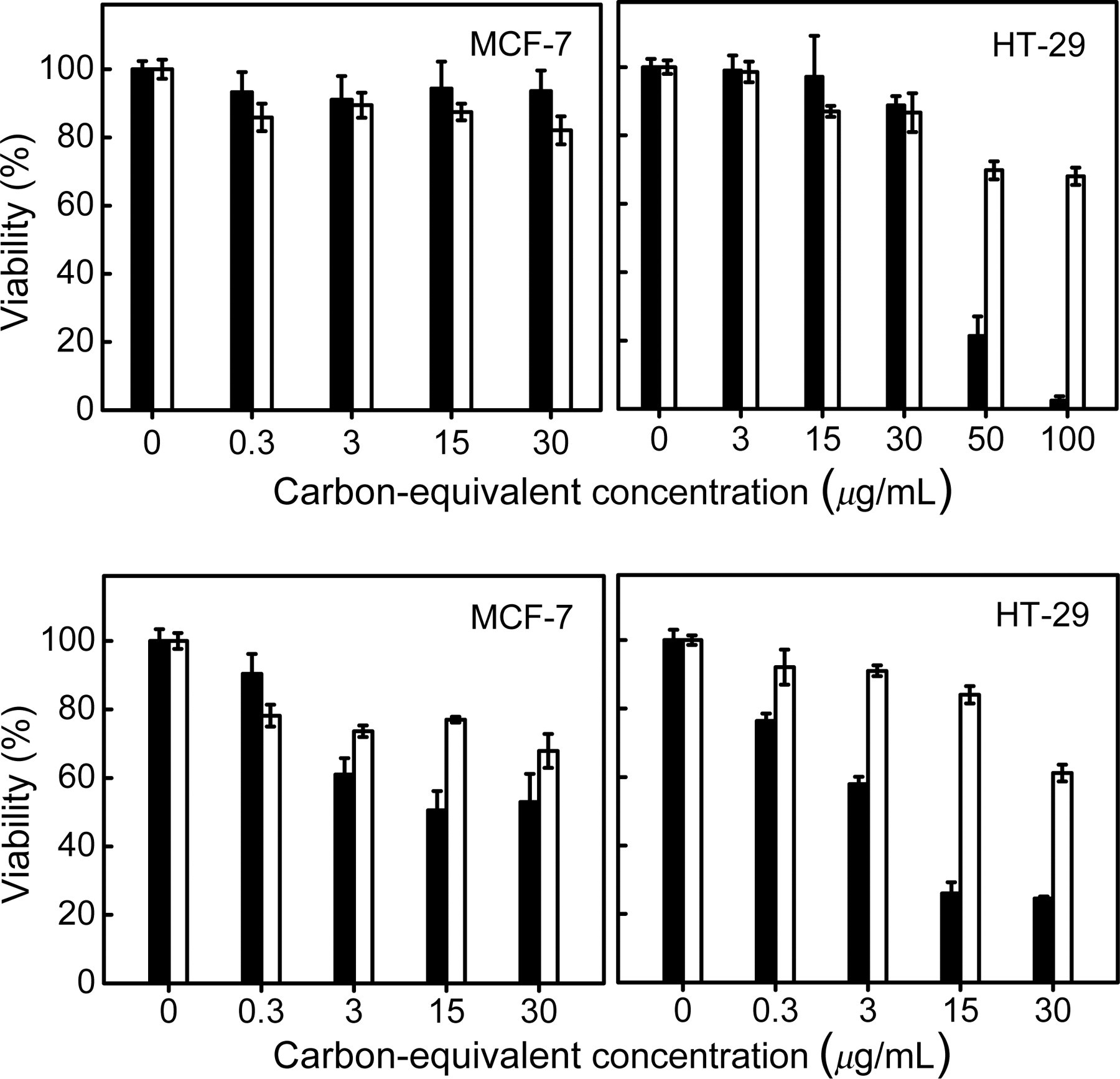
Equivalent to the extreme of high EI mole fraction in PPEI-EI copolymer is the oligomeric PEI homopolymer (EI only without PPEI units, Scheme 1). PEI has found many biomedical applications, for which there have been a number of toxicity evaluations of PEI based on different cell lines and assays. 23–26 According to the MTT assay (3-(4,5-dimethylthiazol-2-yl)-2,5-diphenyltetrazolium bromide reduced to purple formazan in living cells) in this study on free PEI in aqueous solution, the sample was apparently non-toxic to HT-29 cells even at relatively high concentrations (Figure 4), with the cytotoxicity profile compared favorably to those in the relevant literature reports. 23–26
The functionalization chemistry for the surface passivation of carbon nanoparticles with PEI was similar to that for PPEI-EI, except for a lower reaction temperature, (110°C) because at that temperature the oligomeric PEI is already a melt. The PEI-functionalized carbon dots (Figure 1) were also evaluated in terms of the MTT assay, and the results suggested their being more cytotoxic than PPEI-EI-functionalized carbon dots (Figure 4). The increased cytotoxicity is consistent with the trend observed in the study of various PPEI-EI-functionalized carbon dots, namely that more EI units are associated with lower concentration thresholds for the dots to become cytotoxic, 17 as PEI is the homopolymer corresponding to PPEI-EI at the extreme EI fraction of 100% (Scheme 1).
The EI units in PPEI-EI and PEI are secondary amine moieties (Scheme 1). A structurally analogous oligomeric aminopolymer bearing primary amines (PAA, Scheme 1) was used in the functionalization of carbon nanoparticles for carbon dots. The functionalization chemistry was rather similar to that for PEI discussed above, and so were the absorption and fluorescence properties of the resulting PAA-functionalized carbon dots (Figure 1). However, free PAA in aqueous solution was found to be harmful to cells even at relatively low concentrations (on the order of 50 μg/mL). As shown in Figure 5, the PAA-functionalized carbon dots were generally comparable to free PAA at the same carbon particle core-equivalent concentrations, both obviously more toxic to the cells with an exposure time of 24 h, but less so when the exposure time was shortened to four hours.
Results from cytotoxicity evaluations of PAA-functionalized carbon dots (black) and free PAA (white) with MCF-7 and HT-29 cell lines at different incubation times (left: 4 h, and right: 24 h). Data presented as mean±SD (n = 4). PAA, polyallyl amine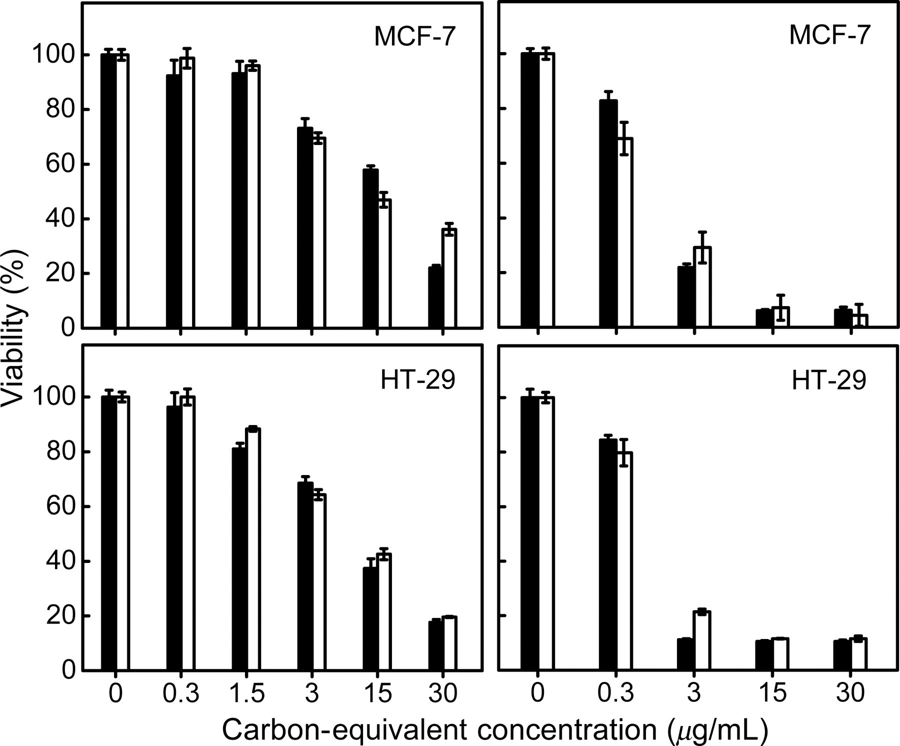
Overall, the results presented above suggest that the cytotoxicity of carbon dots is dependent on the selection of the surface passivation agent. The oligomeric aminopolymers are generally more cytotoxic than PEG1500N, and somewhat more so are their corresponding carbon dots. Except for PAA, however, the threshold concentrations for significant cytotoxicity are still relatively high, thus limiting any negative impact on the eventual use of carbon dots (such as for the optical imaging of live cells).
For potential cell imaging applications,
27
carbon dots of the various surface functional groups (hydrodynamic sizes significantly less than 50 nm even for those functionalized with the polymers) could be internalized into cells (HT-29 and MCF-7) for ready detection in confocal fluorescence microscopy. Since the PEG1500N-functionalized carbon dots are generally benign to cells up to relatively high concentrations, there should not be concerns in terms of cytotoxicity issues for their uses in live cell imaging. For carbon dots with PPEI-EI or especially PEI functionalization, it is probably necessary to keep concentrations of the dots below the threshold corresponding to significant cytotoxicity (Figure 4). As shown in Figure 6, the internalized PPEI-EI-functionalized carbon dots were very bright. Even at a rather low concentration (used for the incubation with cells), the PEI-functionalized carbon dot uptaken by the cells could still be readily detected under a confocal fluorescence microscope (Figure 6).
Results from confocal fluorescence imaging (458 nm excitation) of PPEI-EI-functionalized (upper) and PEI-functionalized (lower) carbon dots internalized in HT-29 cells (incubation for 24 h). PPEI-EI, poly (propionylethyleneimine-co-ethyleneimine); PEI, polyethyleneimine (A color version of this figure is available in the online journal)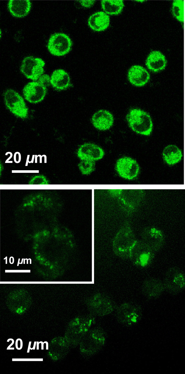
The PAA-functionalized carbon dots being more cytotoxic was manifested in the cell imaging results, with obvious cell damage by the dots at concentrations near the threshold for significant cytotoxicity (Figure 7). However, similarly guided by the cytotoxicity results (Figure 5), the use of lower concentrations for the same incubation time (24 h) or higher concentrations for a much shorter incubation time (1 h) caused no apparent cell damage, and the internalized carbon dots could still be readily detected in confocal fluorescence microscopy imaging (Figure 7). The results seem to suggest that the cellular uptake of PAA-functionalized carbon dots is rather efficient, requiring only a short incubation time.
Results from confocal fluorescence imaging (458 nm excitation) of PAA-functionalized carbon dots internalized in HT-29 cells, corresponding to different carbon core-equivalent concentrations of the dots and incubation times: 50 μg/mL and 24 h (upper), 10 μg/mL and 24 h (middle), and 50 μg/mL and 1 h (lower). PAA, polyallyl amine. (A color version of this figure is available in the online journal)
The results from this study provide some new insights into the cytotoxicity issues on carbon dots as related to their potential cell imaging applications. It remains true that carbon dots in terms of their nanoscale structures and configurations are not intrinsically cytotoxic, with any observed cytotoxicity due exclusively or primarily to the particle surface passivation molecules. For those molecules, their cytotoxicity is largely similar when free versus being functionalized on the particle surface. Therefore, the cytotoxicity consideration in the selection of surface passivation agents for carbon dots targeted for uses with live cells may be readily accomplished by looking at the cytotoxicity profiles of the free agents. According to the results presented above, surface passivation molecules of no or low cytotoxicity even at high concentrations are available for carbon dots of similarly no or low cytotoxicity, suitable for uses in live cell imaging. Even for those molecules that are more cytotoxic, their corresponding carbon dots may still be used with live cells if their concentrations are kept low enough and/or the incubation time is short enough. Therefore, it seems reasonable to conclude that carbon dots are promising fluorescence agents for optical imaging of live cells.
Footnotes
ACKNOWLEDGEMENTS
This work was initially supported by National Institutes of Health and then by National Science Foundation. YW was on leave from the Institute of Nanochemistry and Nanobiology at Shanghai University in China. LC was supported by a Susan G. Komen for the Cure Postdoctoral Fellowship. J-HL was a visiting student from Peking University (the group of Professor Haifang Wang and Professor Yuanfang Liu) in China. PW was affiliated with the Palmetto Academy, an education-training program managed by South Carolina Space Grant Consortium.