
Abstract
Our recent study showed that intravenously administered B-type natriuretic peptide (BNP) decreases gastric emptying and intestinal absorption in mice. We aimed to test whether acute myocardial injury and heart failure have similar effects. Wild-type (WT) and natriuretic peptide receptor type A (NPR-A) knockout (KO) mice underwent cryoinfarction (myocardial infarction [MI]) of the left ventricle (LV) versus sham. LV dysfunction was confirmed by echocardiography. Percent gastric emptying and intestinal absorption were measured and analyzed one and two weeks after infarction, by gavage feeding the mice with fluorescein-isothiocyanate-dextran. Ejection fraction was 48 ± 3% versus 64 ± 2% (P < 0.05) and fractional shortening was 24 ± 2% versus 35 ± 2% (P < 0.01), MI versus sham, respectively. BNP levels (pg/mL) were 4292 ± 276 one week after MI versus 105 ± 11 in sham (n = 5, P < 0.05) and 1964 ± 755 two weeks after MI (n = 5, P < 0.05). Gastric emptying was significantly decreased, 68 ± 6% in MI versus 89 ± 3% in sham (n = 5, P < 0.05) one week after MI and 82 ± 0.5% versus 98 ± 0.4%, MI versus sham (n = 5, P < 0.05), two weeks post-MI. Absorption, measured in relative plasma fluorescence units in WT mice, was 350 ± 79 in MI versus 632 ± 121 in sham (n = 6, P < 0.05). KO mice did not show a significant difference in emptying or absorption compared with sham. These findings suggest that MI and LV dysfunction decrease gastric emptying and absorption in mice through a mechanism that involves NPR-A.
Introduction
Heart failure and acute coronary syndrome are frequently associated with symptoms of impaired gastrointestinal (GI) function such as dyspepsia, indigestion, nausea and vomiting in humans. 1,2 Prior studies that have searched for a physiological explanation of this observation have described mechanisms including passive venous congestion in the GI tract and possible chemoreceptive reflex mediated by the central nervous system. 3–5 However, the discovery and functional characterization of the cardiac hormones over the past two decades has led to the recognition of a direct humoral link between the heart and other organ systems. 6–8 For instance, one such interorgan communication between the heart and the kidney prominently features the release of cardiac hormones atrial natriuretic peptide (ANP) and B-type natriuretic peptide (BNP) as conveyors of a humoral message from the distressed heart to the kidney whereby diuresis and natriuresis by the kidney maintains volume homeostasis. 9,10 Natriuretic peptides and their receptors (the plasma membrane guanylyl cyclase enzymes) also are expressed in various regions of the stomach and intestine 11–15 where there is strong evidence that they are involved in paracrine regulation of secretory functions in the GI tract. 16–22 Moreover, recent studies have confirmed that at least one of the natriuretic peptides, ANP, is secreted from the enterochromaffin cells of the gastric mucosa into the local micro capillaries. 20 Prior studies have shown that natriuretic peptides relax gastric and intestinal smooth muscle cells in vitro 23,24 and also have an effect on absorption of water and electrolytes across various regions of the intestinal epithelium. 25–27 A study from our laboratory also showed that BNP has a functionally significant effect on gastric emptying and intestinal absorption in mice. 28 In our recent study, BNP versus vehicle was administered intravenously to anesthetized mice, following which gastric emptying and intestinal absorption were measured and compared between wild-type (WT) and natriuretic peptide receptor type A (NPR-A) knockout (KO) mice. Significant reduction in both gastric emptying and intestinal absorption were shown in BNP-treated WT mice compared with both vehicle-treated WT and NPR-A KO mice. 28 This finding led to our current premise that the expression of natriuretic peptides and their receptors in the GI tract indicates that the cardiac hormones convey a message from the heart to the GI tract. As such, BNP and the other members of the natriuretic peptide family could be part of a network of ‘communication’ between the heart and the GI tract as has been shown for the heart and kidney. We postulate that the ultimate objective of such interorgan communication could be modulation of volume status through the effect of natriuretic peptides on water and electrolyte absorption from the GI tract. Since conditions that are associated with elevated plasma BNP, heart failure and acute coronary syndromes are also accompanied by signs and symptoms of perturbed GI function, we found it of interest to test whether acute myocardial injury and the consequent heart failure would produce measurable physiological or pathophysiological changes on gastric emptying and intestinal absorption.
The objective of this study was to examine the effect of myocardial injury and left ventricular dysfunction on gastric emptying and absorption. We used a whole animal mouse model. Based on previous findings, we hypothesized that elevation of plasma BNP following acute myocardial injury and heart failure would decrease gastric emptying by acting through the NPR-A and lead to decreased contractility of gastric and intestinal smooth muscle cells. GI smooth muscle relaxation in turn would lead to decreased gastric emptying and also impair downstream absorptive function. NPR-A KO mice were used to rule out a non-specific response.
Materials and methods
Experimental animals
NPR-A KO mice were obtained from our resident colony that was founded with pathogen-free breeding pairs and were genetically monitored by polymerase chain reaction of tail-snip DNA. The generation of NPR-A KO mice has previously been described in detail. 27 WT (C57BL/6) mice were purchased from commercial sources.
Induction of myocardial infarction by cryoinjury method
WT and NPR-A KO mice were anesthetized with 2% isoflurane-oxygen flow at 500 mL/min. A 2-mm incision was made through the skin, between the fourth and fifth intercostal space. Blunt dissection through all thoracic musculature, using a fine-tipped dissecting forceps, was performed to accommodate passage of a probe induction catheter, so that only the tip of the cooled probe would be in contact with the mouse's heart. The distal tapered end tip of the catheter was then manually passed through the muscle wall with a cap in place. Prior to removing the cap, the operator pinched the proximal tip of the catheter shut (to prevent pneumothorax). A bronze wire cooled in liquid nitrogen was then quickly threaded through the catheter to bring the cooled probe in direct contact with the left ventricle (LV) of the heart. Correct placement of the probe, with subsequent cardiac thermal injury, was confirmed as the super-cooled probe ‘grabbed’ the warmer tissue of the heart, and remained affixed until the probe had warmed enough for release. After passive release, the probe and catheter were quickly withdrawn and the ribs were approximated and kept in position with tissue glue (VetBond™; 3M Corporation, St Paul, MN, USA) delivered from a preloaded 1/2 cc syringe. The gastric emptying and intestinal absorption experiments were performed at one and two weeks after this surgical procedure. At the end of the experiments, microscopic (histological) confirmation of the infarctions (or no infarction) was made in all experimental and sham animals.
Measurement of gastric emptying
At one and two weeks after the cryoinjury procedure, conscious WT and NPR-A KO mice (n = 5 in each group) were gavaged with 0.1 mL of 0.5 millimole (mmol) 70-kDa fluorescein-isothiocyanate (FITC)-dextran. The 70-kDa FITC-dextran is known to be non-diffusible across the intestinal membrane, thus suitable for measurement of emptying. 29 Thirty minutes after the gavage meal, the mice were euthanized and the stomach was separated from the intestine, each flushed with 3 mL of phosphate-buffered saline and centrifuged for 10 min. Fluorescence of the supernatant fluid was measured and the percent gastric emptying was quantified by subtracting the dextran remaining in the stomach from the total dextran (in the stomach and small intestine) and dividing this value by the total dextran. The percent gastric emptying was compared in myocardial infarction (MI) versus sham mice for both WT and NPR-A KO mice. This method of evaluating gastric emptying has been previously established. 28,30
Measurement of absorption
Conscious WT and NPR-A KO mice (n = 5 in each group) were gavaged with 0.01 mL/g of a solution containing 22 mg/mL of 4-kDa FITC-dextran. One hour after gavage, blood was collected via cardiac puncture under pentobarbital anesthesia. Fluorescence was quantified using relative fluorescence units (RFUs) in the plasma. The plasma fluorescence measured one hour after gavage in WT and NPR-A KO mice was compared for MI versus sham. The group differences were analyzed using a t-test with P < 0.05 considered the significant level for statistical difference. This method of evaluating gastric absorption has been previously established. 28,30 We also compared the fluorescence of a 50-μL plasma sample taken 1 h after intravenous administration of 100 μL of 0.5 mmol 4-kDa FITC-dextran in BNP-treated versus vehicle WT mice. The purpose of this test was to control for the possibility that the differences in plasma fluorescence were produced by other actions of BNP such as increased excretion, redistribution or metabolism of the dextran, as this effect of BNP has been previously reported. 29
Measurement of fluorescence
The concentration of fluorescein was determined using a fluorimeter (FLUOstar Galaxy, BMG Lab Technologies, Ortenberg, Germany) with an excitation wavelength at 485 nm and an emission wavelength of 520 nm using serially diluted samples of the marker as standard.
Plasma BNP measurement by radioimmunoassay
BNP levels were measured on 0.2 mL of extracted plasma samples by radioimmunoassay (Phoenix Pharmaceuticals, Burlingame, CA, USA) using Kit RK-011-17 with an antibody designed to measure rat BNP-45. The antibody has a cross-reactivity of 41% with mouse BNP. Values presented were corrected for extraction ratio (78%) and cross-reactivity.
Echocardiography
Measurements were taken two weeks after infarction. The animals were anesthetized using isoflurane inhalation (3–4% for induction and 1–2% for maintenance) and the mice placed on a warming pad under a heat lamp. The fur overlying the acoustic window was wet with 70% alcohol and acoustic gel was generously applied. After the position of beating heart was identified, the sample volume of the pulsed Doppler was placed over the entire heart to obtain 2-D views and blood velocities and evaluated from different angles of insonation to obtain the maximal flow velocities. Each animal was interrogated by the same technician. The echocardiograms were performed at our core facility using the Visual Sonics Vevo 770 system (Visual Sonics, Toronto, Canada) equipped with a 35-MHz transducer. Measurements of interventricular septum, left ventricular posterior wall thickness and the diameter of the LV at the end-diastole and end-systole were obtained. Images were digitally recorded and analyzed using the Visual Sonic software Vevo 770 (Version 2.30).
Statistics
Data are presented as means±SEM. Statistical significance was determined by Student's t-test (two-tailed) for two groups or one-way analysis of variance (ANOVA) for multiple groups with Fisher's least significant difference (LSD) test as a post hoc test; a P value of P < 0.05 was considered statistically significant.
Results
Cryoinjury model of myocardial infarction
The cryoinjury model used in this study produced histological changes that were consistent with MI. Histological sections from each mouse were microscopically analyzed and showed similar changes; a representative sample is shown in Figure 1.
Representative histological appearance of the myocardium 14 days after cryoinfarction. Stain: hematoxylin & eosin, (a) ×4; (b) ×20 of the boxed area of infarct. LV, left ventricular. (A color version of this figure is available in the online journal)
Echocardiogram measurements confirmed significantly reduced LV function
Echocardiography results are shown in Figure 2. Measurements of LV function were significantly reduced in the MI group compared with sham. LV ejection fraction (%) was 48 ± 3 versus 64 ± 2 (P = 0.001), fractional shortening (%) was 24 ± 2 versus 35 ± 2 (P = 0.001), LV systolic volume (μL) was 51 ± 7 versus 26 ± 3 (P = 0.005), LV diastolic volume (μL) was 96 ± 8 versus 70 ± 6 (P = 0.023), LV posterior wall thickness systolic (mm) was 0.76 ± 0.10 versus 1.15 ± 0.10 (P = 0.014), LV posterior wall thickness diastolic (mm) was 0.60 ± 0.05 versus 0.80 ± 0.04 (P = 0.007), interventricular septum thickness (mm) was 0.69 ± 0.08 versus 1.12 ± 0.08 (P = 0.0019) and LV internal diameter systolic (mm) was 3.44 ± 0.20 versus 2.61 ± 0.13 (P = 0.003), MI versus sham, respectively.
Echocardiography measurements of ejection fraction, fractional shortening, left ventricular (LV) volume (systolic) and left ventricular posterior wall (LVPW) dimension confirm significant reduction of ventricular function in infarcted mice compared with sham surgery mice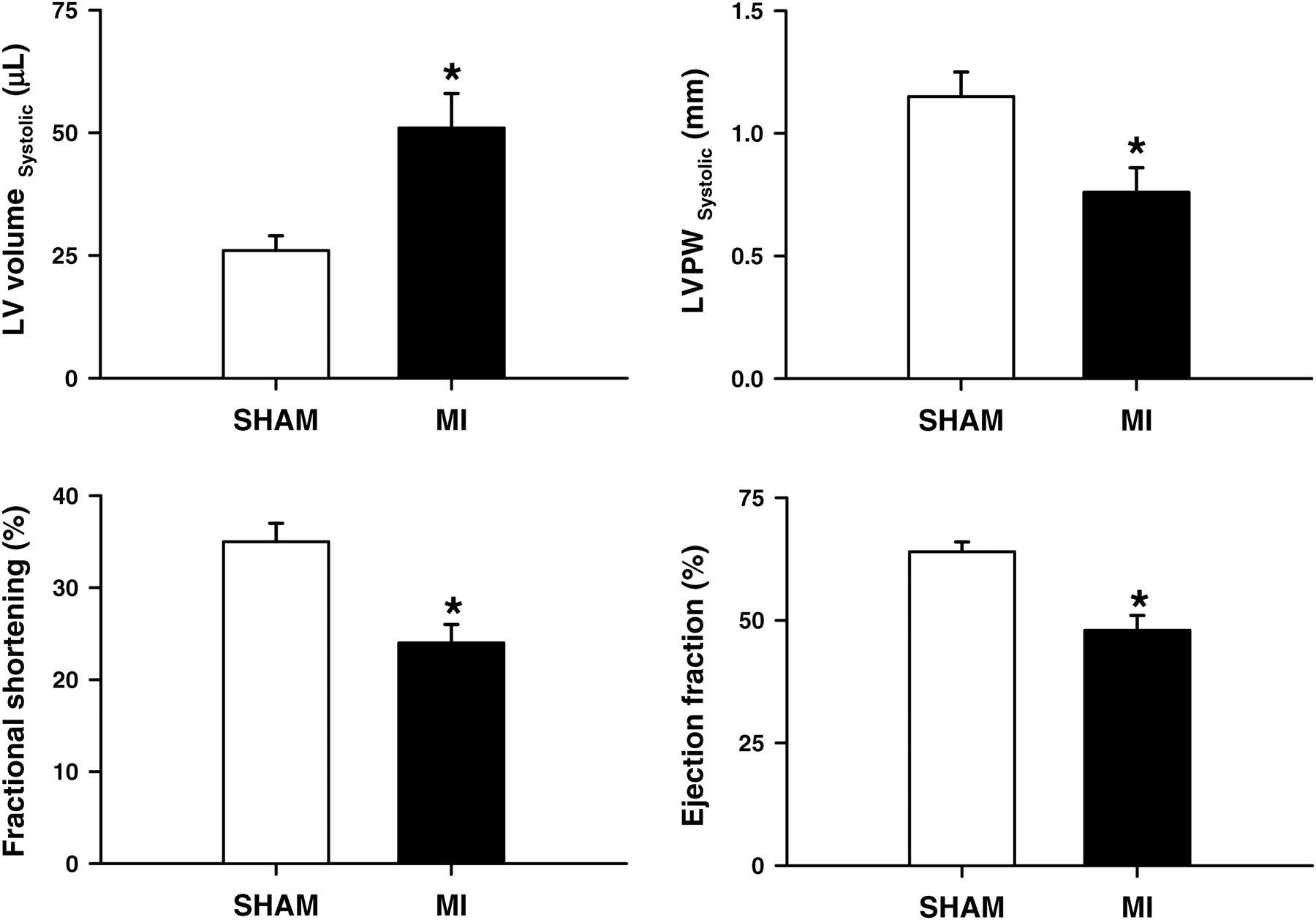
Plasma BNP levels
BNP levels were 4292 ± 277 pg/mL one week after MI versus 105 ± 11 pg/mL in sham mice (n = 5, P < 0.05, ANOVA). BNP levels were 1965 ± 755 pg/mL two weeks after MI (n = 5, P < 0.05, one-way ANOVA, Fisher's LSD test compared with one week post-MI, as shown in Figure 3).
Plasma BNP levels in sham mice, one week postinfarction and two weeks postinfarction (*P < 0.01, sham compared with MI one and two weeks postinfarction). BNP, B-type natriuretic peptide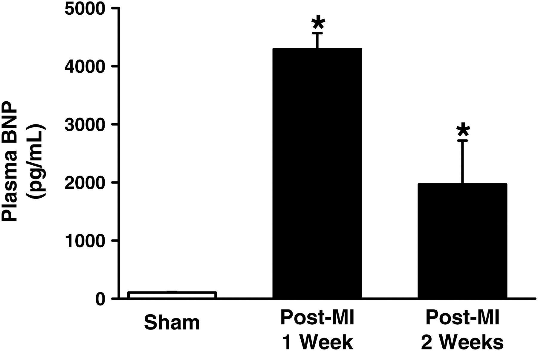
Decreased gastric emptying and intestinal absorption
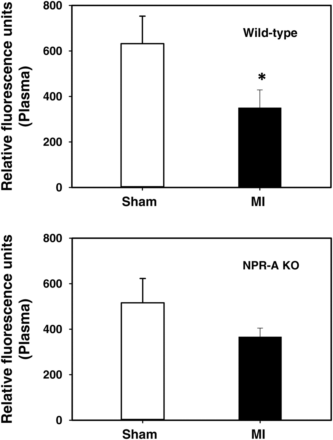
In the WT mice, gastric emptying was measured one and two weeks after MI. One week after MI, gastric emptying was significantly decreased in the MI group compared with the sham mice; percent gastric emptying was 68 ± 6% for the MI group versus 89 ± 3% for the sham group (P < 0.05, t-test). Two weeks after infarction, percent gastric emptying was also significantly lower in the MI group as compared with the sham group, 82 ± 1% for MI versus 98 ± 0.4% for sham (n = 5, P < 0.05) (shown in Figure 4). In NPR-A KO mice, the percent gastric emptying was identical between the MI versus the sham group, 85 ± 1% for mice with MI versus 88 ± 1% for sham (n = 6, P > 0.05) (Figure 4). There was also a statistically significant difference in the degree of absorption measured in RFUs in the plasma following a gavage meal of a 22 mg/mL 4-kDa FITC dextran solution given at 22 mL/kg body weight. In WT mice, absorption was 350 ± 79 (RFU) for the MI group versus 632 ± 121 (RFU) for the sham group; n = 6, P < 0.05. In the NPR-A KO group, absorption was decreased for the MI group compared with sham even though it was not statistically significant (Figure 5).
Gastric emptying, measured in the amount of fluorescence that emptied the stomach as a percentage of the total fluorescence measured in the entire gastrointestinal tract, 30 min after gavage feeding of 70-kDa fluorescein-isothiocyanate dextran (n = 5, *P < 0.05). Measurements were taken two weeks after myocardial infarction Degree of absorption of 4-kDa fluorescein-isothiocyanate (FITC)-dextran (measured in relative plasma fluorescence units) one hour after gavage feeding of 4-kDa FITC-dextran. n = 6 for each group. * P < 0.05 for WT but not significant in KO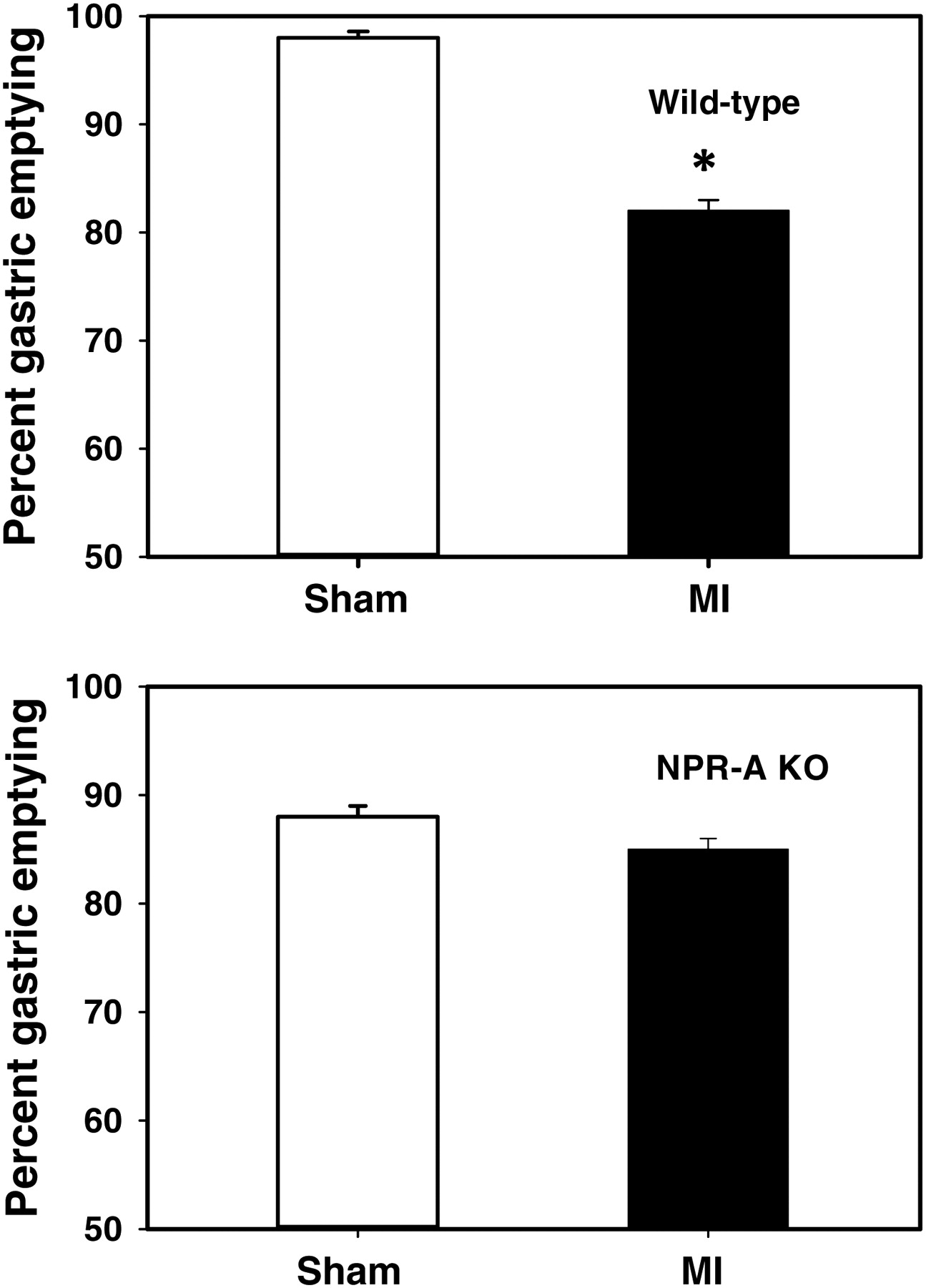
Discussion
The results clearly demonstrate that mice with myocardial injury and heart failure exhibit marked increases in plasma BNP, reduced gastric emptying and intestinal absorption compared with the sham-operated mice. Since these differences were absent in NPR-A KO mice, the results indicate that natriuretic peptides (specifically BNP and/or ANP) released during MI and heart failure have an effect on GI function through a mechanism that involves NPR-A. As shown in Figure 1, the surgical model we used produced histological changes consistent with MI. Two weeks following MI by the cryoinjury method, left ventricular volume was increased, left ventricular wall thickness was decreased and ejection fraction and fractional shortening was decreased (Figure 2). These echocardiographic measurements confirmed heart failure in the infarct group as compared with the sham group (Figure 2). We also measured BNP levels by radioimmunoassay and established that the levels were significantly elevated in the MI group compared with the sham group (Figure 3) at one and two weeks postinfarction. The highest levels of plasma BNP observed (Figure 3) were at one week post-MI but levels remained markedly elevated at two weeks compared with the sham animals. The high levels of BNP observed at one week could be at least partially due to acute myocardial injury; however, the fact that the levels remained elevated at two weeks in the MI group is most likely due to heart failure, which was also confirmed by echocardiography. Gastric emptying (Figure 4) and intestinal absorption (Figure 5) showed striking decreases in the MI mice and both these effects were attenuated or absent in the NPR-A KO mice (Figures 4 and 5). These results support the hypothesis that natriuretic peptides contribute to impaired gastric and intestinal function in this myocardial injury and heart failure model. Our findings show that gastric emptying was significantly reduced in WT mice one week and two weeks postmyocardial injury compared with sham-operated mice. Since we also compared the mean plasma BNP levels between the groups using receptor KO mice as additional experimental controls, the findings suggest that the reductions in gastric emptying corresponded to the degree of plasma BNP elevation and that these effects are mediated by NPR-A. It is interesting to note that even the sham mice had a slightly lower degree of gastric emptying at one week compared with baseline gastric emptying values we previously established. 28 Since the mean BNP level we found for sham-operated WT mice (105.4 ± 11.3 pg/mL) is higher than the undetectable level we previously found in WT mice (WT mice without any surgery), this finding further supports our hypothesis that BNP levels corresponded with the degree of reduction in gastric emptying. As shown in Figure 4, at two weeks' postinfarction, the sham-operated mice had a percent gastric emptying statistically identical to controls whereas infarcted mice continue to show a markedly lower degree of gastric emptying, which again indicates that the BNP levels significantly correspond to the degree of gastric emptying, adding evidence that BNP may be the major (or one of the major) reasons for the observed differences. Our findings validate that the effect of MI and heart failure on gastric emptying and absorption are specific and probably mediated by the effect of BNP on gastric and intestinal smooth muscle cells. This premise is supported by previous studies that showed that not only are natriuretic peptides, including ANP and BNP, expressed in the GI tract, but they also reduce smooth muscle contractility in the GI tract.
The detection of ANP and BNP in the GI tract of the rat has been reported using various techniques starting soon after the discovery of the natriuretic peptides in the 1980s. The expression of the ANP gene in the rat GI tract was confirmed soon after, showing that not only are natriuretic peptides detectable in the GI tract, but they are also regionally expressed and regulated. 13–15 The presence of ANP in the human GI tract was also later demonstrated in endoscopically obtained human mucosal tissues from the stomach, duodenum, jejunum, colon and rectum. 16 The NPRs (membrane-bound guanylyl cyclase enzymes) were also shown to be expressed in the various regions of the GI tract, 13,14 suggesting a functional significance for these peptides in the GI tract. More recent studies have shown that natriuretic peptides relax both animal and human GI smooth muscle cells in vitro. 22,23 Moreover, ANP and BNP were shown to have an effect on intestinal transit and epithelial absorptive and secretory functions in vitro in various animal models. 25–27 Our recent study confirmed these findings in conscious mice where we showed that intravenously administered BNP resulted in a significant reduction of gastric emptying and intestinal absorption in a dose-dependent manner. 28
Heart failure is accompanied by physiological and pathophysiological changes in several organ systems and it is a common clinical experience that symptoms of perturbed GI function ranging from dyspepsia to malabsorption are frequently encountered in patients with acute MI and heart failure. 31–35 However, there has so far been a very limited number of studies that investigated the underlying mechanisms for this age-old observation. Interestingly, the few previous studies that investigated this relationship between heart failure, gastric emptying and absorption have shown that these effects appeared to be mediated by cyclic guanylyl monophospahate-coupled pathways, 36,37 a pathway through which the ANP and BNP effect are mediated. As shown in Figures 4 and 5, NPR-A KO mice with MI did not show significant difference in gastric emptying compared with sham mice in our study. This indicates that the observed difference in gastric emptying between the WT and KO mice is due to the difference in NPR status and thus further supports that this effect is mediated by the NPR-A receptor. Our absorption data for WT mice is consistent with our central hypothesis and infarcted mice did show a significantly lower degree of absorption as compared with sham. The data in the KO mice are less conclusive, since the KO mice with MI did show a reduced absorption compared with the sham mice. This finding would not be totally unexpected, however, since myocardial injury is known to activate a series of systems other than the natriuretic peptides such as the renin−angiotensin−aldosterone system 38–40 as well as the release of inflammatory cytokines such as tissue growth factor β and tumor necrosis factor released from injured myocardium 41 that in themselves could impair intestinal absorption. 8,35,38 Therefore, the observed slight reduction in emptying and modest reduction in absorption (even though not statistically significant) in the NPR-A KO mice could be a manifestation of activation of systems other than natriuretic peptides that would be expected to remain intact in the NPR-A KO mice.
Together with our recently published study which showed that intravenously administered BNP significantly reduced gastric emptying and intestinal absorption in conscious mice, the confirmation of these findings in an acute MI/heart failure model provides a strong evidence that plasma BNP elevation during acute MI and/or heart failure is at least partly responsible for the observed GI effects. This finding strengthens the case for a direct humoral link between the heart and the GI tract and possibly opens up new avenues for heart failure research. Maintaining volume homeostasis is a major challenge in heart failure patients and we believe that further investigation in this area could lead to the identification of potential therapeutic targets for the treatment of heart failure.
Footnotes
ACKNOWLEDGEMENTS
This study was supported by a Veterans Administration Merit award to WRG and a National Science Foundation grant DGE # 0221681 to AA.